Anatomical Landmarks Edentulous Maxillary Arch Introduction
Maxillary landmarks are divided into three categories as supporting areas (stress bearing area), valve forming areas (limiting areas), and relief areas. All landmarks are discussed in this chapter with their anatomical position, identication, and clinical signicance. Each landmark is shown on dental cast as well as in oral cavity for easy identication.
Table of Contents
Anatomical Landmarks Edentulous Maxillary Arch Classification
Supporting Areas (Stress Bearing Areas):
Primary Supporting Areas (Primary Stress Bearing Areas):
- Hard palate (on either side of palatal raphe)
- Firm maxillary tuberosity
Read and Learn More: Preclinical Prosthodontics Notes
Secondary Supporting Areas (Secondary Stress Bearing Areas):
- Palatine rugae
- Maxillary alveolar ridge
Valve Forming Areas (Limiting Areas):
- Labial frenum
- Labial vestibule
- Buccal frenum
- Buccal vestibule
- Hamular notch
- Posterior palatal seal area
- Coronomaxillary pouch.
Relief Areas:
- Mid palatine raphe
- Incisive papilla
- Torus palatinus*
- Canine eminence*
- Bony protuberance on alveolar ridge*
- Opening of greater palatine foramen in severely resorbed cases*
Primary Stress Bearing Areas (Primary Supporting Areas)
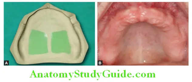
Hard Palate:
The horizontal portion of the palate lateral to midline is primary stress bearing area. Trabecular pattern in the bone is perpendicular to the direction of force thus withstanding masticatory force. Serve as main supporting area for maxillary denture.
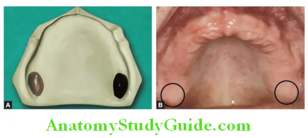
Maxillary Tuberosity:
This is bulbous extension of residual ridge in last molar region. It is covered with dense brous connective tissue which has minimal
compressibility. Sometimes they show overgrowth and need surgical correction before fabrication of denture. Considered as primary stress bearing area as resorption in this area is very less.
Secondary Stress Bearing Areas (Secondary Supporting Areas)
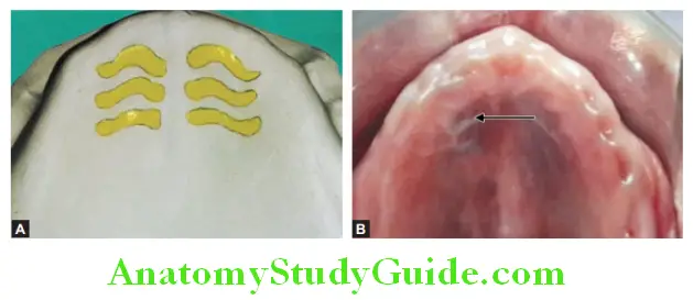
Palatine Rugae:
Mucosal folds located in the anterior region of palatal mucosa. It is firmly attached to the bone and is keratinized. These folds are important for proper speech. They should not be distorted during impression making. They should be reproduced in denture. Rugae area is covered with mucosal folds and established at an angle with residual alveolar ridge, these two features prevent anterior displacement of denture hence considered as secondary stress
bearing area.
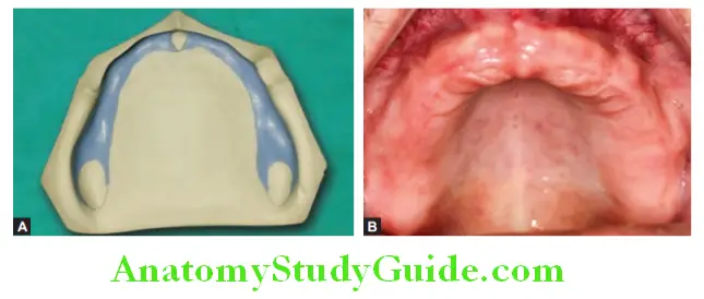
Residual Alveolar Ridge:
Portion of alveolus remaining after loss of teeth is called residual alveolar ridge. Alveolar ridge has firmly attached mucosa over it which is resilient enough to support denture. Mainly the posterolateral portion of residual ridge is the primary stress bearing area.
Relief Areas
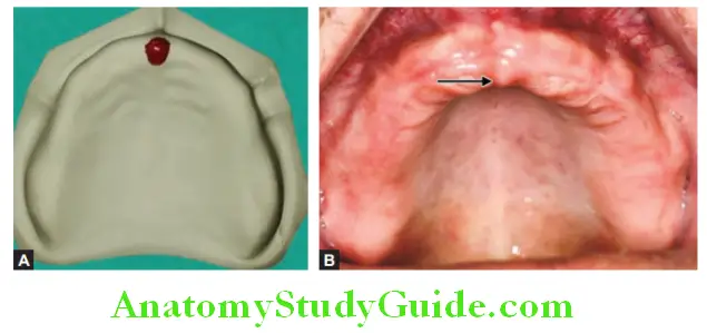
Incisive Papilla:
It is a pad of fibrous connective tissue. It is located on midline (8–10 mm) behind the maxillary central incisor and so serves as guide during teeth arrangement. It covers incisive foramen
which is exit point of nasopalatine nerve and vessels. If not relieved, the denture will compress the vessels and nerve and may lead to pressure ischemia and necrosis of distributing area and paresthesia of anterior palate.
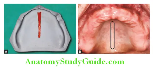
Median Palatine Raphe:
This is median structure area extending from incisive papilla till posterior part of palate. It represents area of bony suture formed by two halves of palatine process and median fusion of two maxillary processes. As covered by thin mucosa, it is the most sensitive part of palate to pressure. The submucosa in this region is very thin or absent, hence relief should be provided. In some cases, the mid palatine raphe is prominent.
Torus Palatinus:
It is hard bony enlargement in midline region of palate. It is seen in 20% of population and covered by thin mucosa. It may need surgical removal if extremely large in size.
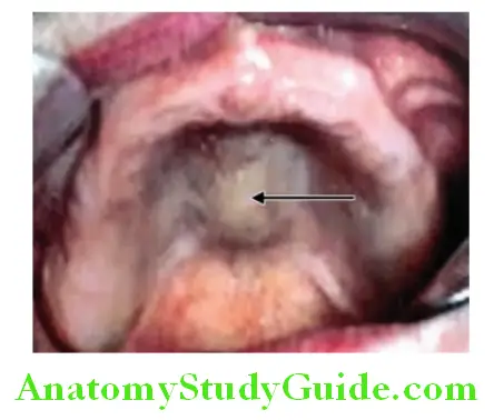
However, if small, relief should be confirmed accurately to the shape of the torus to prevent discomfort in that area. Excess relief may reduce support for denture.
Valve Forming Areas (Limiting Areas)
Labial Frenum:
It is fold of fibrous band covered by mucous membrane at the midline. It contains no muscle fiber so performs no particular action. It is fan-shaped and attached on labial side of ridge. During impression, labial frenum position should be recorded correctly to produce a proper notch to house the frenum. If not relieved properly it may get traumatized due to denture impingement,
results in ulceration, pain and denture dislodgment.
Labial Vestibule:
That part of oral cavity bound on one side by alveolar ridge and on the other side by lip. Vestibule is covered by nonkeratinized lining
mucosa. Submucosa is thick and contains loose areolar tissue and elastic fibers. Labial vestibule extends from labial frenum to buccal frenum (bilaterally). Labial flange corresponding to labial vestibule provide lip support and aids in peripheral seal.
Buccal Frenum:
It divides labial and buccal vestibule. It may be a single or double fold of mucous membrane. It is broad and fan-shaped and moves with muscles during speech and mastication thus active frenum. The muscles
attached with it are orbicularis oris (OO), levator anguli oris (LAO), and buccinator (B). Adequate clearance should be provided in denture to allow muscle movements in buccal frenum area without compromising denture retention.
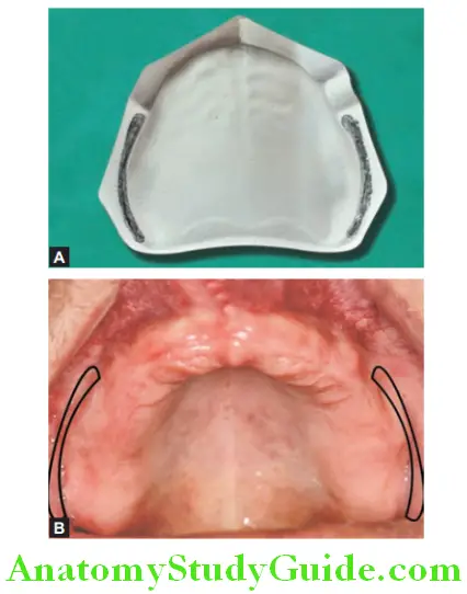
Buccal Vestibule:
It extends from buccal frenum to hamular notch posteriorly. Its size varies with contraction of the buccinator, position of mandible, and amount of bone lost in maxilla in that area.
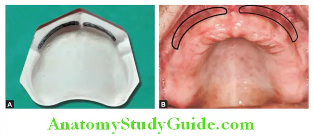
Hamular Notch:
A depression between the maxillary tuberosity and hamulus of medial pterygoid plate is hamular notch. It is soft area of loose areolar tissue. The distolateral border of denture rests in the hamular notch. If denture is over extended in this area, the forward pulling of pterygomandibular raphe (on wide opening of mouth) may dislodge the denture.
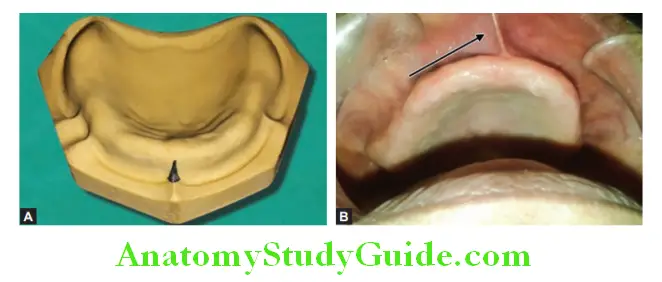
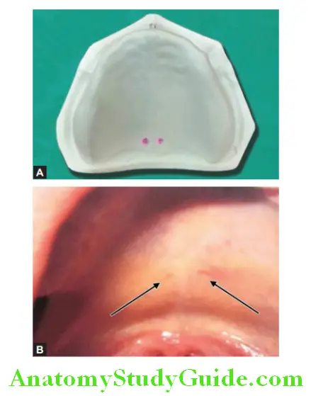
Fovea Palatine:
It is unique feature of humans. It is formed by the coalescence of ducts of several minor
mucous glands. It may guide to locate posterior border of denture. In patient with thick and ropy saliva, fovea palatine should be left relieved to prevent displacement of denture due to collection of thick saliva in between denture and mucosa
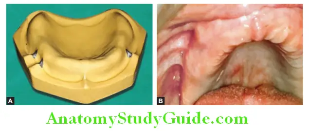
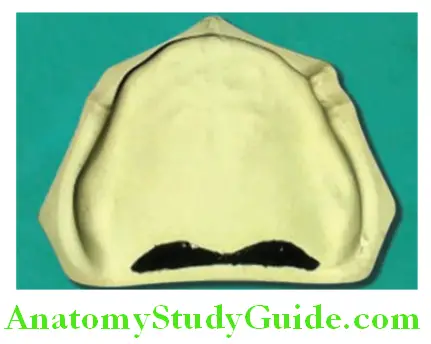
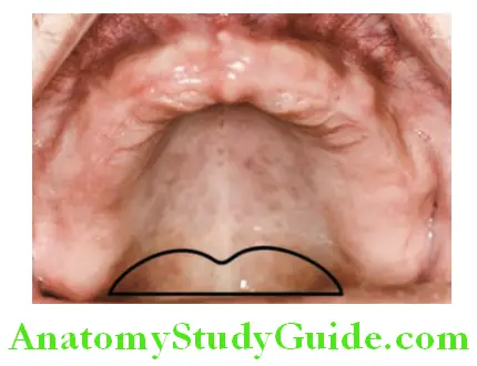
Posterior Palatal Seal Area:
It is junction of hard and soft palate on which pressure within physiological limits can be applied by denture to aid in its retention. It is area bounded anteriorly and posteriorly by two imaginary lines namely anterior vibrating line and posterior vibrating line.
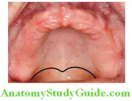
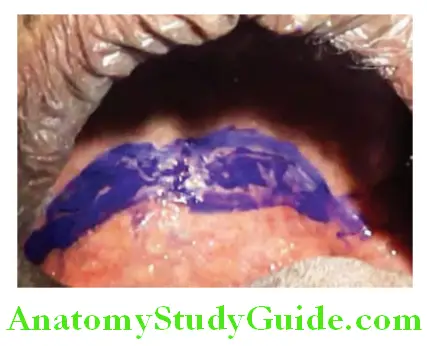
Anterior vibrating line can be located by asking patient to perform Valsalva maneuver. Close patients’ nostrils and ask him to blow slowly through nose, look at the posterior part of the palate you can see elevation of mucosa in cupid bow shape. This elevation is attachment of soft tissue with palatal bone indicating anterior vibrating line. From this point soft palate start its movements but in very slow burst (rhythms).
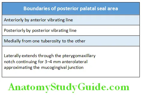
As we go even posterior from this line we can observe clear demarcation of areas, i.e. one showing slow movement and the other one is showing vigorous or more movement of soft palate. The demarcation between slow and vigorous movement is done by posterior vibrating line, which runs from one hamular notch to the other one. Posterior vibrating line decides posterior limit of maxillary complete denture. Area bounded by anterior and posterior vibrating lines is posterior palatal seal area.
Leave a Reply