Cutaneous Nerves Superficial Veins And Lymphatic Drainage
Question-1:Describe The Origin And Termination Of The Cephalic Vein
Table of Contents
Answer:
1. The Cephalic Vein Origin:
The cephalic vein begins at the lateral end of the dorsal venous arch.
2. The Cephalic Vein Termination:
It ends in an axillary vein in the deltopectoral groove.
Read And Learn More: Anatomy Notes And Important Question And Answers
Question 2: Describe The Cephalic Vein Under The Following Heads
1. Cephalic Vein Origin,
2. Cephalic Vein Draining areas,
3. Cephalic Vein Course,
4. Cephalic Vein Relations,
5. Cephalic Vein Tributaries,
6. Cephalic Vein Features,
7. Cephalic Vein Termination, and
8. Cephalic Vein Applied anatomy.
Answer:
The Cephalic Vein Introduction:
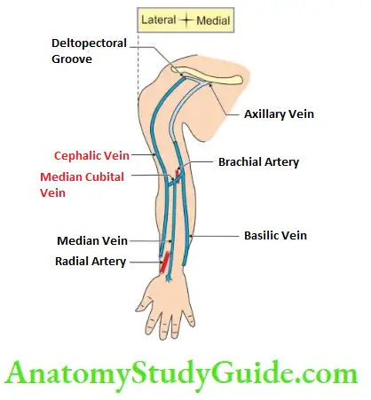
It is a pre-axial superficial vein on the lateral side of the upper limb.
1. The Cephalic Vein Origin
It begins at the lateral end of the dorsal venous arch in the subcutaneous tissue.
2. The Cephalic Vein Draining areas
It drains the lateral side of
- Hand,
- Forearm,
- Arm, and
- Shoulder.
3. The Cephalic Vein Course
It runs on the preaxial border of the upper limb. It forms
1. Roof of
- Anatomical Snuffbox, and
- Cubital fossa.
2. Divides the flexor and extensor muscles of the forearm,

3. Ascends in front of the elbow superficial to a groove between the brachioradialis and biceps,
4. Courses between biceps and brachialis in the arm,
5. Runs at the lateral border of the pectoralis major,
6. Enters the deltopectoral groove, and
7. Pierces costocoracoid part of clavipectoral fascia
4. The Cephalic Vein Relations
- In the beginning, it lies medial to the radial nerve and superficial to the extensor retinaculum.
- It lies posterior and lateral to the styloid process of the radius.
- In the forearm, it lies superficial to the lateral cutaneous nerve of the forearm,
- In the deltopectoral groove, it is accompanied by a deltoid branch of the acromiothoracic artery, and
- It runs along infraclavicular nodes.
5. The Cephalic Vein Tributaries
1. At the formation: Dorsal digital veins of the thumb.
2. During the course:
- The central part of the dorsal venous arch,
- The accessory cephalic vein on the lateral side of the forearm,
- Median cubital vein, and
- Deep veins.

3. Before piercing clavipectoral fascia: (ABCD)
Acromial vein,
Breast or pectoral vein,
Clavicular vein, and
Deltoid vein.
6. The Cephalic Vein Features
1. It is the largest tributary of the axillary vein,
2. It is one of the important structures that pass between the pectoralis minor and subclavius,
3. In the embryo,
- It crosses in front of the clavicle and
- Ends in the external jugular vein.
- It may continue in postnatal life.
- It may be injured in fracture of the clavicle, and
4. Most of the radial lymphatics accompany the cephalic vein.
7. The Cephalic Vein Termination
- Cephalic Vein Termination terminates into the axillary vein at a right angle.
- A homologous vein in the lower limb is a great saphenous vein.
8. The Cephalic Vein Applied anatomy
1. Cephalic Vein Practical significance
- Cephalic Vein is the largest vein of the upper limb that remains open in the distal part.
- Cephalic Vein serves as a useful guide to the 1st part of the axillary artery. This is for the following reasons.
- The axillary artery is completely concealed by the axillary vein.
- Follow the cephalic vein and displace the axillary vein anteromedially.
- Cephalic Vein is available for the surgeon to pass a rubber catheter to the axillary vein, subclavian veins, and the heart to withdraw samples of blood.
2. Please note, that the catheter passed along with the cephalic vein may be impeded by the acute angulation at the deltopectoral groove.
Hence, the cephalic vein is not chosen for cardiac catheterization.
3. Cephalic Vein is frequently used for intravenous therapy. It is easily accessible in the anatomical snuffbox. This site has the following advantages.
- Cephalic Vein is selected for indwelling cannula when a long period is anticipated.
- The position of the hand, forearm, and arm are optimal in this position.
4. While ligating the branch of the radial artery at the anatomical snuffbox, care is taken not to ligate the cephalic vein. The cephalic vein lies superficial and the artery lies deep in it.
5. For dialysis, the cephalic vein is arterialized. Arterialization of the cephalic vein is done as follows: A subcutaneous arteriovenous fistula is created at the wrist by anastomosing the cephalic vein to the radial artery.
The wall of the vein is thickened as the pressure in the vein is increased. Such structural change in the wall is called arterialization. This helps repeated puncture in it with a wide-bored needle.
6. During dialysis, the venous and arterial lines of the cannula are introduced in the cephalic vein. The impure blood is drawn in the dialysis machine via the venous line and the purified blood is returned to the cephalic vein via the arterial line.
7. Cephalic Vein is interesting to know that during the surgery for the cancer of the breast, the removable of the clavicular head is spared. It is done to preserve the cephalic vein.
In case of blockage of the axillary vein, the blood passes through a cephalic vein.
8. In the old days, barbers used cephalic veins for letting the blood.
9. The cephalic vein is preferred in the deltopectoral groove for superior vena cava infusion.
Median Cubital Vein-Importance
The median cubital vein is the vein of choice for intravenous injections, for withdrawing blood from donors, and for cardiac catheterization.
1. The vein is chosen for the following reasons. It is
- Fixed by the perforator, and
- Does not slip away during piercing

Median Cubital Vein
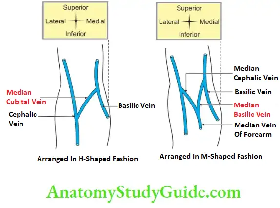
It is a large communicating vein, which shunts blood from the cephalic vein to the basilic vein.
1. Median Cubital Vein Origin:
From the cephalic vein, this is present 1″ below the bend of the elbow. It runs obliquely upward and medially.
2. Median Cubital Vein Termination:
It ends in a basilic vein, 1″ above the medial epicondyle.
3. Median Cubital Vein Relations:
Superficial to deep
- Skin and fascia,
- Bicipital aponeurosis, and
- Brachial artery.
4. Median Cubital Vein Tributaries:
Median antebrachial vein.
5. Median Cubital Vein Applied Anatomy:
1. Median Cubital Vein is a commonly used vein for the withdrawal of blood for
- Investigation, and
- Therapeutic purposes (intravenous injection, intravenous fluid, blood).
2. This vein is fixed by a perforator vein.
Bicipital Aponeurosis Introduction:
The medial expansion of biceps tendon is called bicipital aponeurosis.
1. Bicipital Aponeurosis Course and Relations:
- Bicipital Aponeurosis descends across the brachial artery and fuses with deep fascia.
- The tendon gives off an extension called bicipital aponeurosis. It separates the median cubital vein from the brachial artery.
- Bicipital Aponeurosis extends to the ulna.
- The cubital fossa lies in front of the median nerve.
2. Bicipital Aponeurosis Features:
- Bicipital Aponeurosis forms the roof of the cubital fossa.
- Bicipital Aponeurosis protects the brachial artery.
- Bicipital Aponeurosis separates the median cubital vein from the brachial artery.
3. Bicipital Aponeurosis Palpation:
Bicipital Aponeurosis Palpation is palpable and conspicuous when the elbow is flexed against resistance.
Leave a Reply