Osteology
Question 1. Describe the hip bone in brief.
Answer:
Brief:
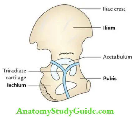
The hip bone (pelvic bone) is a large irregular bone of the pelvic girdle. It is constructed in the center and expanded above and below. The outer aspect of constricted middle part presents a cup-shaped hollow called the acetabulum.
Before puberty, it consists of 3 parts, viz. ilium, ischium, and pubis but in the adult, they fuse in the region of the acetabulum to form a single bone.
Question 2. Write a short note on the iliac crest.
Answer:
Iliac crest:
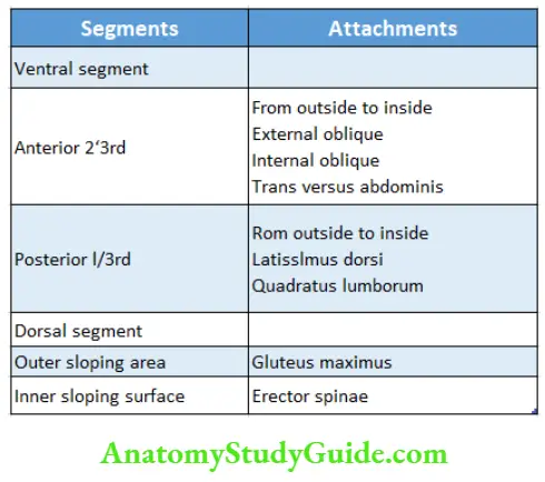
- It is S-shaped, flattened upper border of the ilium
- It is subdivided into two segments
- Larger ventral segment (anterior 2/3rd)
- Smaller dorsal segment (posterior 1/3rd).
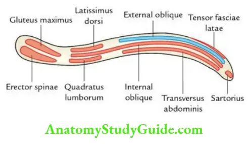
Iliac crest Attachments:
Iliac crest Applied anatomy:
Iliac crest Bone grafting:
The iliac crest is often used for bone grafting.
Iliac crest Bone marrow examination:
The iliac crest is the preferred site for bone marrow aspiration in children.
Question 3. Write a short note on the acetabulum.
Answer:
Acetabulum:
The acetabulum (L. shallow vinegar cup) is a large cup-shaped hollow on the lateral aspect of the hip bone, above the obturator foramen. The primary bones of the hip bone i.e., ilium, ischium, and pubis contribute to the formation of the acetabulum. It articulates with the large round head of the femur to form a large synovial hip joint.
Acetabulum Applied Anatomy: Fractures of acetabulum though rare but if occur they can be serious due to involvement/damage of nerves and vessels in the region.
Question 4. Write a short note on a greater sciatic notch.
Answer:
Greater sciatic notch:
It is a large bony notch on the posterior border of the ilium, above the ischial spine. It is divided into upper and lower parts by the piriformis muscle.
Greater sciatic notch Structures passing through the greater sciatic notch:
Piriformis passes through the middle of the notch
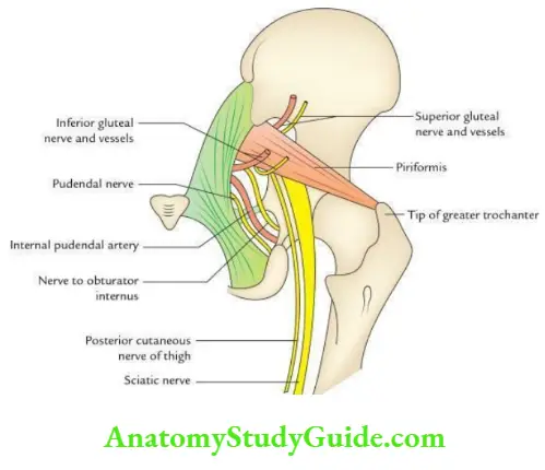
Greater sciatic notch Structures passing above piriformis:
- Superior gluteal vessels
- Superior gluteal nerve
Greater sciatic notch Structures passing below piriformis:
- Sciatic nerve
- Pudendal nerve
- Internal pudendal vessels
- Nerve to obturator internus
- Inferior gluteal nerve and vessels
- Posterior femoral cutaneous nerve
Question 5. Write a short note on the lesser sciatic notch.
Answer:
Lesser sciatic notch:
It is a small bony notch on the posterior border of the ilium below the ischial spine.
Structures passing through the lesser sciatic notch:
- Tendon of obturator internus
- Pudendal nerve
- Internal pudendal vessels
- Nerve to obturator internus
Lesser sciatic notch Mnemonic: PIN.
Question 6. Write a short note on the ischial tuberosity.
Answer:
Ischial tuberosity:
It is a rough tuberosity present on the lower end of the dorsal surface of the ischium.
Ischial tuberosity Subdivisions:
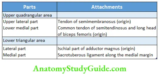
It is divided by a transverse ridge into upper quadrilateral and lower triangular areas. The upper quadrilateral area is further subdivided by an oblique ridge into upper lateral and lower medial parts. The lower triangular area is subdivided by a longitudinal ridge into lateral and medial parts.
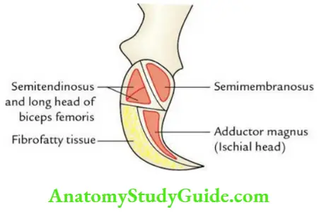
Ischial tuberosity Attachments:
Question 7. Enumerate muscles attached to the greater trochanter.
Answer:
Greater trochanter:
Six muscles attached to the greater trochanter are:
- Gluteus minimus: On the anterior surface.
- Gluteus medius: On the lateral surface into an oblique ridge.
- Obturator internus: On the medial surface.
- Obturator externus: On the trochanteric fossa.
- Piriformis: On the apex.
- Quadratus femoris: On the quadrate tubercle.
Question 8. Enumerate structures attached to the linea aspera.
Answer:
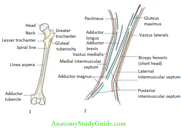
Linea Aspera:
It is a thick posterior border of the femur presenting medial and lateral lips. It provides attachment to 9 structures: 2 intermuscular septa and 7 muscles.
Question 9. Write a short note on the adductor tubercle.
Answer:
Adductor tubercle:
It is a conical bony projection at the lower end of the femur posterosuperior to the medial epicondyle.
Adductor tubercle Attachments:
- Tendon of ischial/hamstring part of the adductor magnus
- Tibial collateral ligament
Adductor tubercle Applied anatomy:
The lower epiphyseal plate of the femur in children passes through the adductor tubercle. The growth in length of the femur is essentially due to activity at this plate. Therefore, any interference with it in children will affect the growth of the femur in length causing shortening of the limb.
Question 10. Avascular necrosis of the head of the femur is common in intracapsular fracture neck of the femur. Give the anatomical basis.
Answer:
Anatomical basis:
The head of the femur is supplied in 3 sets of vessels:
- Artery of the head of the femur, a branch of the ascending branch of the medial circumflex femoral artery
- Nutrient artery, a branch of the 2nd perforating branch of profunda femoral artery
- Retinacular arteries, derived from medial and lateral circumflex femoral arteries (the most important source of blood supply)
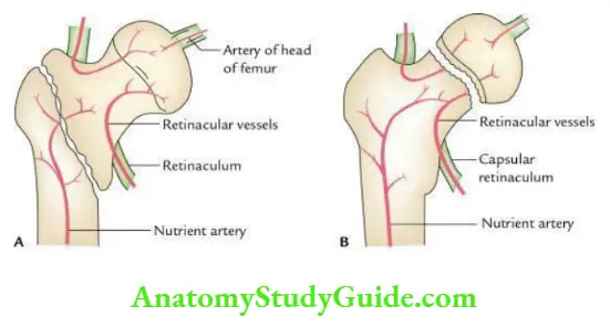
The Avascular necrosis of the head of a femur commonly occurs in the intracapsular fracture neck of the femur due to involvement/damage of the retinacular vessels.
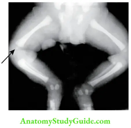
Question 11. Write a note on the ossification center at the lower end of the femur.
Answer:
Ossification center:
The ossification center of the lower end of the femur occurs at birth (9 months). Therefore, the presence of this center in the radiograph of a newly born child found dead suggests that the baby was full-term and viable.
Question 12. Enumerate the sites of sesamoid bones in the lower limb.
Answer:
Sesamoid bones:
- Patella: In the tendon of quadriceps femoris.
- Fabella: In the lateral head of the gastrocnemius.
- A sesamoid bone: In the tendon of peroneus longus where it winds around the cuboid.
- Two sesamoid bones below the head of the 1st metatarsal; one in each half of the tendon of flexor hallucis brevis.
Sesamoid bones Note:
Patella is the largest sesamoid bone in the body.
Question 13. Write a short note on the patella.
Answer:
Patella:
The patella is the largest sesamoid bone (also called knee cap) situated in front of the lower end of the femur with which it articulates to form a saddle type of patella-femoral joint. It develops in the tendon of the quadriceps femoris muscle to prevent its attrition.
Patella Applied Anatomy:
- Dislocation of the patella: The patella has an inherent tendency to dislocate laterally especially in females due to a large angle.
- Patellar fracture: It is common and often occurs due to sudden forceful contraction of the quadriceps femoris muscle. Patellar fractures do not heal due to the absence of periosteum in the patella as it is a sesamoid bone.
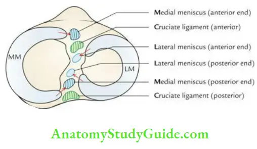
Question 14. Write a note on the intercondylar area of the tibia.
Answer:
Tibia:
- The intercondylar area is a rough area that separates the two articular surfaces on the upper aspect of the upper end of the tibia.
- This area provides attachment to
- Anterior and posterior cruciate ligaments and
- Anterior and posterior ends of medial and lateral menisci.
- The sequence of attachments of these structures can be easily remembered by a Mnemonic; Medical College Lucknow Lucknow Medical College.
Tibia Applied anatomy
Knowledge of the attachment of these structures is important to surgeons as injury of these structures is common.
Leave a Reply