Hepatitis B
- Hepatitis B (serum hepatitis) caused by HBV infection has a longer incubation period (30-180 days) and is transmitted parenterally such as in recipients of blood and blood products, intravenous drug addicts, patients treated by renal dialysis and hospital workers exposed to blood, and by intimate physical contact such as from mother to child and by sexual contact.
- The disease may occur at any age. HBV infection causes a more severe form of illness that includes acute hepatitis B, chronic hepatitis, progression to cirrhosis, fulminant hepatitis and an asymptomatic carrier stage.
- HBV plays some role in the development of hepatocellular carcinoma as discussed later.
Read And Learn More: Systemic Pathology Notes
Hepatitis B Virus (HBV): The etiologic agent for hepatitis B, HBV, is a DNA virus which has been extensively studied. Electron microscopic studies on the serum of patients infected with HBV show 3 forms of viral particles of 2 sizes: small (spheres and tubules/filaments) and large (spheres) as under:
- Small particles are most numerous and exist in two forms— as 22 nm spheres, and as tubules 22 nm in diameter and 100 nm long.
- These are antigenically identical to the envelope protein of HBV and represent the excess of viral envelope protein referred to as hepatitis B surface antigen (HBsAg)

- Large particles, 42 nm in diameter, are double-shelled spherical particles, also called Dane particles.
- These are about 100 to 1000 times less in number in serum compared to small 22 nm particles and represent intact virion of HBV.
- The genomic structure of HBV is quite compact and complex. The HBV DNA consists of 4 overlapping genes which encode for multiple proteins:
1. S gene codes for the surface envelope protein, hepatitis B surface antigen (HBsAg); this product is a major protein. HBsAg is present on the outer surface of the large spherical particles as well as in small spherical and tubular structures.
- Pre-S1 and pre-S2 regions of the genome are upstream of the S gene and code for pre-S gene protein products that include receptors on the HBV surface and for hepatocyte membrane proteins.
- The protein product of S-gene plus the adjacent pre-S2 region is the middle protein, while the protein products of pre-S1 plus pre-S2 regions are the large protein. Large protein coming from both pre-S proteins is rich in complete virions.
2. P gene is the largest and codes for DNA polymerase.
3. C gene codes for two nucleocapsid proteins, HBeAg and a core protein termed HBcAg.
4. X gene codes for HBxAg which is a small non-particulate protein. HbxAg has a role in transactivating the transcription of both viral and cellular genes.
- The processes transactivated by X-genes include signal-transduction pathways, increased replication of HBV DNA, replication of other viruses including HIV, enhanced susceptibility of HBV-infected hepatocytes to cytolytic T cells, and pro-apoptotic pathway.
- The expression of HBxAg and its antibodies associated with enhanced HBV DNA replication has been implicated in hepatocellular carcinoma in patients with chronic hepatitis.
Pathogenesis: There is strong evidence linking immune pathogenesis with hepatocellular damage:
- Since a carrier state of hepatitis B without hepatocellular damage exists, it means that HBV is not directly cytopathic.
- It has been observed that individuals with defects or deficiency of cellular immunity have more persistent hepatitis B disease instead of clearing HBV from their blood.
- Support for cell-mediated mechanism in hepatocellular damage by HBV comes from the observation that viral antigens (in particular nucleocapsid proteins HbcAg and HBeAg) are attacked by host cytotoxic CD8+T lymphocytes.
- The host response of CD8+T lymphocytes by elaboration of antiviral cytokines is variable in different individuals and that determines whether an HBV-infected person recovers, develops mild or severe disease, or progresses to chronic disease.
Serologic and viral markers: In support of immune pathogenesis is the demonstration of several immunological markers in the serum and in hepatocytes indicative of the presence of HBV infection. These are as under.
1. HBsAg: In 1965, Blumberg and colleagues in Philadelphia found a lipoprotein complex in the serum of a multiple-transfused haemophiliac of Australian aborigine which was subsequently shown by them to be associated with serum hepatitis.
- This antigen was termed Australia antigen by them (In 1977, Blumberg was awarded the Nobel prize for his discovery).
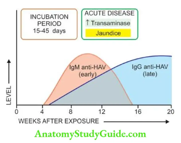
- The term Australia antigen is now used synonymously with hepatitis B surface antigen (HBsAg). HBsAg appears early in the blood after about 6 weeks of infection and its detection is an indicator of active HBV infection.
- It usually disappears in 3-6 months. Its persistence for more than 6 months implies a carrier state.
- In carriers and in chronic hepatitis patients, HBsAg expression is observed at an intracytoplasmic and membranous location in the hepatocytes by Orcein staining (orange positivity) and anti-HBs-antibody immunohistochemical stain, indicating active HBV replication and correlates well with serum HBV-DNA.
2. Anti-HBs: Specific antibody to HBsAg in serum called anti-HBs appears late, about 3 months after the onset.
- Anti-HBs response may be both IgM and IgG type. The prevalence rate of antiHBs ranges from 10-15%. In these individuals, it persists for life providing protection against reinfection with HBV.
3. HBeAg: HBeAg derived from core protein is present transiently (3-6 weeks) during an acute attack. Its persistence beyond 10 weeks is indicative of the development of chronic liver disease and carrier state.

4. Anti-HBe: An antibody to HBeAg called anti-HBe appears after the disappearance of HBeAg. Seroconversion from HBeAg to anti-HBe during the acute stage of illness is a prognostic sign for the resolution of infection.
5. HBcAg: HBcAg derived from core protein cannot be detected in the blood.
- But HBcAg can be demonstrated in the nuclei of hepatocytes in the carrier state and in chronic hepatitis patients by Orcein staining and by anti-HBc-antibody immunohistochemical stain but not in the liver cells during the acute stage.
6. Anti-HBc: Antibodies to HBcAg called anti-HBc can, however, be detected in the serum of acute hepatitis B patients during the pre-icteric stage.
- In the initial period, it is an IgM class antibody which persists for 4-6 months and is followed later by IgG anti-HBc.
- Thus, the detection of a high titre of IgM anti-HBc is indicative of recent acute HBV infection, while an elevated level of IgG antiHBc suggests HBV infection in the remote past.
7. HBV-DNA Detection of HBV-DNA by molecular hybridisation using the Southern blot technique is the most sensitive index of hepatitis B infection.
- It is present in the pre-symptomatic phase and transiently during the early acute stage.
Leave a Reply