Describe the Ovary under the following headings:
- Ovary Introduction
- Ovary External features
- Ovary Relations
- Ovary Arterial supply
- Ovary Venous drainage
- Ovary Lymphatic drainage
- Ovary Nerve supply and
- Ovary Applied anatomy.
Answer:
1. Ovary Introduction:
- They are a pair of female gonads, which produce ova and female sex hormones viz. estrogen and progesterone.
- Each ovary is almond-shaped and situated in the ovarian fossa in the lateral wall of the lesser pelvis below the pelvic brim.
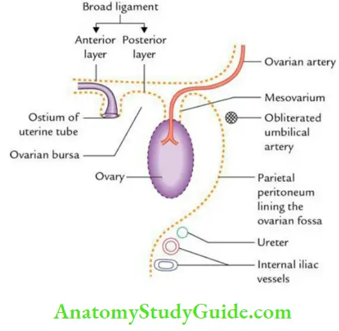
- It lies on each side of the uterus and is attached to the posterior layer of the broad ligament by a short peritoneal fold called mesovarium.
2. Ovary External features:
- Two surfaces: medial and lateral.
- Two borders: anterior (mesovarian) and posterior (free).
- Two poles: upper (broader) and lower (narrower).
3. Ovary Relations:
Read And Learn More: Anatomy Question And Answers
Peritoneal relations:
The ovary is covered by a single layer of low cuboidal epithelium called – germinal epithelium (modified peritoneum). Along the mesovarian border peritoneum forms mesovarium.
Visceral relations:
- Poles:
- Upper or tubal pole: It is directed upward and is related to the distal end of the uterine tube.
- Lower or uterine pole: It is directed downward and is connected to the lateral angle of the uterus by the ligament of the ovary.
- Borders:
- Anterior or mesovarian border: It is straight and is related to a uterine tube and the obliterated umbilical artery. It presents hilum.
- Posterior or free border: It is convex and is related to the uterine tube in the upper part and the ureter in the lower part.
- Surfaces:
- Lateral surface: It is convex and lies in the ovarian fossa. It is related to obturator vessels and nerves separated by a peritoneum.
- Medial surface: It is related to the terminal part of the uterine tube separated by the ovarian bursa, a peritoneal recess between the mesosalpinx and the ovary.
4. Ovary Arterial supply:
- The ovarian artery (main artery), is a branch of the abdominal aorta which reaches the ovary through the suspensory ligament of the ovary.
- The uterine artery is a branch of the internal iliac artery which reaches the ovary via the mesovarium.
5. Ovary Venous drainage: The right ovarian vein drains into the inferior vena cava, while the left ovarian vein drains into the left renal vein.
6. Ovary Lymphatic drainage: Lymphatics from the ovary drain into the preaortic and paraaortic nodes along the side of the origin of the ovarian artery.
7. Ovary Nerve supply: By both sympathetic and parasympathetic fibers.
- Sympathetic fibers: Derived from T10–T11 segments: These are vasoconstrictors and form afferent pathways to pain; hence, ovarian pain is referred to as the loin and groin.
- Parasympathetic fibers: Derived from S2, S3, and S4 segments. These are vasodilators.
8. Ovary Applied Anatomy:
- Oophoritis:
- It is an inflammation of the ovary.
- It may produce localized peritonitis of the ovarian fossa and an eventual irritation of the obturator nerve, which may lead to pain that is referred to the medial aspect of the thigh.
- Ovarian tumors:
- The ovary is a common site for carcinoma, teratoma, and secondaries.
- Carcinoma of the ovary is common and accounts for 15% of all cancers and 20% of gynecological cancers.
- The commonest secondary tumor of the ovary is Krukenberg’s tumor, which occurs via trans-coelomic migration of cancer cells from a carcinoma breast.
- Ovarian cysts: These are common and occur as a result of the developmental arrest of ovarian follicles.
- Prolapse of the ovary: Ovary is frequently displaced into the recto-uterine pouch (pouch of Douglas).
Give the histological features of the ovary.
Answer:
Histological features of the ovary:
The ovary is a solid ovoid organ covered by cuboidal epithelium (germinal epithelium). Beneath, this is a thin layer of connective tissue called tunica albuginea.
Part deep to tunica albuginea is demarcated into 2 zones:
- Outer cortex and
- Inner medulla
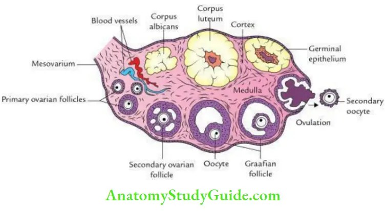
1. Cortex:
- It contains numerous ovarian follicles in various stages of development:
- Primordial follicles consist of primary oocytes covered by a single layer of flat cells.
- Primary follicles consist of primary oocytes covered by a single layer of cuboidal cells.
- Secondary follicles consist of oocytes covered by zona pellucida and membrane granulosa.
- Graafian follicles are fluid-filled follicles with an ovum at one side embedded in a mass of cells called cumulus oophorus.
2. Medulla:
It consists of connective tissues presenting a swirly appearance. It contains several blood vessels (mostly veins) and smooth muscle fibers.
Leave a Reply