Abdominal Cavity and Peritoneum.
Peritoneal Reflection
It is divided into:
Table of Contents
1. Parietal peritoneum: Peritoneum covering anterior, posterior and lateral walls of abdomen and pelvis is called parietal peritoneum.
2. Visceral peritoneum: The peritoneum covering the organs is called visceral peritoneum. It is named as extent of folds of visceral peritoneum.
Read And Learn More: General Histology Question And Answers
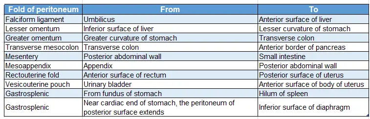
Extent of folds of visceral peritoneum:
Foramen of Winslow (aditus to lesser sac, epiploic foramen)
Introduction: It is an opening, communicating between the lesser sac and the greater sac.
1. Size: 3cm
2. Disposition: It is vertically displayed.
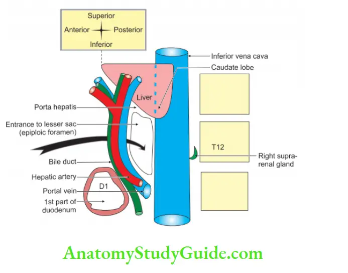
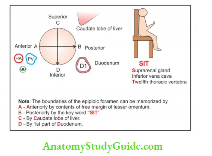
3. Boundaries:
Anterior: Right free margin of the lesser omentum. It contains from left to right
- Hepatic artery.
- Portal vein (more posteriorly).
- Bile duct.
Posterior: Structures forming the posterior boundary of foramen of Winslow
(SIT)
- Suprarenal gland (right).(S)
- Inferior vena cava. (I)
- Twelfth thoracic vertebra.(T)
Superior: Caudate process of liver.
Inferior:
- Peritoneum extending from inferior vena cava to 1st part of duodenum.
- Horizontal part of hepatic artery.
4. Applied anatomy:
- The foramen of Winslow cannot be enlarged since there are important structures situated in the anterior wall.
- The intestines may herniate through the epiploic foramen.
- The infection spreads from the greater sac to lesser sac or vice versa through the foramen of Winslow.
- After cholecystectomy due to biliary leakage, if patient sits in left lateral position, the fluid tends to permeate through the epiploic foramen and distend the lesser sac.
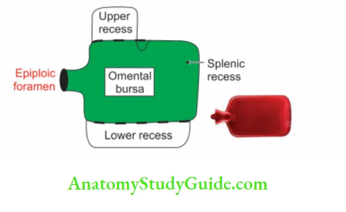
Lesser sac (omental bursa)
Introduction: It is the large recess of peritoneal cavity present behind the stomach.
1. Gross:
- Location: It lies behind the stomach and the lesser omentum.
- Extends: Into greater omentum.
- Communication: To greater sac through epiploic foramen.
2. Boundaries: Rule of 3.
There are 3 structures in each boundary:
Anterior wall—for legend: From the given diagram
- The posterior layer of lesser omentum — Aa
- Peritoneum on the posterior layer of stomach — Ab
- Posterior of the anterior two layers of greater omentum — Ac
Posterior wall:
1. Below transverse colon: Anterior of the posterior two layers of greater omentum — Ba
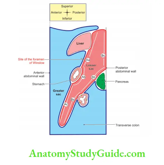
2. Between transverse colon and pancreas: Posterior most layer of greater omentum fused with superior layer of transverse mesocolon — Bb
3. Above pancreas: Anterior layer of posterior two layers continues as parietal peritoneum of posterior abdominal wall — Bc
Inferior wall: Fusion of inner layer of greater omentum up to the level of transverse colon raises the inferior margin.
Superior wall:
- Reflection of the peritoneum from esophagus to diaphragm.
- Upper end of fissure for ligamentum venosum.
- Upper border of caudate lobe of liver.
Right margin (from below upwards):
1. Right margin of greater omentum.
2. Right free margin of lesser omentum containing from left to right.
- Hepatic artery (left)
- Portal vein more posteriorly
- Bile duct
3. Floor of aditus to lesser sac
Left margin of lesser sac (from below upwards):
- Left margin of greater omentum
- Gastrosplenic
- Lienorenal ligament
3. Functions:
- It supports and facilitates the movements of the stomach.
- Acts as a bursa.
- It is used by surgeons for operative procedures since it is a bloodless area.
- The strangulated internal hernia is approached through greater omentum.
Morison’s pouch
It is also called left extraperitoneal space, right subhepatic space or hepatorenal pouch of Morison.
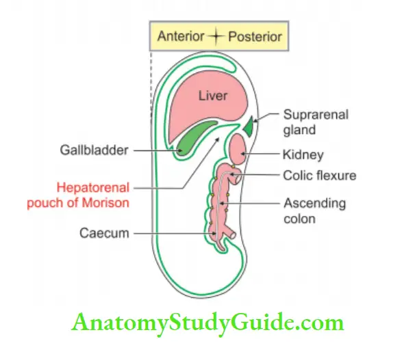
1. It is intraperitoneal space:
2. Boundaries: It is bounded by
Anteriorly:
- The inferior surface of the right lobe of the liver
- Gallbladder
Posteriorly:
- Right suprarenal gland
- The anterior surface of upper part of the right kidney
- 2nd part of the duodenum
- Hepatic flexure
- Transverse mesocolon
- Head of pancreas
Superiorly: Inferior layer of coronary ligament
Inferiorly: Peritoneal cavity
Left: Communicated to omental bursa
Right: Limited by diaphragm
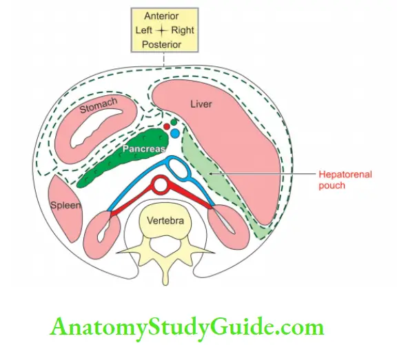
3. Communication on:
- Left: Foramen of Winslow
- Right: Abdominal wall
4. Applied anatomy:
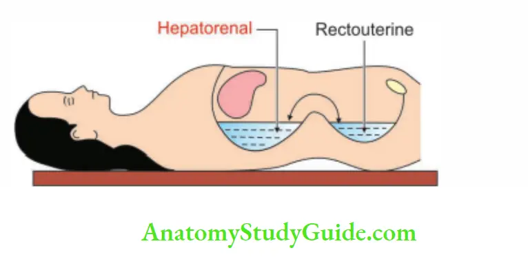
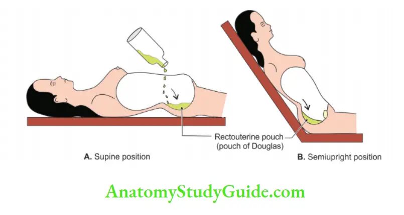
- It is most dependent part in supine position.
- There may be accumulation of pus, blood or fluid in this space.
- Liquids and bile from the lesser peritoneal cavity pass in this pouch.
- It can be drained by a tube inserted through a “stab wound” made through the abdominal wall just outside the right kidney.
Douglas’ pouch/pouch of Douglas (rectouterine pouch)
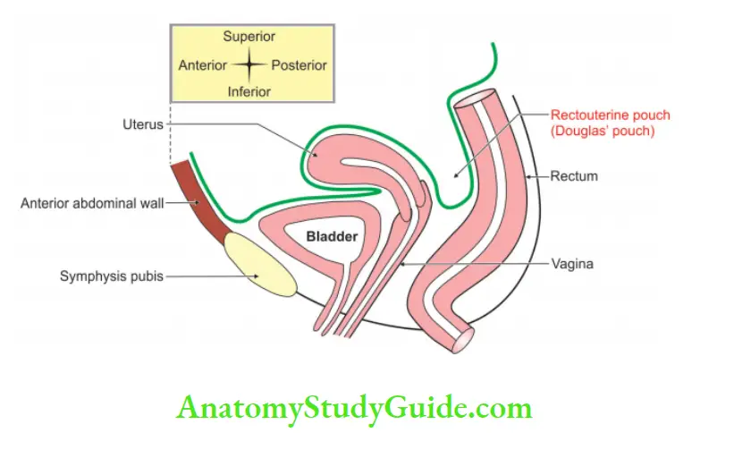
Introduction: It is the peritoneal space present between the anterior surface of rectum and the posterior surface of the uterus. It is most dependent part in female ♀.
1. Features:
- It is deeper than the male ♂ rectovesical pouch.
- It extends laterally and posteriorly to form pararectal fossae on each side of rectum.
- The lateral extensions on each side of the rectum, the pararectal fossae are deeper than the pouch of Douglas.
2. Contents:
- Loops of small intestine, and
- Anterior surface of rectum.
3. Functions: The fascia uniting rectum and uterus resists increased intra-abdominal pressure.
4. Applied anatomy:
- Prolapse of the ovary in the Douglas’ pouch do occur in normal woman.
- Abscess in the Douglas’ pouch is painful.
- Culdocentesis: The incision is made in the posterior part of the vaginal fornix. It is done for the drainage of a pelvic abscess in the rectouterine pouch and a fluid in the peritoneal cavity Example: Blood.
- Pouch of Douglas may contain abnormal structures like descended inflamed appendix. It may be detected by per rectal examination.
Leave a Reply