Abdominal Part of Oesophagus and Stomach
Blood supply of the stomach
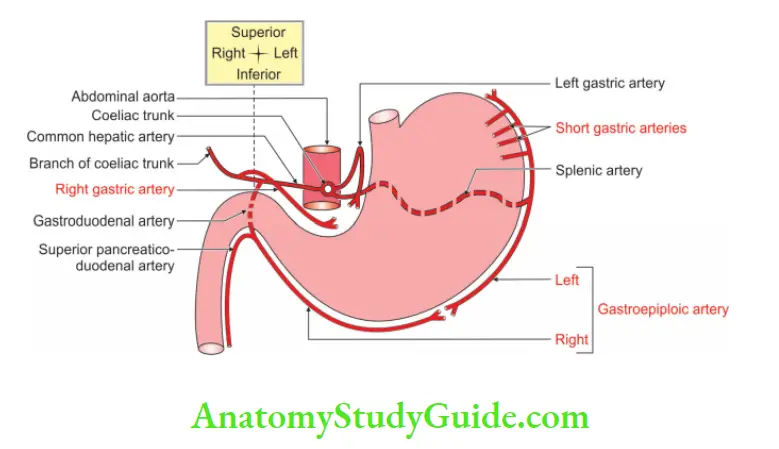
1. Arterial supply: It is supplied by
Table of Contents
Left gastric artery: The smallest branch of the coeliac trunk and supplies the largest area of the stomach. It supplies the upper 2/3rd of the organ.
Right gastric: A branch of the common hepatic artery. It anastomoses with the left gastric artery within the lesser omentum.
Short gastric arteries: These are branches of the splenic artery and supply the fundus of the stomach.
Left gastroepiploic: It is a branch of the splenic artery and reaches the greater curvature.
Right gastroepiploic: It is a branch of the gastroduodenal artery and anastomoses with the left gastroepiploic artery.
| Body Fluids | Muscle Physiology | Digestive System |
| Endocrinology | Face Anatomy | Neck Anatomy |
| Lower Limb | Upper Limb | Nervous System |
Read And Learn More: General Histology Questions and Answers
2. Venous drainage:
1. Right and left gastric veins are tributaries of the portal vein.
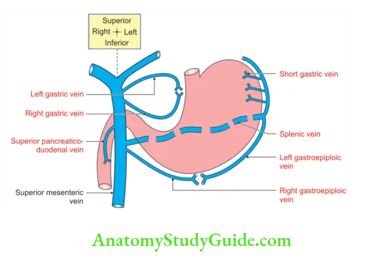
2. Short gastric and left gastroepiploic veins drain into the splenic vein.
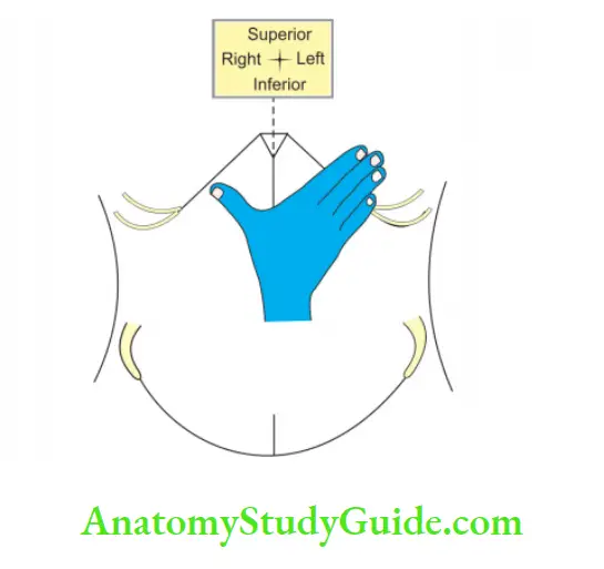
- The right hand on the anterior abdominal wall represents the portal vein
- The thumb represents a right branch of the portal vein
- The index finger represents a left branch of the portal vein
3. Right gastroepiploic vein > superior mesenteric vein.
Bare areas of the stomach
1. Posterior surface of the stomach near cardiac orifice: It is cranial to the gastrophrenic ligament.
2. Area on the lesser curvature of the stomach between anterior and posterior layers of the lesser omentum.
3. Area on the greater curvature of the stomach between anterior and posterior layers of the greater omentum.
4. Area of the fundus of the stomach between the attachments of gastrophrenic ligament
Question -1: What are the surface mucus cells and how do they differ from neck mucus cells in the stomach?
Answer:
1. Surface mucus cells: The epithelial cells secrete mucus. A mucus film is formed. It covers the gastric mucosa. It protects the mucosa of the stomach from acidic gastric juice.
2. Mucous neck cells: They are present in between the parietal cells of the neck of the gland. They secrete mucus. The nucleus of these cells is basal in location and
is round.
Surface mucus and neck mucus cells:

Question – 2: Differentiate between the fundus and pylorus of the stomach histologically
Answer:
Differentiate between the fundus and pylorus of the stomach histologically:

Question – 3: Describe the stomach under the following heads
1. Gross anatomy
2. Histology
3. Development, and
4. Applied anatomy.
Answer:
Introduction: It is the most dilated part of the alimentary system.
1. Gross anatomy
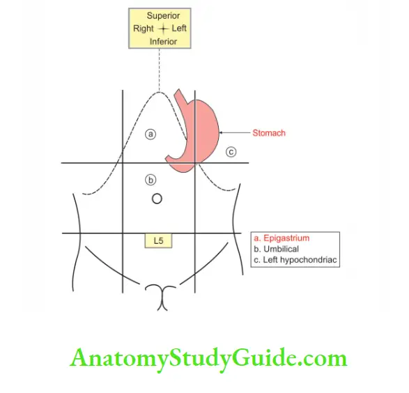
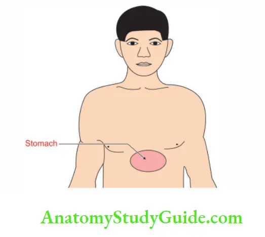
1. Location: The stomach lies in the upper and left part of the abdomen. It occupies
- Epigastrium
- Umbilical, and
- Left hypochondriac regions.
2. External features:
1. Shape:
- Depends upon the degree of distension.
- The physique of the individual.
- Position of adjacent organs
- It is J is shaped when the stomach is empty.
- Pyriform shaped
 when partially distended.
 - Steer horn in obese.
2. Capacity: It varies with age.
- At birth: 30 ml.
- At puberty: 1000 ml.
- In adults: 1.5 litres.
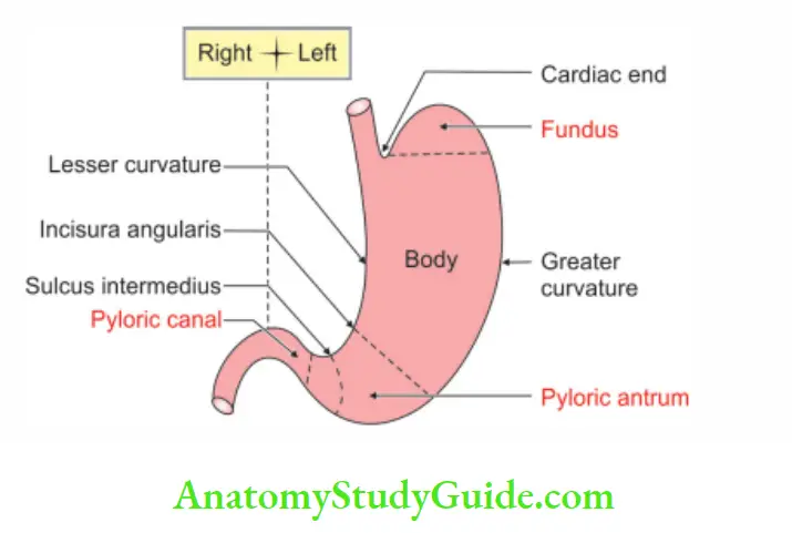
3. Curvatures: It has two curvatures.
1. Lesser curvature: Lesser curvature is concave and forms the right border of the stomach. It provides attachment to the lesser omentum.
2. Greater curvature: Greater curvature: is convex and forms the left border of the stomach. It provides the attachment to the
- Greater omentum
- Gastrosplenic ligament, and
- Gastrophrenic ligament.
4. Surfaces: In a distended position, it has two surfaces.
- Anterosuperior and
- Posteroinferior.
5. Subdivisions: It is divided into two parts by the line extending from incisura angularis (junction of horizontal and vertical parts of the lesser curvature of the stomach) to the greater curvature.
1. The larger part is called the cardiac part and the smaller part is called the pyloric area.
2. The cardiac part is subdivided into the fundus and the body.
- Fundus: The area above the horizontal line extending from the cardiac orifice to greater curvature.
- Body of stomach: It lies between the fundus and pyloric antrum.
- Pyloric antrum: It is 3″ in length.
- Pyloric canal: It is 1″ long. It is narrow and tubular.
Internal features:
1. Orifice: There are two orifices.
- Cardiac orifice: It is present at the lower end of the oesophagus. It is a physiological sphincter. It cannot be demonstrated anatomically.
- Pyloric sphincter (pylorus—gate): It is situated V” to the right of the median plane, at the lower border of the L1 vertebra.
2. Mucous membrane:
- It is thick: Velvety, and has a number of temporary folds or rugae. They are on a long axis. These folds disappear when the stomach is distended.
- Gastric canal (magenstrasse): Gastric canal is a mucous gutter. It is present on lesser curvature. It extends from the cardiac orifice to the pyloric antrum.
3. Relations:
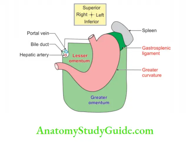
1. Peritoneal relations: The stomach is covered by the peritoneum everywhere except
1. The bare area It is an area behind the cardiac end of the stomach, which is in contact with the diaphragm.
2. At the lesser curvature along the attachment of lesser omentum.
3. At the greater curvature along the attachment of
- Gastrophrenic ligament
- Gastrosplenic ligament
- 1st and 2nd layers of the greater omentum.
2. Visceral relations:
1. Anterior:
- Liver
- Diaphragm, and
- Anterior abdominal wall.
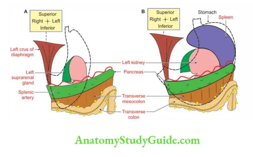
2. Posterior: The surface of the stomach is related to structures forming the stomach bed.
- Left crus of the diaphragm
- Splenic artery
- Transverse mesocolon
- Left colic flexure
- The anterior surface of the left kidney
- Left suprarenal gland, and
- Body of pancreas.
4. Blood supply:
1. Arterial supply: It is supplied by
- Left gastric artery: The smallest branch of the coeliac trunk and supplies the largest area of the stomach. It supplies the upper 2/3rd of the organ.
- Right gastric: A branch of the common hepatic artery. It anastomoses with the left gastric artery within the lesser omentum.
- Short gastric arteries: These are branches of the splenic artery and supply the fundus of the stomach.
- Left gastroepiploic: It is a branch of the splenic artery and reaches the greater curvature.
- Right gastroepiploic: It is a branch of the gastroduodenal artery and anastomoses with the left gastroepiploic artery.

2. Venous drainage:
1. Right and left gastric veins are tributaries of the portal vein.

2. Short gastric and left gastroepiploic veins drain into the splenic vein.

- The right hand on the anterior abdominal wall represents the portal vein
- The thumb represents the right branch of the portal vein
- The index finger represents a left branch of the portal vein
3. Right gastroepiploic vein > superior mesenteric vein.
5. Nerve supply: It is supplied by sympathetic and parasympathetic nerves.
Sympathetic fibres: They arise from the coeliac plexus. The pre-ganglionic motor fibres arise from T6 to T9 segments of the spinal cord and the fibres reach via greater splanchnic nerves. They rely on the coeliac ganglion. The sympathetic fibres have the following functions.
- They are vasomotor in function.
- They stimulate the pyloric sphincter.
- They inhibit the smooth muscles of the stomach.
- Sensory sympathetic fibres convey painful sensations from the stomach.
Parasympathetic fibres: They are derived from both the vagi nerves. They consist of the anterior and posterior gastric nerve. The stimulation of the parasympathetic nerve
- Increases motility of the stomach.
- Secretes gastric juice which is rich in pepsin and hydrochloric acid.
- Inhibits pyloric sphincter.
6. Lymphatic drainage:
1. All the lymphatics from the stomach ultimately drain into a coeliac group of lymph nodes > intestinal lymph trunk > cisterna chyli.
2. It is divided into 4 regions by a vertical line A. It runs in the long axis of the stomach. The line “A” extends from the fundus of the stomach to the pyloric end of the stomach.
3. It divides an area of the stomach into the right 2/3rd and left 1/3rd region.
4. Left 1/3rd is subdivided by a horizontal line into upper 1/3rd and lower 2/3rd.
5. Pyloric region
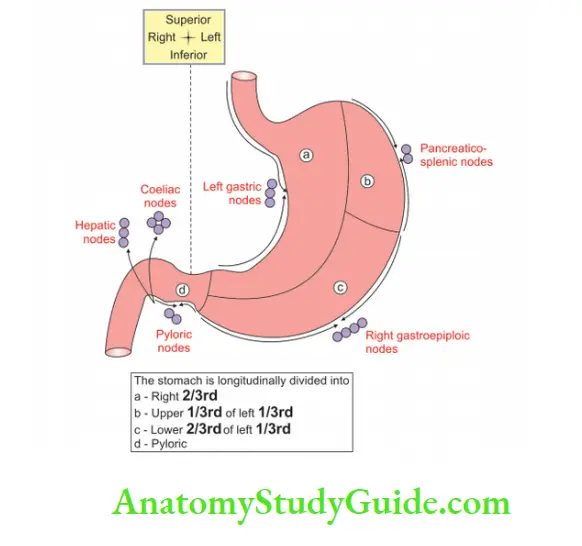
Lymph nodes of the stomach and their afferent and efferent lymphatics:

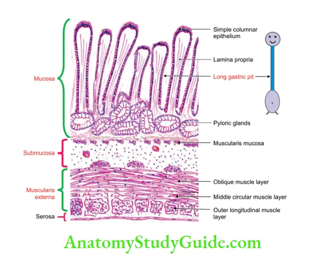
2. Histology
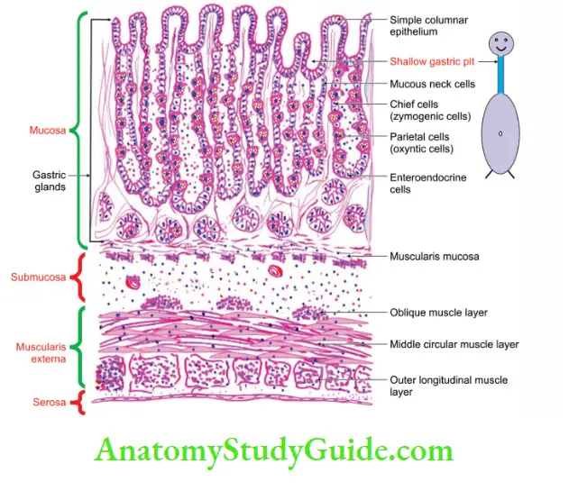
The wall of the stomach presents 4 coats from inside out.
1. Mucous membrane: It consists of epithelium, lamina propria and muscular mucosae.
Epithelium:
1. It is a tall, simple columnar epithelium. It is honeycomb in appearance and presents numerous depressions of gastric pits. They receive ducts of gastric glands.
2. It has
- Thin basement membrane formed by connective tissue.
- Light staining apical cytoplasm.
- Dark staining basal nuclei.
Lamina propria: It contains
- Collagen fibres
- Cells of connective tissue
- Blood vessels
- Various types of gastric glands.
The gastric glands are of three types:
1. Cardiac glands: They are few in number. They are situated close to the cardiac orifice. They secrete mucus.
2. Pyloric glands: They are lined by mucous-secreting cells. Some of the cells secrete the hormone, gastrin.
3. Fundic glands: They are present in the fundus and body. They contain three types of cells.
- Zymogenic or chief cells. These are cubical and possess basophilic cytoplasm.
- Oxyntic cells (parietal cells): These are large polyhedral cells and possess acidophilic cytoplasm. They secrete HCl.
- Mucous cells: Mucous cells are found in the neck of the gland.
Muscularis mucosae: It is well-developed in the stomach. It has two types of smooth muscles
- Inner circular, and
- Outer longitudinal. An additional circular layer may be present outside the circular coat.
2. Submucosa:
1. It consists of loose connective tissue.
2. It has
- Blood vessels
- Lymphatics
- Plexus of autonomic nerves (Auerbach’s or myoenteric plexus).
3. Muscular coat: It consists of smooth muscles, namely
- Outer longitudinal muscles.
- Inner circular muscles: They are thickened at the pylorus and form a ring of muscle known as the pyloric sphincter.
- Oblique muscles.
4. Serous coat: It is lined by simple squamous epithelium of peritoneum
3. Development
It is described under
1. Chronological age: It first becomes apparent in the 4th week. The rotation of the stomach occurs in the 7th week of intrauterine life.
2. Germ layer: Endoderm and mesoderm.
3. Site: Inferior to septum transversum in lateral plate mesoderm.
4. Sources: The epithelium and the glands of the stomach develop as a fusiform dilatation of the foregut distal to the oesophagus.
All the remaining layers of the stomach develop from intra-embryonic splanchnopleuric mesoderm.
5. Anomalies: Congenital hypertrophic pyloric stenosis. It is a congenital defect with a neuromuscular incoordination of the thickened pyloric sphincter. The infant suffers from progressive vomiting within 2 weeks to 2 months of post¬natal life.
4. Applied anatomy:
1. Gastric pain: Gastric pain is felt in epigastrium. The stomach is supplied from segments T6 to T10 of the spinal cord, which also supplies the skin of the upper part of the abdominal wall. The pain is produced by spasms of muscle, or by over-distension.
2. Gastric ulcers: Gastric ulcers usually occur along the lesser curvature of the stomach. This is because of the following factors.
3. Gastric canal: Gastric cana is formed along the lesser curvature of the stomach. During swallowing, the liquid or bolus of the food passes through this canal and thus the gastric mucosa is exposed to irritant liquids and spices in food.
This results in gastritis and ulceration. The vessels to the gastric mucosa along the lesser curvature do not arise from a submucous plexus but directly arise from gastric arteries outside the gastric wall.
4. Truncal vagotomy: Truncal vagotomy involves a section of the main trunks of both vagi. It should always be accompanied by either pyloroplasty or gastrojejunostomy.
5. Selective vagotomy: Selective vagotomy is designed to section the nerves of Latarjet of both vagi.
6. Highly selective vagotomy: Highly selective vagotomy is the operation of choice because it denervates only those small branches on the left side of both nerves of Latarjet which supply the acid-bearing area of the stomach.
Stomach bed
Acronym of stomach bed :
The first letter of these structures is the letter of the weekdays like Monday, Tuesday
The structures present on the posteroinferior surface form the stomach bed. The posteroinferior surface of the stomach is covered with the peritoneum of the lesser sac except the bare area. It forms a shallow fossa upon which the stomach rests in a recumbent supine position.
The bed consists of the following structures:
- The main structure is the left crus of the diaphragm.
- Tortuous splenic artery.
- When the stomach is distended, the gastric surface of the spleen also comes in contact.
- The spleen is separated from the stomach by a recess of the greater sac. W is inverted M.
- Transverse mesocolon.
- Left colic Flexure.
- Anterior Surface of the left kidney.
- Anterior Surface of left suprarenal gland.
- Body of the pancreas.
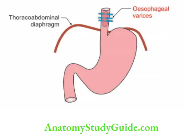
Oesophageal varices
Varices (plural of varix): Varix is an enlarged tortuous vein, artery or lymphatics.
1. Definition: It is varicosities of the branches of the azygos vein which anastomose with tributaries of the portal vein in the lower oesophagus, occurring in patients with portal hypertension.
2. Site: The lower end of the oesophagus.
1. It is one of the important sites of portosystemic anastomoses. The lower end of the oesophagus drains into
- Azygos and hemiazygos > superior vena cava a systemic vein.
- Gastric and splenic veins > portal vein.
2. Because of portal hypertension, the veins dilate and the dilated veins are called varices.
3. Applied anatomy:
- It is diagnosed by barium X-ray which shows a typical cotton wool appearance.
- Endoscopic examination at the lower end of the oesophagus is preferred.
- It is treated by injecting a sclerosing agent on 2 to 3 occasions.
- It can be ligated endoscopically by the application of bands.
- May rupture and cause dangerous or even fatal haematemesis.
Leave a Reply