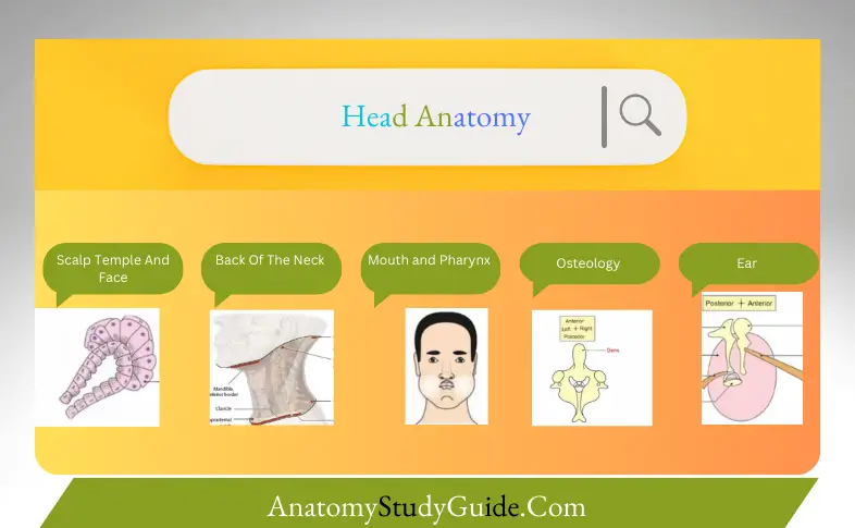
Anatomy of the Scalp, Temple, and Face
The anatomy of the scalp, temple, and face is complex, consisting of multiple layers, muscles, blood vessels, and nerves that are crucial for facial expressions, protection, and sensory perception.
Table of Contents
- Scalp Anatomy: The scalp comprises five layers, often remembered by the mnemonic “SCALP”—Skin, Connective tissue, Aponeurosis, Loose areolar tissue, and Pericranium. These layers provide protection to the skull and underlying brain and facilitate the blood supply and nerve innervation necessary for scalp health.

- Temple Anatomy: The temple is the flat area on the side of the skull, bordered by the forehead and the ear. The temporal muscles, which help in chewing, lie beneath the skin and subcutaneous tissue, anchored by the temporal bone.
- Face Anatomy: The face is one of the most intricate parts of the human body, composed of various muscles responsible for expressions, sensory organs such as eyes and nose, and critical arteries and nerves like the facial nerve. Understanding the anatomy of the face helps in diagnosing facial pain, injuries, and diseases.
Appendix Anatomy
The appendix is a small, tube-like structure attached to the cecum, part of the large intestine. Despite its reputation as a “vestigial organ,” recent studies suggest the appendix may play a role in gut immunity and maintaining a healthy gut flora.
- Location and Structure: Positioned in the lower right quadrant of the abdomen, the appendix is often about 10 cm long, though its size can vary. It has a small lumen, a muscular layer, and lymphoid tissue, indicating its potential role in immune function.
- Clinical Significance: Appendicitis, an inflammation of the appendix, is a common condition requiring surgical intervention. Understanding appendix anatomy is crucial for timely diagnosis and management of appendicitis and other related conditions.
Back of the Neck Anatomy
The back of the neck is composed of muscles, vertebrae, nerves, and blood vessels that support head movement, protect the spinal cord, and connect the skull to the upper body.
- Muscles: The major muscles include the trapezius, splenius capitis, and semispinalis capitis, which facilitate movements like head rotation, extension, and lateral bending.
- Vertebrae: The cervical vertebrae, especially C1 (atlas) and C2 (axis), are crucial for head movements. These bones support the head’s weight and allow nodding and rotational movements.
- Nerves and Blood Vessels: The spinal nerves in this region control sensation and motor functions of the neck and shoulders. Major blood vessels such as the vertebral artery also traverse this area, supplying the brain with oxygenated blood.
Anatomy of the Mouth and Pharynx
The mouth and pharynx play significant roles in digestion, respiration, and speech. Understanding their anatomy helps diagnose and treat conditions related to swallowing, breathing, and oral health.
- Mouth Anatomy: The mouth comprises the lips, teeth, gums, tongue, and palate. Each component has a specific function, from initial food breakdown by teeth to taste sensation by the tongue and the start of digestion with saliva.
- Pharynx Anatomy: The pharynx is a muscular tube that serves as a passageway for both air and food. It is divided into three parts: the nasopharynx, oropharynx, and laryngopharynx, each with a distinct role in breathing and swallowing.
- Clinical Relevance: Conditions like tonsillitis, pharyngitis, and oral cancers necessitate a clear understanding of mouth and pharynx anatomy for proper medical management.
Osteology Notes
Osteology refers to the study of bones, and understanding osteology is fundamental in anatomy, orthopedics, and forensic science.
- Skull Bones: The skull consists of several bones, including the frontal, parietal, temporal, and occipital bones, which protect the brain and support facial structures.
- Spinal Vertebrae: The vertebral column, made up of cervical, thoracic, lumbar, sacral, and coccygeal vertebrae, provides structural support and protects the spinal cord.
- Clinical Implications: Osteology helps in identifying fractures, bone diseases like osteoporosis, and conditions like scoliosis, which can affect posture and movement.
Anatomy of the Temporal and Infratemporal Regions
The temporal and infratemporal regions are key anatomical areas involved in mastication (chewing) and jaw movements.
- Temporal Region: This area houses the temporalis muscle, temporal bone, and crucial neurovascular structures. The temporal fossa serves as the attachment site for the temporalis muscle, which plays a vital role in elevating the jaw.
- Infratemporal Region: Located below the temporal fossa, the infratemporal fossa contains the pterygoid muscles, mandibular nerve, maxillary artery, and other essential structures involved in jaw movements and sensory innervation.
Ear Anatomy
The ear is a highly specialized organ responsible for hearing and balance. It is divided into three main parts: the outer ear, middle ear, and inner ear.
- Outer Ear: Comprising the auricle and ear canal, it captures sound waves and directs them toward the eardrum.
- Middle Ear: Contains the ossicles (malleus, incus, and stapes), which amplify sound vibrations and transmit them to the inner ear.
- Inner Ear: The cochlea and semicircular canals are responsible for hearing and balance, respectively. The vestibulocochlear nerve then transmits these sensory inputs to the brain.
- Clinical Insights: Common ear conditions include infections (otitis media), hearing loss, and balance disorders, which can be better understood through detailed ear anatomy.
Leave a Reply