Describe the femoral artery under the following heads
1. Femoral Artery Origin,
2. Femoral Artery Extent,
3. Femoral Artery Course,
4. Femoral Artery Relations,
5. Femoral Artery Branches, and
6. Femoral Artery Applied anatomy.
1. Femoral Artery Origin: Femoral artery is the continuation of the external iliac artery distal to inguinal ligament.
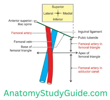
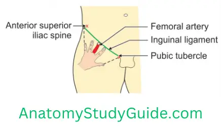
2. Femoral Artery Extent: It extends from mid-inguinal point to the apex of adductor canal.
3. Femoral Artery Course: It passes downward and medially. The upper ½ part is in the femoral and the lower ½ is in the adductor canal. At the apex of the adductor canal, it pierces adductor magnus and continues as popliteal artery.
Read And Learn More: Anatomy Notes And Important Question And Answers
4. Femoral Artery Relations
1. At the base of femoral
- Medially—femoral Vein inside the femoral sheath
- Laterally—femoral nerve outside the femoral sheath and
- Anteriorly in the femoral sheath—femoral branch of genitofemoral nerve.
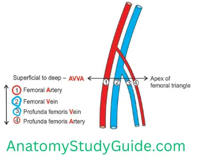
2. At the apex of the femoral, relations from anterior to posterior are AVVA
- Femoral Artery,
- Femoral Vein,
- Profunda femoris Vein, and
- Profunda femoris Artery.
3. Posterior relations from above downward are
- Psoas major,
- Pectineus, and
- Adductor longus.
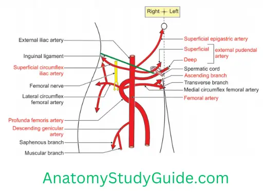
5. Femoral Artery Branches
1. Superficial
- Superficial epigastric artery,
- Superficial circumflex iliac artery, and
- Superficial external pudendal artery.
2. Deep
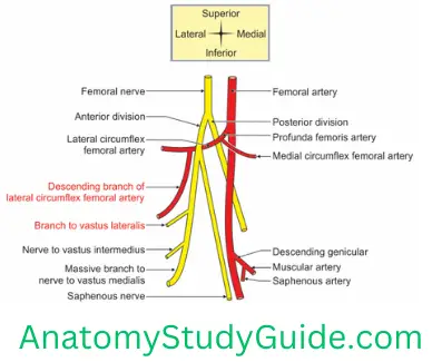
1. Profunda femoris: It is the largest deep branch of femoral artery. It is equal to the size of femoral artery. It arises 4 cm below the inguinal ligament. It originates from posterolateral aspect of femoral artery. It spirals deep to femoral artery and passes downwards deep to adductor longus. It soon disappears through the gap between the pectineus and adductor longus. It gives following branches.
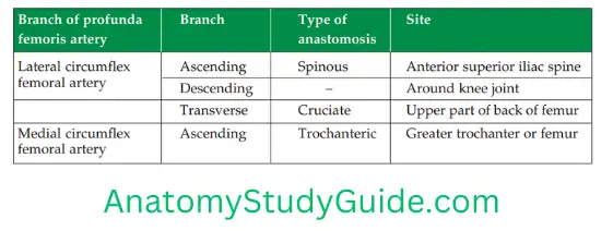
1. Lateral circumflex femoral artery. It gives
- Ascending branch: It takes part in spinous anastomosis.
- Descending branch: It takes part in anastomosis around knee joint.
- Transverse branch: It takes part in cruciate anastomosis.
2. Medial circumflex femoral artery. It gives
- Ascending branch: It takes part in trochanteric anastomosis. It is chief artery of head and neck of femur.
- Transverse branch: It takes part in cruciate anastomosis.
3. Perforating: There are four perforating arteries. They perforate adductor magnus. One of the perforating arteries gives a large nutrient branch to the femur.
2. Deep external pudendal artery supplies blood to the scrotum in male and labium majus in female
3. Muscular branches to all the deep muscles of thigh.
4. Descending genicular artery: It gives the following branches
- Saphenous artery, and
- Muscular artery
3. Termination: It continues as popliteal artery through an opening present in the adductor magnus.
6. Femoral Artery Applied anatomy
The pulsations of femoral artery can be felt just below the inguinal ligament over the head of femur, i.e. midway between the anterior superior iliac spine (ASIS) and pubic symphysis. It may be easily demonstrated using ultrasound.
1. Position of femoral vein can be localized as it lies just medial to the femoral artery.
2. Femoral artery can be compressed against the head of femur to control haemorrhage.
3. The circulation of the limb is not affected in case of the blockage of the femoral artery.
4. In case of the blockage proximal part of the femoral artery, the circulation is maintained through cruciate and trochanteric anastomoses.
5. In case of the blockage of the arteries of the distal part of the thigh, the circulation is maintained through perforating branches of profunda femoris artery and its anastomosis with the branches of the popliteal artery.
6. Severe narrowing of arteries of the lower limb, with an inadequate collateral circulation, can lead to pain in muscles. The pain appears on walking and disappears with rest. The pain appears as the person takes a few steps. It is called intermittent claudication (claudication, limping).
Name the branches of femoral nerve
1. Muscular branches
1. In pelvis. To
- Iliacus
- Pectineus
2. In thigh
1. Sartorius
2. Quadriceps femoris
- Rectus femoris
- Vastus medialis
- Vastus lateralis
- Vastus intermedius
2. Cutaneous branches
- Saphenous nerve
- Medial CUTaneous nerve of thigh
- Intermediate CUTaneous nerve of thigh
3. Articular branches to hip and knee joint
Describe the femoral nerve under the following heads
1. Femoral Nerve Root value,
2. Femoral Nerve Branches,
3. Femoral Nerve Course and relations, and
4. Femoral Nerve Applied anatomy.
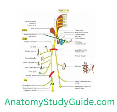
1. Femoral Nerve Root value: It is formed by dorsal division of ventral rami of.
2. Femoral Nerve Branches
1. From the trunk
- Branch to iliacus,
- Branch to pectineus, and
- Vascular branches to the femoral artery.
2. From the divisions
1. Anterior division (superficial branches)
It has one muscular and two cutaneous branches.
Muscular: SARTorius.
CUTaneous
- Medial CUTaneous nerve of thigh. It carries sensations of the skin of the medial side of thigh.
- Intermediate CUTaneous nerve of thigh. It carries sensations of the skin of front of thigh and takes part in the patellar plexus.
2. Posterior division. It gives the following branches
1. Muscular branches to QUADRIceps femoris
- Rectus femoris,
- Vastus medialis,
- Vastus intermedius, and
- Vastus lateralis.
2. Cutaneous branch is the SAPHenous nerve. It supplies skin over
- Upper part of leg, and
- Medial part of leg
- Medial part of sole of foot.
3. Articular branch to Joints.
- Hip joint, and
- Knee joint.
4. Femoral Nerve Vascular: To the femoral artery and its branches.
3. Course and relations
1. In the pelvis: The nerve is formed in the substance of psoas major. It runs on the lateral border of psoas major between psoas major and iliacus.
2. In the thigh: It enters thigh by passing deep to inguinal ligament.
- It lies on the lateral side of the femoral artery and outside the femoral sheath.
- The nerve runs 2.5 cm below the inguinal ligament. It breaks like a “cauda equina” into numerous motor and sensory twigs.
- The two divisions of the femoral nerve pass anterior and posterior to the lateral circumflex femoral artery.
3. Relations
- Anterior: Skin, superficial fascia and deep fascia.
- Posterior and medial: Psoas major.
- Posterior and lateral: Iliacus.
4. Femoral Nerve Applied anatomy
Disease of the hip joint may be referred to the knee joint.
Femoral nerve
- Neuroma of the femoral nerve or its branches is one of the differential diagnoses of lump in femoral
- Accumulation of pus or blood in the psoas sheath may compress the femoral nerve.
- Injury to femoral nerve causes paralysis of quadriceps femoris and affects extension of knee joint.
Femoral nerve neuropathy: The main trunk of the femoral nerve may be compressed by
- Entrapment of femoral nerve
- Compression by retroperitoneal tumour.
- Manifestations
- Wasting and weakness of quadriceps which results in
- Difficulty in walking.
- A localized neuropathy of the femoral nerve may occur in diabetes mellitus.
- Following are the characteristic clinical features
- Wasting and weakness of quadriceps leading to considerable difficulty in walking.
- Pain and paraesthesia on the anterior and medial aspects of the thigh. It extends along the medial aspect of the leg and foot.
- The femoral nerve block can be easily achieved in the femoral . The anaesthetic solution is injected one-finger breadth lateral to the point of femoral pulse. It is just below the mid-inguinal point.
- Injury to nerve results in atrophy of extensor muscles of thigh. It presents as weakness in extension of knee joint. There is loss of knee jerk.
- Effect of femoral nerve injury in gunshot wounds results in
- Motor loss: Inability to extend the knee. It is due to paralysis of the quadriceps femoris muscle.
- Sensory loss medial aspect of the thigh, leg and medial border of the foot as far as the ball of the big toe. It is due to the involvement of
- Intermediate and lateral cutaneous nerves of the thigh, and
- Saphenous nerve.
- The saphenous nerve may be entrapped at the site where it pierces the roof of the adductor canal. This will cause pain and paraesthesia along the medial side of the knee and leg.
Name the nerves forming the sub sartorial plexus
Subsartorial plexus is formed by
- Posterior branch of the medial femoral cutaneous nerve,
- Anterior division of obturator nerve, and
- Saphenous nerve.
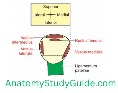
Attachments to patella
- Vastus medialis,
- Vastus intermedius,
- Vastus lateralis,
- Rectus femoris, and
- Ligamentum patellae.
Leave a Reply