• Skip to main content
  • Skip to secondary menu
  • Skip to primary sidebar
  • Skip to footer
  • Anatomy
    • Anatomy Question And Answers
    • Face Anatomy
    • Neck Anatomy
    • Head Anatomy
    • Oral Anatomy
    • Lower Limb
    • Upper Limb
  • Endodontics
    • Paediatric Dentistry
  • General Histology
    • Oral Histology
    • Genetics
  • Pediatric Clinical Methods
  • Complete Dentures
    • Pharmacology for Dentistry
  • Medical Physiology
    • Body Fluids
    • Muscle Physiology
    • Digestive System
    • Renal Physiology
    • Endocrinology
    • Nervous System
    • Respiratory System
    • Cardiovascular System
    • Reproductive System
    • Oral Physiology
  • General Medicine
  • General Pathology
    • Systemic Pathology
    • Oral Pathology
    • Neoplasia
    • Homeostasis
    • Infectious Diseases
    • Infammation
    • Amyloidosis Notes
  • Periodontology
  • General Surgery
    • Basic Principles Of Surgery
    • General Surgery

Anatomy Study Guide

Anatomy Study Guide

  • About Us
  • Contact Us
  • Privacy Policy
  • Terms of Use
  • Disclaimer
  • Sitemap
Home » Adaptation Of Spacer On Primary Cast Notes

Adaptation Of Spacer On Primary Cast Notes

November 8, 2023 by Joankessler parkland Leave a Comment

Adaptation Of Spacer On Primary Cast Armamentarium

  • Maxillary and mandibular primary casts
  • Spacer wax or modeling wax
  • Red and blue pencils
  • Rubber bowl with hot water
  • BP Blade No. 15 with handle.

Guiding Lines:

Table of Contents

  • Adaptation Of Spacer On Primary Cast Armamentarium
  • Adaptation Of Wax Spacer On Mandibular Primary Cast

Read and Learn  More: Preclinical Prosthodontics Notes

Step 1:

Mark the midline using the frenum as the reference point. Mark the outline of the sulcus on the primary cast. The Sulcus line is marked in the deepest part of the sulcus. A red color pencil can be used for marking the sulcus line. The Sulcus line is a continuous line. In a posterior aspect of the maxillary cast, a red line is drawn 1 mm beyond the posterior vibrating line.
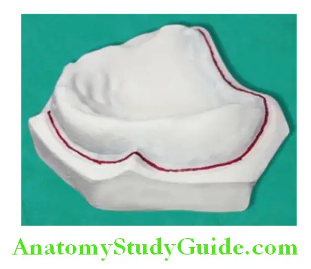
Adaptation Of Spacer On Primary Cast marking the sulcus outline on the cast

Step 2:

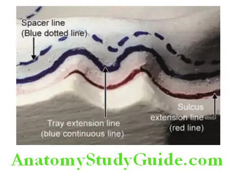
Place scale in the sulcus and mark several points 2 mm away from the sulcus line at different areas. Join these points as a bold line 2 mm away from the sulcus on the labial and buccal side of the cast. Use a blue color pencil for this line. This line (continuous blue line) is 2 mm short of the sulcus line. It indicates an extension of the custom tray border. The blue line is not marked 2 mm short in the posterior aspect of casts.

Step 3:

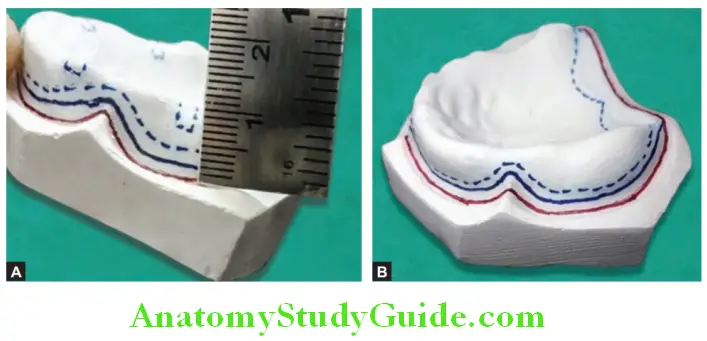
Draw a spacer outline. This line is drawn as a dotted line. Draw it at a level 2 mm short of the tray outline. In the posterior part of the maxillary cast, this line is limited till the anterior vibrating line of the posterior palatal seal area.

Step 4:

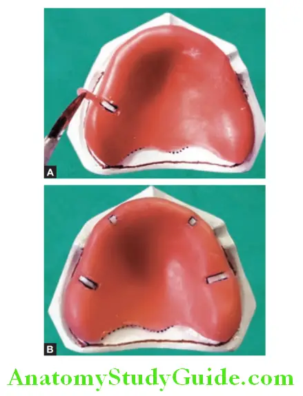
Outline of tissue stops: Four tissue stops are marked on the maxillary cast, two anteriorly (canine region) and two posteriorly (molar region). On the mandibular cast, two tissue stops are marked in the canine region.
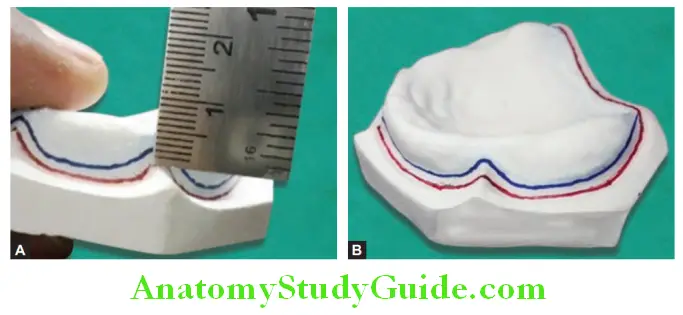
Adaptation Of Spacer On Primary Cast marking tray outline 2 mm short of sulcus line

Adaptation Of Spacer On Primary Cast draw spacer line 2 mm short of tray extension line

Adaptation Of Spacer On Primary Cast showing guidelines on casts

Adaptation of Spacer:

Step 1:

Take half a sheet of modeling wax (or spacer wax) and soften it over the Bunsen burner. Place it over the maxillary cast and adapt it without any wrinkles.
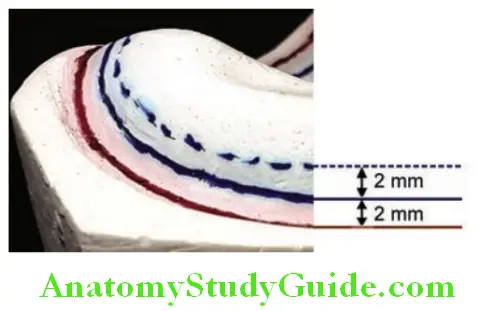
Adaptation Of Spacer On Primary Cast distance betwween different guidelines

Step 2:

Put a cut on wax at the level of the spacer line (dotted line) with a No. 15 BP blade. Take care not to scratch the cast with the blade. Remove excess wax beyond the dotted line.
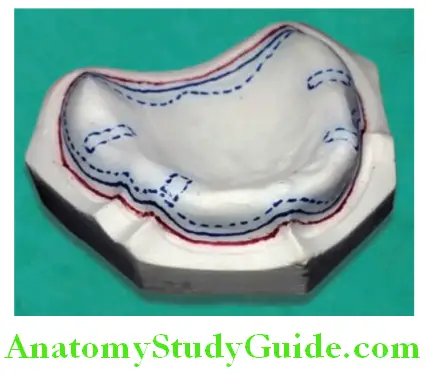
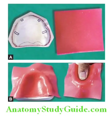
Adaptation Of Spacer On Primary Cast guidelines with tissue stops

Adaptation Of Spacer On Primary Cast adaptation of modeling wax on cast

Adaptation Of Spacer On Primary Cast cut the modeling wax along the spacer line

Step 3:

Cut the tissue stop area according to the outline to make a window in spacer wax. The spacer is 4 mm short of the sulcus and limited to the anterior vibrating line in the posterior palatal seal area.

Adaptation Of Spacer On Primary Cast creating window for fabrication of tissue stops

Note: Wax is cut and removed from the spacer according to the tissue stop outline. This will create a window in the spacer wax. This is not the tissue stop. The tissue stop is the extension of a custom tray (through this window) that is in contact with the cast.

Adaptation Of Wax Spacer On Mandibular Primary Cast

Guiding Lines:

Step 1:

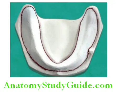
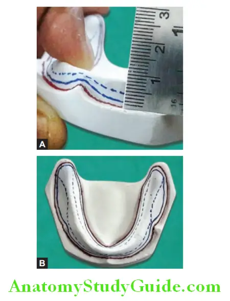
Mark the outline of the sulcus on the mandibular primary cast as done for the maxillary cast.

Adaptation Of Spacer On Primary Cast drawing the sulcus line

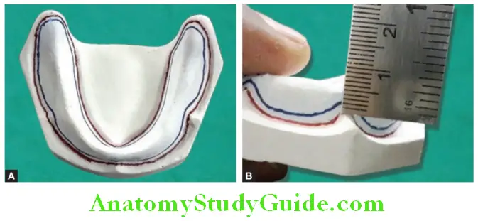
Adaptation Of Spacer On Primary Cast marking tray extension line 2 mm short of sulcus line

Step 2:

Mark custom tray extension line (continuous blue line). It is 2 mm short of the sulcus line. It indicates an extension of the custom tray border.

Step 3:

Draw a spacer outline. This line is drawn as a dotted line. Draw it at a level 2 mm short of the tray outline. In the case of a mandibular cast, the buccal-shelf area is not covered with a spacer as it is the primary stress-bearing area. Mark outline for two tissue stops (2 × 2 mm) on mandibular cast at canine region.

Adaptation Of Spacer On Primary Cast all guidelines draw on mandibular cast

Note: The buccal-shelf area itself acts as a tissue stop. So no separate tissue stops are made in the posterior region of the mandibular cast.

Adaptation of Spacer:

Step 1:

Take half a sheet of modeling wax roughly shape it according to arch shape, and soften it over the Bunsen burner. Place it over the mandibular cast and adapt it without any wrinkles.

Step 2:

Put a cut on wax at the level of the spacer line (dotted line) with the No. 15 BP blade. Take care not to scratch the cast with the blade. Remove excess wax beyond the dotted line. Remove spacer wax from the buccal-shelf area.
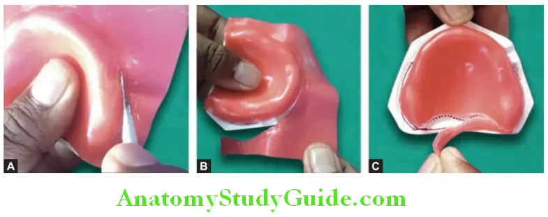
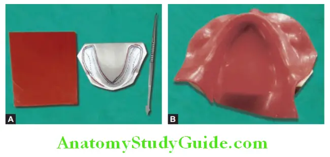
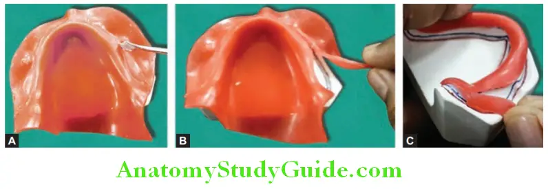
Adaptation Of Spacer On Primary Cast adaptation of modeling wax to create spacer

Adaptation Of Spacer On Primary Cast cut modeling wax at the level of spacer line

Step 3:

Cut the tissue stop area according to the outline to make a window in spacer wax. The spacer is 4 mm short of the sulcus. Make sure that there is no spacer on the buccal shelf area.
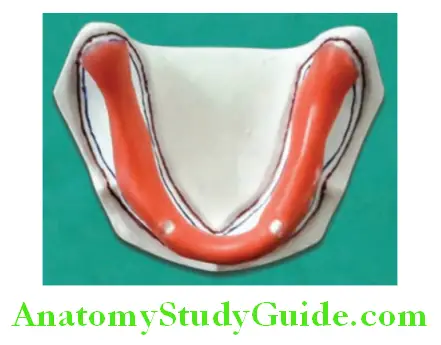
Adaptation Of Spacer On Primary Cast spacer adapted on mandibular cast

Filed Under: Preclinical Prosthodontics

Reader Interactions

Leave a Reply Cancel reply

Your email address will not be published. Required fields are marked *

Primary Sidebar

Recent Posts

  • Esophagus Anatomy
  • Lacrimal Apparatus: Anatomy, Parts & Function
  • Scalp Temple And Face Question and Answers
  • Orbicularis Oculi Muscle Anatomy
  • Extraocular Muscles Anatomy
  • Ciliary Ganglion Anatomy
  • Femoral sheath Anatomy
  • Femoral Artery – Location and Anatomy
  • Adductor Canal: Anatomy And Function
  • Ankle Joint: Anatomy, Bones, Ligaments And Movements
  • Risk Factors For Breast Cancer
  • Cervical Tuberculous Lymphadenitis Notes
  • Carbuncles: Causes, Symptoms, and Treatments
  • Sinuses And Fistulas Notes
  • Cellulitis: Treatments, Causes, Symptoms
  • Pyogenic Liver Abscess: Causes, Symptoms, and Diagnosis
  • Acid Base Balance Multiple Choice Questions
  • General Surgery Multiple Choice Questions
  • Hypertrophic Scarring Keloids Multiple Choice Questions
  • Surgical Site Infection Multiple Choice Questions
  • Facebook
  • Pinterest
  • Tumblr
  • Twitter

Footer

Anatomy Study Guide

AnatomyStudyGuide.com is a student-centric educational online service that offers high-quality test papers and study resources to students studying for Medical Exams or attempting to get admission to different universities.

Recent

  • Esophagus Anatomy
  • Lacrimal Apparatus: Anatomy, Parts & Function
  • Scalp Temple And Face Question and Answers
  • Orbicularis Oculi Muscle Anatomy
  • Extraocular Muscles Anatomy

Search

Copyright © 2026 · Magazine Pro on Genesis Framework · WordPress · Log in