Physiological Anatomy Of Respiratory Tract Introduction
Respiration is the process by which oxygen is taken in and carbon dioxide is given out. The first breath takes place only after birth.
Table of Contents
- Fetal lungs are nonfunctional. So, during intrauterine life, the exchange of gases between fetal blood and mother’s blood occurs through the placenta.
- After the first breath, the respiratory process continues throughout the life. The permanent stoppage of respiration occurs only at death.
Normal respiratory rate in different age:
- Newborn: 30-60/minute
- Early childhood: 20-40/minute
- Late childhood: 15-25/minute
- Adult: 12-16/minute
Types Of Respiration:
Respiration is often classified into two types:
- External respiration that involves exchange of respiratory gases, i.e. oxygen and carbon dioxide between lungs and blood.
- Internal respiration which involves exchange of gases between blood and tissues.
Phases Of Respiration:
Respiration occurs in two stages:
- Inspiration during which the air enters the lungs from the atmosphere
- Expiration during which the air leaves the lungs.
- While passing through the lungs, the atmospheric air (inspired air) delivers oxygen to the blood in the pulmonary capillaries and in exchange, it takes away carbon dioxide from the blood.
- During normal breathing, inspiration is an active process and expiration is a passive process.
Functional Anatomy Of Respiratory Tract
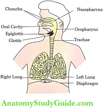
Respiratory tract is the anatomical structure through which air moves in and out. It consists of nose, pharynx, larynx, trachea, bronchi, and lungs.
Pleura:
Each lung is enclosed by a bilayered serous membrane called pleura or pleural sac. The two layers of pleura are the visceral and parietal layers.
- Visceral (inner) layer is attached firmly to the surface of the lungs. At hilum, it is continuous with parietal (outer) layer, which is attached to the wall of the thoracic cavity.
- The narrow space in between the two layers of pleura is called intrapleural space or pleural cavity.
- This space contains a thin film of serous fluid called pleural fluid. It is secreted by the visceral layer of the pleura.
- It functions as a lubricant to prevent friction between two layers. It is involved in the creation of negative pressure called intrapleural pressure within intrapleural space.
- In some pathological conditions, the pleural cavity expands with an accumulation of air (pneumothorax), water (hydrothorax), blood (hemothorax), or pus (pyothorax).
Read And Learn More: Medical Physiology Notes

Tracheobronchial Tree:
The trachea and bronchi are together called the tracheobronchial tree. It forms a part of the air passage.
- The trachea bifurcates into two main or primary bronchi called right and left bronchi.
- Each primary bronchus enters the lungs and divides into lobar or secondary bronchi. The secondary bronchi divide into segmental or tertiary bronchi.
- In the right lung, there are ten tertiary bronchi, and, in the left lung, there are eight tertiary bronchi.
- The tertiary bronchi divide several times with a reduction in length and diameter into many generations of bronchioles.
- When the diameter of bronchioles becomes 1 mm or less, it is called terminal bronchiole. The terminal bronchiole continues or divides into the respiratory bronchiole, which has a diameter of 0.5 mm.
- Generally, the respiratory tract is divided into two parts upper respiratory tract and lower respiratory tract.
Upper Respiratory Tract:
- The upper respiratory tract includes all the structures from the nose up to the vocal cords.
- Vocal cords are the folds of mucous membranes within the larynx that vibrate to produce the voice.
Lemur Respiratory Tract:
The lower respiratory tract includes the trachea, bronchi, and lungs.
Respiratory Unit
- Lung parenchyma is formed by a respiratory unit that forms the terminal portion of the respiratory tract.
- The respiratory unit is defined as the structural and functional unit of the lung.
- The exchange of gases occurs only in this part of the respiratory tract.
Structure Of Respiratory Unit:
- The respiratory unit starts from the respiratory bronchioles.
- Each respiratory bronchiole divides into alveolar ducts. Each alveolar duct enters an enlarged structure called the alveolar sac.
- The space inside the alveolar sac is called the antrum. The alveolar sac consists of a cluster of alveoli. Few alveoli are present in the wall of the alveolar duct also.
Thus, the respiratory unit includes:
- Respiratory bronchioles
- Alveolar ducts
- Alveolar sacs
- Antrum
- Alveoli.
Each alveolus is like a pouch with a diameter of about 0.2-0.5 mm. It is lined by epithelial cells.

Alveolar Cells or Pneumocytes: The epithelial lining of the alveoli consists of two types of cells called type I alveolar cells and type II alveolar cells. Alveolar cells are also called pneumocytes.
Type 1 alveolar cells:
- Type I alveolar cells are squamous epithelial cells forming about 95% of the total number of cells.
- These cells form the site of gaseous exchange between the alveolus and blood.
Type 2 alveolar cells:
- These alveolar cells are cuboidal in nature and form about 5% of alveolar cells.
- These cells are also called granular pneumocytes. Type II alveolar cells secrete the alveolar fluid and surfactant.
Respiratory Membrane:
The respiratory membrane is the membranous structure through which the exchange of gases occurs.
- The respiratory membrane separates air in the alveoli from the blood in the capillary. It is formed by the alveolar membrane and capillary membrane.
- The respiratory membrane has a surface area of 70 sq. meters and a thickness of 0.5 microns. The structure of the respiratory membrane is explained.
Nonrespiratory Functions Of Respiratory Tract
Besides the primary function of gaseous exchange, the respiratory tract is involved in several nonrespiratory functions of the body. Particularly, the lungs function as a defense barrier and metabolic organs, which synthesize some important compounds. The non-respiratory functions of the respiratory tract are:
Olfaction: Olfactory receptors present in the mucous membrane of the nostril are responsible for olfactory sensation.
Vocalization: Along with other structures, the larynx forms the speech apparatus. However, the larynx alone plays a major role in the process of vocalization. Therefore, it is called a sound box.
Prevention Of Dust Particles:
The dust particles, which enter the nostrils from the air, are prevented from reaching the lungs by the filtration action of the hairs in the nasal mucous membrane.
- The small particles, which escape the hairs, are held by the mucus secreted by the nasal mucous membrane.
- Those dust particles, which escape the nasal hairs and nasal mucous membrane, are removed by the phagocytic action of the macrophages in the alveoli.
- The particles which escape the protective mechanisms in the nose and alveoli are thrown out by the cough reflex and sneezing reflex.
Defense Mechanism:
- Lungs play an important role in the immunological defense system of the body. The defense functions of the lungs are performed by their own defenses and by the presence of various types of cells in the mucous membrane lining the alveoli of lungs.
- These cells are leukocytes, macrophages, mast cells, natural killer cells, and dendritic cells.
Lung’s Own Defenses:
- The epithelial cells lining the air passage secrete some innate immune factors called defensins and cathelicidins.
- These substances are the antimicrobial peptides which play an important role in lung’s natural defenses.
Defense through Leukocytes:
- The leukocytes, particularly the neutrophils and lymphocytes present in the alveoli of lungs provide a defense mechanism against bacteria and viruses.
- The neutrophils kill the bacteria by phagocytosis. Lymphocytes develop immunity against
Defense through Macrophages:
Macrophages engulf the dust particles and the pathogens, which enter the alveoli and thereby act as scavengers in the lungs.
- Macrophages are also involved in the development of immunity by functioning as antigen-presenting cells.
- When foreign organisms invade the body, the macrophages, and other antigen-presenting cells kill them.
- Later, the antigen from the organisms is digested into polypeptides.
- The polypeptide products are presented to T lymphocytes and B lymphocytes by the macrophages.
- Macrophages secrete interleukins, tumor necrosis factors (TNF), and chemokines.
- Interleukins and TNF activate the general immune system of the body.
- Chemokines attract the white blood cells toward the site of any inflammation.
Defense through Mast Cell: A mast cell is a large tissue cell resembling the basophil. The mast cell produces the hypersensitivity reactions like allergy and anaphylaxis. it secretes heparin, histamine, serotonin, and hydrolytic- enzymes.
Defense through Natural Killer Cell:
- A natural killer (NK) cell is a large granular cell, considered as the third type of lymphocyte.
- Usually, NK cell is present in the lungs and other lymphoid organs. Its granules contain hydrolytic enzymes, which destroy the microorganisms.
- NK cell is said to be the first line of defense in specific immunity particularly against viruses.
- It destroys the viruses and the viral infected or damaged cells, which may form tumors.
- It also destroys malignant cells and prevents the development of cancerous tumors. The NK cells secrete interferons and tumor necrosis factors.
Defense through Dendritic Cells: Dendritic cells in the lungs play an important role in immunity. Along with macrophages, these cells function as antigen-presenting cells.
Maintenance Of Water Balance: The respiratory tract plays a role in the water loss mechanism. During expiration, water evaporates through the expired air and some amount of body water is lost by this process.
Regulation Of Body Temperature: During expiration, along with water, heat is also lost from the body. Thus, the respiratory tract plays a role in the heat loss mechanism.
Regulation Of Acid-Base Balance:
Lungs play a role in the maintenance of the acid-base balance of the body by regulating the carbon dioxide content in the blood.
- Carbon dioxide is produced during various metabolic reactions in the tissues of the body.
- When it enters the blood, carbon dioxide combines with water to form carbonic acid. Since carbonic acid is unstable, it splits into hydrogen and bicarbonate ions
CO2 + H2O → H2CO–3 → H+ + HCO–3
The entire reaction is reversed in the lungs when carbon dioxide is removed from the blood into the alveoli of the lungs.
H+ + HCO–3 → H2CO–3 → CO2 + H2O
- As carbon dioxide is a volatile gas, it is practically blown out by ventilation.
- When metabolic activities are accelerated, more amount of carbon dioxide is produced in the tissues and me concentration of hydrogen ions is also increased leading to a reduction in pH.
- The increased hydrogen ion concentration causes increased pulmonary ventilation, i.e. hyperventilation by acting through various mechanisms like chemoreceptors in aortic and carotid bodies and in the medulla of the brain.
- Due to hyperventilation, the excess of carbon dioxide is removed from the body fluids, and the pH is brought back to normal.
Anticoagulant Function:
Mast cells in the lungs secrete heparin. Heparin is an anticoagulant and it prevents intravascular clotting.
Secretion Of Angiotensin Converting Enzyme:
- Endothelial cells of the pulmonary capillaries secrete the angiotensin-converting enzyme (ACE).
- It converts the angiotensin I into active angiotensin II which plays an important role in the regulation of ECF volume and blood pressure.
Synthesis Of Hormonal Substances:
- Lung tissues are also known to synthesize the hormonal substances, prostaglandins, acetylcholine, and serotonin which have many physiological actions in the body including the regulation of blood pressure.
Respiratory Protective Reflexes
- Respiratory protective reflexes are the reflexes that protect the lungs and air passage from foreign particles.
- The respiratory process is modified by these reflexes in order to eliminate foreign particles or to prevent the entry of these particles into the respiratory tract. The respiratory protective reflexes are:
Cough Reflex:
- Cough is a modified respiratory process characterized by forced expiration. It is the protective reflex that occurs because of irritation of the respiratory tract and some other areas such as the external auditory canal (see below).
Cough Reflex Causes:
- Cough is produced mainly by irritant agents. It is also produced by several disorders such as cardiac disorders (congestive heart failure), pulmonary disorders (chronic obstructive pulmonary disease – COPD), and tumors in the thorax which may exert pressure on the larynx, trachea, bronchi, or lungs.
Cough Reflex Mechanism:
The cough begins with a deep inspiration followed by forced expiration with the closed glottis. This increases the intrapleural pressure above 100 mm Hg.
- Then, the glottis opens suddenly with the explosive outflow of air at a high velocity. The velocity of the airflow may reach 960 km/ hour. It causes the expulsion of irritants out of the respiratory tract.
Reflex Pathway:
- The receptors that initiate the cough are situated in several locations such as the nose, paranasal sinuses, larynx, pharynx, trachea, bronchi, pleura, diaphragm, pericardium, stomach, external auditory canal, and tympanic membrane.
- Afferent nerve fibers pass via the vagus, trigeminal, glossopharyngeal, and phrenic nerves. The center for the cough reflex is in the medulla oblongata.
- The efferent nerve fibers arising from the medu llary center pass through the vagus, phrenic, and spinal motor nerves. These nerve fibers activate the primary and accessory respiratory muscles.
Sneezing Reflex: Sneezing is also a modified respiratory process characterized by forced expiration. It is a protective reflex caused by irritation of the nasal mucous membrane.
Sneezing Reflex Causes: Irritation of the nasal mucous membrane occurs because of dust particles, debris, mechanical obstruction of the airway, and excess fluid accumulation in the nasal passages.
Sneezing Reflex Mechanism: Sneezing starts with deep inspiration, followed by forceful expiratory effort with opened glottis resulting in the expulsion of irritant agents out of the respiratory tract.
Reflex Pathway: Sneezing is initiated by the irritation of the nasal mucous membrane, the olfactory receptors, and trigeminal nerve endings present in the nasal mucosa.
- Afferent nerve fibers pass through the trigeminal end olfactory nerves. The sneezing center is in the medulla oblongata.
- It is located diffusely in the spinal nucleus of the trigeminal nerve, the nucleus solitarius, and the reticular formation of the medulla.
- The efferent nerve fibers from the medullary center pass via trigeminal, facial, glossopharyngeal, vagus, and intercostal nerves. These nerve fibers activate the pharyngeal, tracheal, and respiratory muscles.
Swallowing (Deglutition) Reflex:
Swallowing is a respiratory protective reflex that prevents the entrance of food particles into the air passage during swallowing.
- While swallowing the food, respiration is arrested for a while. The temporary arrest of respiration is called apnea. The arrest of breathing during swallowing is called swallowing apnea or deglutition apnea.
- It takes place during the pharyngeal stage, i.e. II stage of deglutition, and prevents entry of food particles into the respiratory tract.
Leave a Reply