Describe Interossei Under The Following Heads
Table of Contents
1. Interossei Insertion,
2. Interossei Axis,
3. Interossei Strength,
4. Interossei Relation,
5. Interossei Action,
6. Interossei Nerve supply, and
7. Interossei Testing.
Answer:
Interossei Introduction:
These are the small muscles of the hand present in between the metacarpals. They are divided into palmar and dorsal interossei. They are discussed as given in Table.
Palmar and dorsal interossei
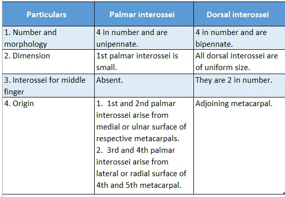
1. Interossei Insertion:
Both are inserted on
- Dorsal digital expansion, and
- The base of the proximal phalanx of the respective finger.
2. Interossei Axis:
Passes through middle finger.
3. Interossei Strength:
Palmar interossei are less strong than dorsal interossei.
Palmar And Dorsal Interossei
Read And Learn More: Anatomy Notes And Important Question And Answers
4. Interossei Relations:
- They are related posteriorly to deep transverse metacarpal ligaments.
- The radial artery passes between the gap produced by two heads of 1st dorsal interosseous.
- Proximal perforating arteries pass between the gap produced by two heads of the 2nd, 3rd, and 4th dorsal interossei.
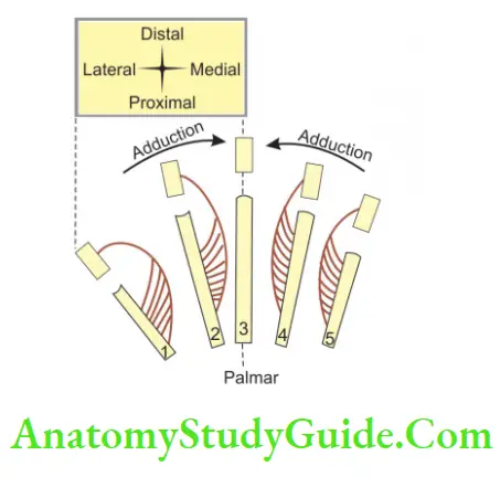
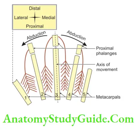
5. Interossei Actions:
(PAD, DAB. P=palmar, AD=adductor; D=Dorsal, AB=abductor, B-bipennate)
- Palmar interossei are adductors, dorsal interossei are abductors.
- They are powerful flexors of the metacarpophalangeal joint (MP) because their tendons are placed ventrally and distally with respect to the metacarpophalangeal joint.
- They produce an extension of the interphalangeal joint. Both these movements are useful for precision work.
6. Interossei Nerve supply:
They are supplied by a deep branch of the ulnar nerve.
7. Interossei Testing:
Interossei can be tested in the following ways.
- Dorsal interossei: By asking to spread the fingers against resistance.
- Palmar interossei: By asking to hold a piece of paper between the testing finger and the normal finger.
Nerve Supply Of Lubricants
- 1st and 2nd lubricants are supplied by the median nerve.
- 3rd and 4th is supplied by the ulnar nerve.
Actions Of Lubricants
- Flexion of metacarpophalangeal joint of 2nd to 5th digits.
- Extension of interphalangeal joint of 2nd to 5th digits.
Lumbricals
1. Lumbricals Features:
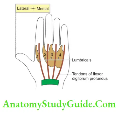
- These are worm-like muscles that arise from tendons of flexor digitorum profundus.
- They are 4 in number and are present on the palmar aspect.
- They are counted from lateral to medial.
- 1st and 2nd lumbricals are unipennate.
- 3rd and 4th lumbricals are bipennate.
- They act as link muscles between the deep flexor and extensor tendons of the hand.
2. Lumbricals Proximal attachments:
- 1st and 2nd lumbricals arise from the radial side of profundus tendon, and
- 3rd and 4th lumbricals arise between neighboring tendons of flexor digitorum profundus.
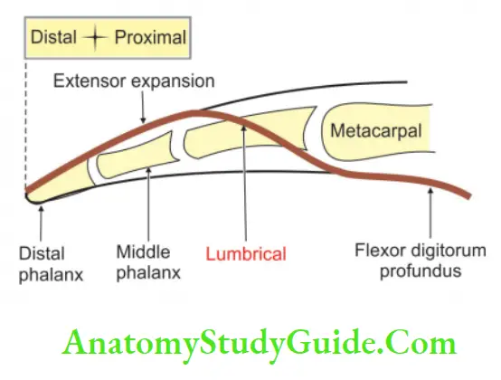
3. Lumbricals Distal attachments:
They are inserted on the dorsal surface of the base of the middle and distal phalanx, through the lateral border of dorsal digital expansion.
4. Lumbricals Action:
They are flexors of metacarpophalangeal joints and extensors of interphalangeal joints.
Palmar And Dorsal Interossei
5. Lumbricals Nerve supply:
Medial two lumbricals are supplied by the deep branch of the ulnar nerve and lateral two lumbricals are supplied by the median nerve.
6. Lumbricals Relations:
They are dorsal to digital vessels and nerves.
7. Lumbricals Functions:
They produce up-and-down strokes of fingers for skilled work. They produce flexion at the metacarpophalangeal joint and extension of interphalangeal joints.
8. Lumbricals Test:
The muscles are best tested by asking the patient to hold a piece of paper between the sides of two adjacent fingers. If the muscles are acting, the paper will be firmly held and some resistance will be offered to its withdrawal.
Branches Of Superficial Palmar Arch
There are 4 digital branches that supply medial 3 1/2 fingers. Out of these 4 digital branches, lateral 3 digital branches are connected with the deep palmar arch by the palmar metacarpal arteries.
Leave a Reply