Describe ankle joint (talocrural) under following heads
1. Ankle Joint Bones taking part,
2. Ankle Joint Classification,
3. Ankle Joint Ligaments,
4. Ankle Joint Movements and muscles producing movements,
5. Ankle Joint Relations,
6. Ankle Joint Arterial supply,
7. Ankle Joint Nerve supply, and
8. Ankle Joint Applied anatomy.
1. Ankle Joint Bones taking part
- Lower end of fibula with lateral malleolus,
- Medial malleolus of tibia, and distal part of tibia, and
- Superior surface of talus.
2. Ankle Joint Classification
1. Ankle Joint Structural:
- Axis: Uniaxial, modified hinge (since the axis of movement is basically transverse with a slight downward inclination on the lateral side) variety of synovial joint. Number of bones: Articulating bones in the formation of ankle joint are three and they are tibia, fibula and talus. Hence it is compound synovial joint.
2. Ankle Joint Functional: Diarthrosis (freely mobile).
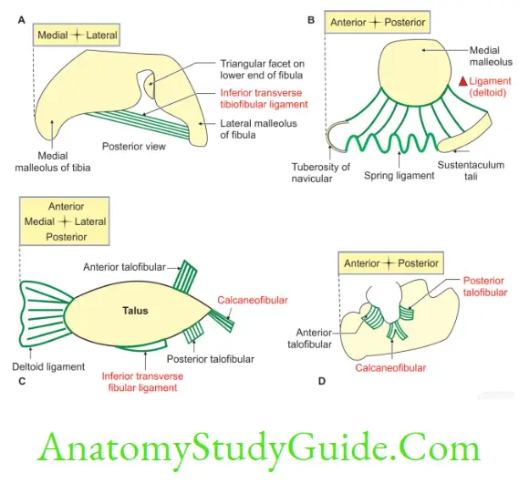
3. Ankle Joint Ligaments
1. Fibrous capsule covers the joint completely
1. Ankle Joint Attachments
- It is attached superiorly to the peripheral margins of the articular surfaces of lower end of tibia and fibula.
- It is attached inferiorly to the peripheral margin of Articular surface of the superior surface of talus. Comma-shaped facet present on the medial surface of talus. lar facet present on the lateral surface of talus.
2. Ankle Joint Variations in thickness
- Thin in front, thin behind.
- Thick laterally, thick medially.
3. Ankle Joint Fate: It blends with collateral ligament.
2. Ankle Joint Synovial membrane
- It lines the inner surface of the fibrous capsule.
- It stops at the periphery of the articular cartilage.
- A small synovial recess extends upwards in the inferior tibiofibular joint.
3. Ankle Joint Deltoid ligament: It is a strong lar ligament present on the medial side of the ankle joint. It has superficial and deep parts.
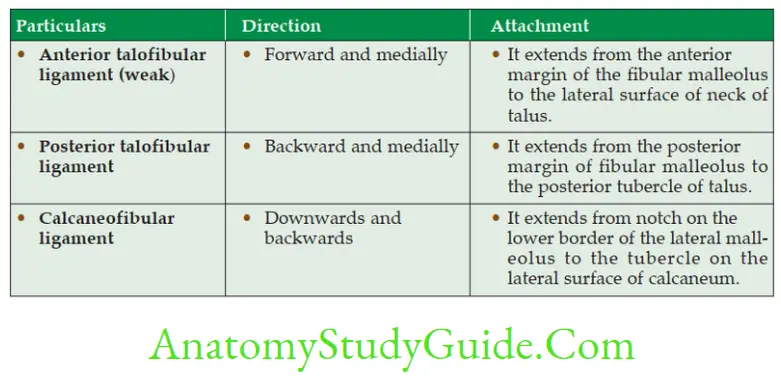
4. Ankle Joint Lateral ligament: It is a ligament present on the lateral side of ankle joint. It connects talus and calcaneum with fibula.
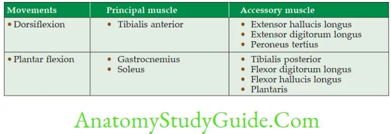
4. Movements and muscles producing movements
1. Dorsiflexion
1. Range of movement—10°–20°
2. All muscles of anterior or extensor compartment of the leg bring dorsiflexion of the ankle joint, i.e.
- Tibialis anterior,
- Extensor hallucis longus, and
- Extensor digitorum longus.
2. Plantar flexion
1. Range of movement is 20°–40°.
2. All the muscles of flexor compartment of the leg are plantar flexors of ankle joint.
3. The gastrocnemius with soleus acts as prime mover.
4. The peroneus longus and brevis come into play in extreme plantar flexion. The muscles bringing plantar flexion are
- Peroneus tertius,
- Tibialis posterior,
- Flexor digitorum longus,
- Flexor hallucis longus,
- Tendo Achilles.
3. In symmetrical standing, the line of gravity passes slightly in front of the ankle joint, therefore, there is a natural tendency for forward dislocation which is prevented by
- Broader anterior part of trochlear surface of talus, and
- Tonic contraction of triceps surae.
5. Ankle Joint Relations
1. Anterior (From medial to lateral)
Tibialis anterior
Extensor Hallucis longus
Anterior tibial Artery
Anterior tibial Nerve
Extensor Digitorum longus
Peroneus tertius
2. Posterior: Behind tibial malleolus.
(From anterior to posterior)
Tendon of tibialis posterior,
Flexor digitorum longus,
Posterior tibial artery,
Tibial nerve, and
Tendon of flexor hallucis longus.
3. Behind the fibular malleolus: Tendon of peroneus brevis and peroneus longus. The peroneus brevis is situated deep and peroneus longus is situated superficially.
6. Ankle Joint Arterial supply: Malleolar branches of anterior tibial and peroneal arteries.
7. Ankle Joint Nerve supply: Branches from deep peroneal and tibial nerves.
8. Ankle Joint Applied anatomy
- Ankle sprain: It is due to over inversion of foot, which is one of the common manifestations. The anterior talofibular and calcaneofibular ligaments are sometimes torn and in severe cases the anterior part of the capsule of the ankle joint is ruptured.
- Pott’s fracture: The forceful eversion of the foot causes avulsion of the deltoid ligament, fracture of the tibia and fibula. This is described in 3 degrees.
- In 1st degree there is only an Avulsion (tearing) of deltoid ligament.
- In 2nd degree
- Avulsion (tearing) of deltoid ligament
- Fracture of medial malleolus of tibia.
- In 3rd degree
- Avulsion fracture of the deltoid ligament,
- Fracture of medial malleolus of tibia, and
- Oblique fracture of the lower part of the fibula.
- The ankle sprains usually occur when the foot is plantar flexed.
- The sprains of the ankle joint are almost always abduction sprains of the subtalar joints.
Deltoid Ligament
Deltoid Ligament Introduction: It is a strong lar ligament present on the medial side of the ankle joint and it has superficial and deep parts.
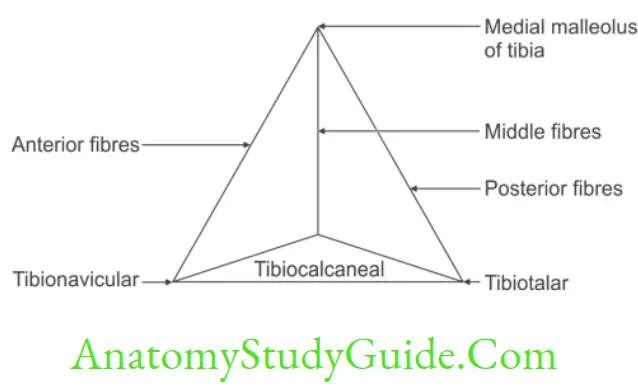
1. Superficial part
1. It is lar.
2. The base of the is attached to tarsal bones, namely navicular, calcaneum and talus.
3. The apex is attached to the tip of medial malleolus. They are subdivided into anterior, middle and posterior fibres.
1. Anterior fibres extend from medial malleolus of tibia to
- Tuberosity of navicular bone, and to the
- Medial margin of spring ligament.
- These fibres are present anteriorly; hence they are called anterior fibres of deltoid ligament or tibionavicular ligament.
2. Middle fibres extend from medial malleolus of tibia to sustantaculum tali of calcaneum. They are called tibiocalcanean ligament.
3. Posterior fibres extend from medial malleolus of tibia to the medial tubercle of talus. They are also called superficial tibiotalar ligament.
2. Deep part blends with fibrous capsule. It connects tibial malleolus to anterior part of medial surface of talus below comma shaped facet. This is also called deep tibiotalar ligament.
Movements of ankle joint
Lower Extremity Joints
Lateral ligaments of ankle joint
Describe inversion under following heads
1. Inversion Bones taking part,
2. Inversion Joints and classification of joints,
3. Inversion Axis,
4. Inversion Combination of action,
5. Inversion Range of movements,
6. Inversion Functions, and
7. Inversion Applied anatomy.
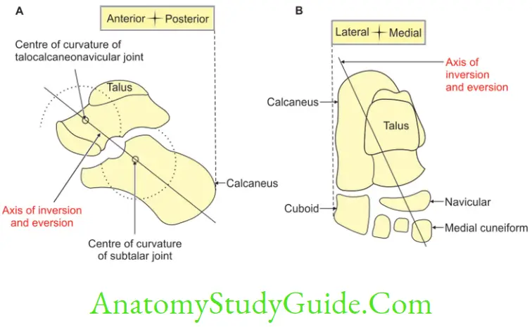
Inversion Introduction: It is a movement in which the medial border of foot is elevated so that the sole or plantar surface of the foot faces medially. Inversion and eversion are best demonstrated when the foot is off the ground.
When the foot is on the ground typical inversion and eversion are not observed.
The malleoli lock the talus and suspended foot inverts and everts around it.
1. Bones taking part: Talus, calcaneus and navicular.
2. Joints and classification of joints
- Talocalcaneonavicular: Ball and socket variety of synovial joint.
- Subtalar: Plane synovial joint.
3. Inversion Axis: It is an oblique axis. Its direction is forwards, upwards and medially. It passes from the back of lateral tubercle of calcaneus, through sinus tarsi. It emerges at the superomedial aspect of neck of talus. This axis corresponds to abduction, adduction, plantar flexion and dorsiflexion and medial and lateral rotations of calcaneus.
4. Inversion Combination of movements: Adduction, plantar flexion, rotation and gliding movements.
5. Inversion Range of movements: It is freer as compared to the eversion.
6. Inversion Functions
- The inversion movement helps to walk on slippery and uneven surfaces.
- It helps to maintain an efficient shift of weight distribution among the head of metatarsal bones during locomotion.
7. Inversion Applied anatomy
Inversion sprain is the most common ankle injury. It involves the tearing of lateral collateral ligaments. Usually, the anterior talofibular ligament is torn first, then the calcaneofibular ligament and in severe cases, the posterior talofibular ligament.
Leave a Reply