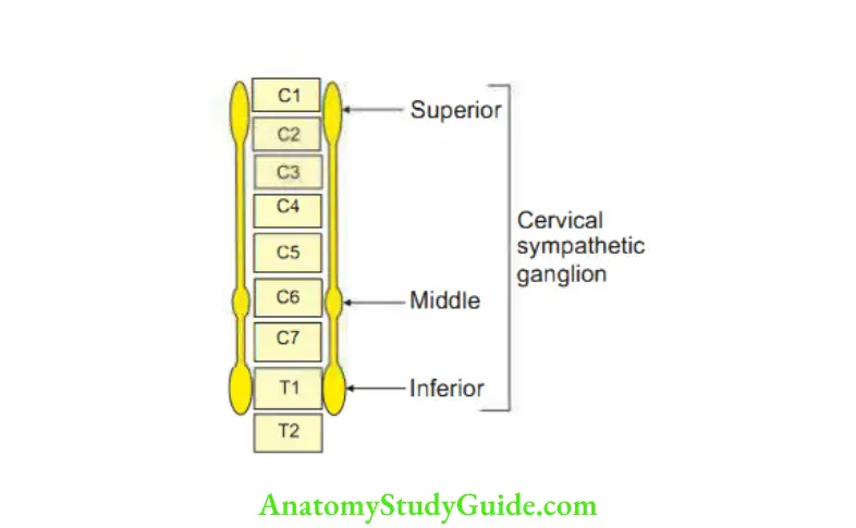
Cervical sympathetic ganglion
They are three in number.
Table of Contents
1. Superior cervical sympathetic ganglion.
2. Middle cervical sympathetic ganglion.
3. Inferior cervical sympathetic ganglion.
1. Superior cervical sympathetic ganglion
Read And Learn More: Head Anatomy Notes And Important Questions With Answers
1. Superior cervical sympathetic ganglion Features
- It consists of 1 million cell bodies.
- Length: 3 cm.
- Situation: In front of the transverse process of C2 and C3 vertebrae.
2. Superior cervical sympathetic ganglion Branches
- Gives grey rami to 1st four cervical nerves.
- The upper left ganglion gives a branch to the superficial cardiac plexus.
- It gives vascular branches to
1. Internal carotid artery and forms plexus which runs along the
- Branches to the internal carotid artery,
- Pterygopalatine ganglion, and
- Dilator pupillae muscle of the eyeball.
2. External carotid artery forms external carotid plexus and distributes along the
- Branches to the external carotid artery.
- Sympathetic fibers to pharyngeal plexus
- Glandular branch to
- Submandibular ganglion, and
- Otic ganglion.
2. Middle cervical sympathetic ganglion: It is a small, inconstant ganglion.
1. Middle cervical sympathetic ganglion Situation: Medial to carotid tubercle.
2. Middle cervical sympathetic ganglion Branches to
- Grey rami of 5th and 6th cervical nerves.
- Branches to deep cardiac plexus.
3. Middle cervical sympathetic ganglion Connection: It is connected by two or more strands to the inferior cervical sympathetic ganglion, one of which passes in front of the subclavian artery called ansa sub Clavia.
3. Inferior cervical sympathetic ganglion
1. Inferior cervical sympathetic ganglion Formation: It is formed by the fusion of the 7th and 8th cervical ganglia at the C7 vertebra.
It forms a stellate ganglion with Tl ganglion (cervicothoracic ganglion)
2. Inferior cervical sympathetic ganglion Relations
- Anterior: 8th cervical spinal nerve.
- Posterior: 3rd part of vertebral artery.
3. Inferior cervical sympathetic ganglion Branches
1. Vascular branches:
- Vertebral artery, and
- Subclavian artery.
2. Visceral branches: To the heart by deep cardiac plexus.
3. Other branches: Grey rami communicantes to the ventral rami of C7 and CS nerves.
5. cervical sympathetic ganglion Applied anatomy
Horner’s syndrome
- Homer’s syndrome: It is due to the involvement of the sympathetic nerve, which is contributed by Tl.
It is due to injury at the root of brachia! plexus. - HORNER. The letters of the word “Homer” give the information about clinical manifestations of Homer’s syndrome.
Hyperhidrosis (hypo-less, hydrolysis-sweating) is due to the involvement of sympathetic nerves, which arise from the first thoracic nerve.
These are secretomotor fibers supplying the sweat glands of the skin of the face and forehead.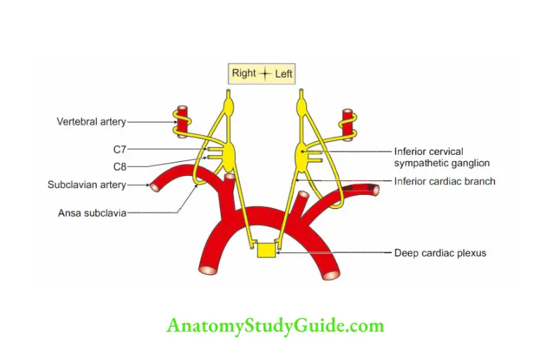
- Opening of the eye is lost due to ptosis (drooping of the upper eyelid). It is caused by paralysis of Muller’s muscle (smooth muscle of levator palpebrae superioris).
In fact, it is pseudoptosis. - Argyll-Robertson pupil [constricted pupil] is due to paralysis of dilator pupillae (unopposed action of sphincter pupillae).
- Narrowing of palpebral fissure.
- Elevation of the lower eyelid.
- Retraction of eyeball (sunken eyeball): Enophthalmos is due to the involvement of the orbitalis muscle.
- Absence of ciliospinal reflex.
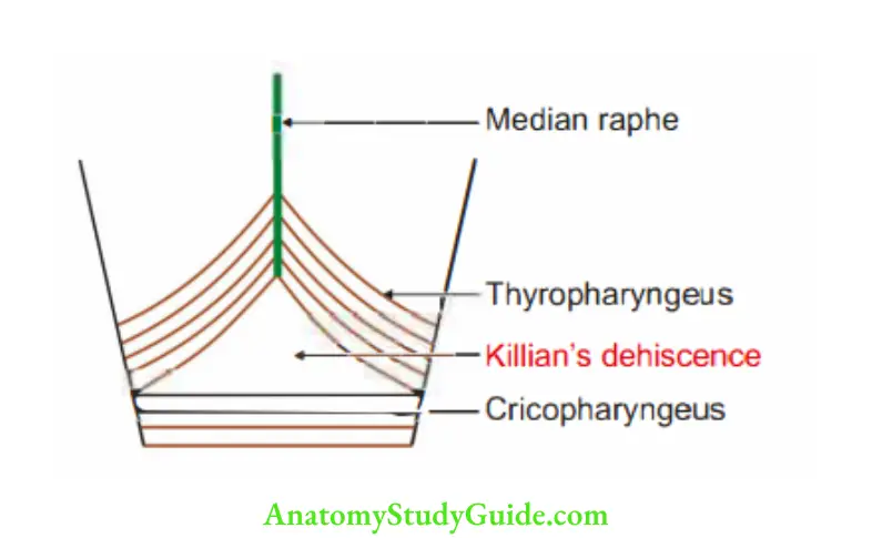
Killian’s dehiscence
Killian’s dehiscence: Part of the posterior wall of the pharynx between the lower part of vocal folds and cricopharyngeus is weak and is not covered by the muscle.
This weak area is called Killian’s dehiscence.
Pharyngeal diverticula are formed by an outpouching of the dehiscence.
The anatomical contributing factor for this condition is the neuromuscular incoordination of the two parts of the inferior constrictor.
The propulsive thyropharyngeus is supplied by the pharyngeal plexus and sphincter of the cricopharyngeus by the recurrent laryngeal nerve.
Leave a Reply