Back Of The Neck Anatomy Notes And Important Questions With Answers
Question 1. What is the cause of neck rigidity in meningitis?
Table of Contents
Neck rigidity is seen in cases with meningitis.
It is due to spasms of the extensor muscles.
This is caused by irritation of the nerve roots.
The nerve roots pass through the subarachnoid space which is infected.
The passive flexion of the neck and straight leg raising test cause pain as the nerves are stretched.
Read And Learn More: Head Anatomy Notes And Important Questions With Answers
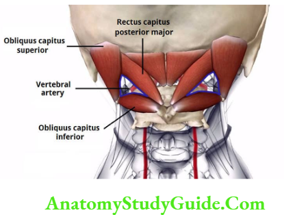
Question 2. Describe the suboccipital triangle under the following heads:
1. Boundaries,
2. Floor,
3. Roof,
4. Contents, and
5. Applied anatomy.
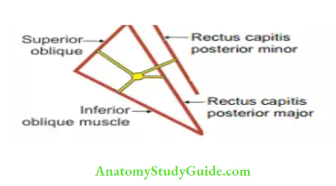
1. Boundaries
- Superolaterally: Superior oblique. It extends from the back of the lateral mass of the atlas to the lateral part of the occipital bone between the superior and inferior nuchal line.
- Superomedially
Rectus capitis posterior major
- It is wrongly named.
- It is not vertical.
- It arises from the outer surface of the bifid spinous process of the 2nd cervical vertebra.
- It is attached to the lateral part of the area below the inferior nuchal line.
- It extends and rotates the head toward the same side.
Rectus capitis posterior minor
- It is the only muscle attached to the posterior arch of the atlas.
- It arises from small fossa near the mid. ine and passes vertically upwards to be inserted into th medial part of th area below th inferior nuchal line.
- It extends the head.
Inferiorly by oblique capitis inferior. It is attached to the
- The outer surface of the bifid spine of the axis, and
- Back of the lateral mass of the atlas.
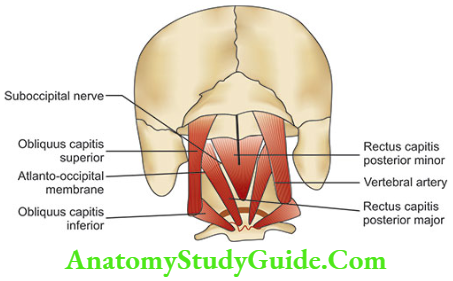
2. Floor
- Posterior arch of the atlas, and
- Posterior atlanto-occipital membrane.
3. Roof is formed by
- Semispinalis capitis medially, and
- Longissimus capitis laterally.
Both muscles are separated by dense fibrous tissue.
The structures crossing the roof are - Greater occipital nerve which crosses inferomedially.
- Occipital artery which crosses superolaterally.
4. Contents
- Third part of vertebral artery runs across the floor of the triangle.
- Dorsal ramus of 1st cervical nerve (suboccipital nerve) and its muscular branches emerge through the floor of suboccipital triangle.
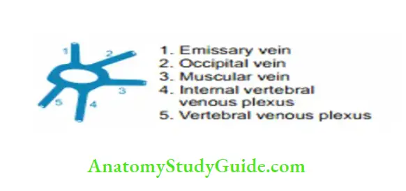
- Suboccipital venous plexus.
- Lymphatic plexus.
Fibrofatty tissue.
5. Applied anatomy
- Neck rigidity is an important sign of meningitis. It is due to spasm of extensor muscles (muscles of suboccipital triangle), caused by irritation of nerve roots, present in the subarachnoid space.
- Cistemal puncture is done through suboccipital triangle. It is done to collect CSF from cisterna magna.
The needle is introduced just above the spine of axis in forward and upward direction. - Posterior cranial fossa can be approached through the suboccipital triangle.
Suboccipital nerve
Introduction: It supplies all the muscles forming boundaries and roof of the suboccipital triangle.
1. Root value: Cl-1st cervical nerve.
2. Peculiarity: It does not have cutaneous branches.
3. Course: Suboccipital nerve is one of the contents of suboccipital triangle. It lies between vertebral artery and bone, i.e. atlas vertebra.
Branches
Ventral ramus: Itwinds around lateral side ofatlanto-occipital joint and passes forward to join the cervical plexus. It forms superior root of ansa cervicalis. It supplies
- Geniohyoid
- Thyrohyoid
Dorsal ramus: It has
Muscular branches supplying two recti, two oblique muscles and semispinalis capitis
- Rectus capitis posterior major,
- Rectus capitis posterior minor,
- Superior oblique (oblique capitis superior),
- Inferior oblique (oblique capitis inferior), and
Semispinalis capitis. - Sensory branch to meninges of brain
4. Applied anatomy: Neck rigidity: It is caused by irritation of the nerve roots that are passing through the infected subarachnoid space.
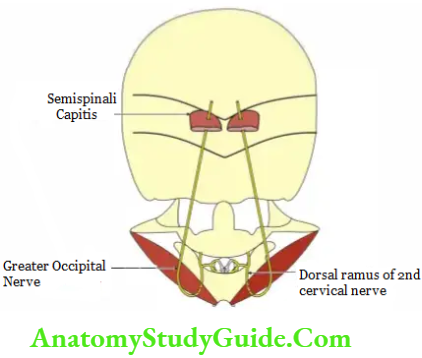
Greater occipital nerve
Introduction: Large medial branch of dorsal ramus of C2.
1. Root value: Dorsal ramus of C2.
2. Peculiarities: Thickest cutaneous nerve of body.
3. Distribution
- Sensory: Skin of scalp over posterior part of ear.
- Motor: Semispinalis capitis.
4. Course and relations
- Crosses suboccipital triangle.
- Pierces semispinalis capitis and trapezius.
- Runs on back of head and reaches vertex.
5. Applied anatomy
- In meningitis, there is irritation of nerve root.
This manifests as neck rigidity. - The nerve is palpable and thick in leprosy.
It is palpated along the posterior border of sternocleidomastoid. - Greater occipital neuralgia: It is a syndrome of pain and paraesthesia felt in the distribution of greater occipital nerve.
The nerve is caught as it pierces the semispinalis capitis and trapezius.
Leave a Reply