Back Of Thigh Muscles
Name the branches of profundal femoris artery
Table of Contents
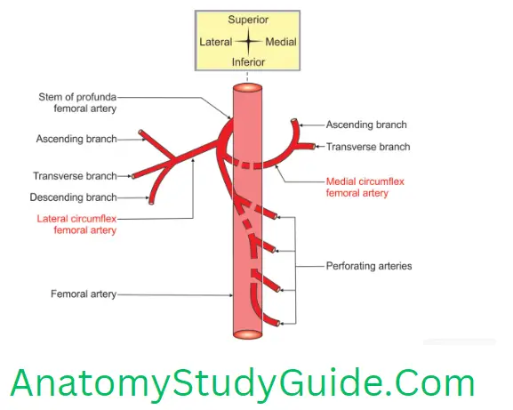
Branches of the profunda femoris artery are
1. Medial circumflex femoral artery. It gives branches
- Ascending branch, and
- Transverse branch.
Read And Learn More: Anatomy Notes And Important Question And Answers
2. Lateral circumflex femoral artery. It gives branches
- Ascending branch,
- Transverse branch, and
- Descending branch.
3. Perforating arteries. They are 4 in number.

Enumerate the hamstring muscles
1. Semimembranosus,
2. Semitendinosus,
3. Ischial fibres of adductor magnus, and
4. Long head of biceps.
Hamstring Muscles
(Ham—posterior aspect of the lower part of thigh and knee; string—thread or rope-like).
Hamstring Muscles Introduction: These are string-like muscles present on the posterior aspect of the thigh.
1. Hamstring Muscles Particulars: They are
1. Semimembranosus
- The upper part of the muscle is membrane-like and measures about 15 cm in length,
- It arises from the smooth facet present on the lateral part of the ischial tuberosity, and
- The tendon is rounded on its lateral margin and sharp at its medial margin. It resembles a hollow ground razor.
2. Semitendinosus: The lower part of the muscle is tendon-like. It arises from a medial
facet of the ischial tuberosity.
3. Biceps femoris: It has two heads. The ischial head is a part of the hamstring muscle.
4. Ischial fibres of adductor magnus.
2. Hamstring Muscles Features
- They are also called muscles of the posterior or flexor compartment of the thigh.
- The hamstring muscles span the femur but gain no attachment to it.
3. Hamstring Muscles Characters
- All the muscles arise from ischial tuberosity. They get inserted into one of the bones of the leg (either the tibia or fibula). The two “semi” muscles are inserted medially and the two heads of the biceps are lateral. When the knee is flexed the alternate contraction of these muscles produces rotation at the knee joint. The “semi” muscles are medial rotators; the biceps are lateral rotators of the tibia on the femur.
- They are supplied by the tibial division of the sciatic nerve.
- They are essentially flexors of the knee joint but they have extensor action of the hip joint.
4. Hamstring Muscles Blood supply
- Perforating branches of profunda femoris artery.
- The upper part of the hamstring is supplied by the inferior gluteal artery branch of the anterior division of the internal iliac artery.
- Popliteal artery.
5. Hamstring Muscles Testing Of Muscles
- Position of subject. Prone position with the knee extended.
- Activity. The subject is asked to flex the knee against resistance.
- Feel the posterior aspect of the thigh.
6. Hamstring Muscles Applied anatomy
- The extension of the hip and flexion of the knee are essential for runners. These muscles are liable for overstraining.
- Professional runners are prone to a painful condition known as a pulled hamstring. The attachment of hamstrings from the ischial tuberosity is torn.
- Muscle injuries may occur as a result of direct trauma or as part of an overuse syndrome. Muscle injuries may occur as a minor muscle tear. This is demonstrated as a focal area of fluid within the muscle. Hamstring muscles are usually torn in the thigh.
- If hamstring muscles are paralyzed, the patient tends to stand upright,
- In the old days, the soldiers used to slash the back of the knees of horses of their opponents. This cuts the tendons of the hamstring muscles. This brings the horse and its rider down.
- People also used to cut the hamstring tendons of soldiers so that they could not run. This was termed “hamstringing” the enemy.
Describe the Sciatic Nerve under the following heads
1. Sciatic Nerve Root value,
2. Sciatic Nerve Peculiarites,
3. Sciatic Nerve Course and Relations,
4. Sciatic Nerve Distribution,
5. Sciatic Nerve Applied anatomy
Introduction: The sciatic nerve is the largest in the body and is the continuation of the main part of the sacral plexus. The rami converge at the inferior border of the piriformis to form the sciatic nerve, a thick, flattened band. The sciatic nerve is the most lateral structure emerging through the greater sciatic foramen.
1. Sciatic Nerve Root Value: It arises from ventral rami of and dorsal divisions of ventral rami
2. Sciatic Nerve Perculiarities
- It is the thickest nerve in the body, 2cm wide. It is equal to the little finger.
- The Sciatic nerve supplies the posterior aspect of the thigh and all other structures below the knee EXPECT the skin of the medial leg, which is supplied by the saphenous nerve, a branch of the femoral nerve.
- The sciatic nerve is accompanied by a thin artery, the Sciatic artery, which is part of the axial artery of the lower limb.
3. Sciatic Nerve Course and relations
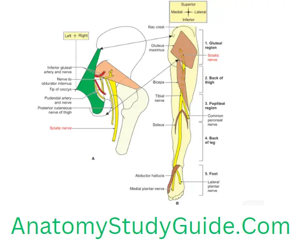
1. Course: The course and relations of the sciatic nerve is as follows
- In the pelvis: It lies in front of the piriformis.
- It enters the gluteal region through the greater sciatic foramen (below the piriformis). It passes between the ischial tuberosity and the greater trochanter. It emerges from the lower border of the gluteus maximus and passes downward deep to the long head of the biceps.
2. Relations
1. Superficial: Gluteus maximus.
2. Superficial and medial: Posterior cutaneous nerve of thigh
3. Deep
- Body of ischium,
- Ascending branch of the medial circumflex femoral artery (branch of profundal femoris),
- Tendon of obturator internus with the gemelli,
- Nerve to quadratus femoris and
- Deep to above structures: Capsule of the hip joint.
4. Sciatic Nerve Medial
- Inferior gluteal vessels branch of the anterior division of internal iliac artery,
- Inferior gluteal nerve,
- Pudendal vessels branch/tributary of the anterior division of the internal iliac artery
- Pudendal nerve and
- The posterior cutaneous nerve of the thigh.
3. Branches
1. Collateral branches
1. Tibial division of sciatic nerve supplies hamstring muscles, namely
- Semimembranosus,
- Semitendinosus,
- Ischial fibres of adductor magnus, and
- The long head of biceps.
2. Common peroneal branch supplies the short head of biceps, and
3. Articular branch to the hip joint.

2. Terminal branches: At the junction of the upper 2/3rd and lower 1/3rd, the sciatic nerve divides into tibial and common peroneal nerves.
4. Sciatic Nerve Distribution: The sciatic nerve supplies no structures in the gluteal regions. It divides into
1. Tibial, and
2. The tibial part of the sciatic nerve supplies
1. The tibial part of the sciatic nerve supplies
1. Medial plantar nerve and
2. Lateral plantar nerve
- 1. The medial plantar nerve supplies all the muscles present on the medial side of the foot except adductor hallucis.
- 2. The lateral plantar supplies all the muscles present on the lateral side of the foot and adductor hallucis.
- The tibial nerve gives articular branches to all joints of the lower limb.
2. Common peroneal nerve
1. Root Value: Dorsal divisions of ventral rami
2. Course: This is one of the smaller terminal branches of the sciatic nerve. It lies in the same superficial plane as the tibial nerve. It extends from the superior angle of the popliteal fossa to the later angle. It runs along the medial border of the biceps femoris. It winds around the posterolateral aspect of the neck of the fibula. It pierces the peroneus longus.
3. Branches
1. Collateral
2. Cutaneous branches are two. They are
- The lateral cutaneous nerve of the calf descends to supply the skin of the upper 2/3rd of the lateral side of the leg, and
- The sural communicating nerve arises in the upper part of the fossa. It runs on the posterolateral aspect of the calf and joints the sural nerve.
3. Articular branches
- The superior lateral genicular nerve accompanies the artery of the same name and lies above the lateral femoral condyle,
- The inferior lateral genicular nerve also runs with the artery of the same name to the lateral aspect of the knee joint above the head of a fibula, and
- Recurrent genicular nerve arises where the common peroneal nerve divides into superficial and deep peroneal nerves It ascends anterior to the knee joint and supplies the tibialis anterior muscle in addition to the knee joint.
- Muscular branches do not arise from this nerve. However, it may give a branch to the short head of biceps femoris.
4. Terminal branches
- Superficial peroneal nerve, and
- Deep Peroneal nerve.
5. Sciatic Nerve Blood supply: Arterial supply of the sciatic nerve is by artery of the sciatic nerve. It is a branch of the inferior gluteal artery. It represents the axis artery in the gluteal region and may at times be quite large. It is companion artey of the sciatic nerve (arteria comitans nervi ischiadici). It is a long branch that descends for some distance with the sciatic nerve. It anastomoses with the perforating branches of the profunda femoris artery.
6. Sciatic Nerve Applied anatomy
1. A pain in the buttock may result from compression of the sciatic nerve by the piriformis muscle (piriformis syndrome). Individuals involved in sports that require excessive use of the gluteal muscles (e.g. ice skaters, cyclists, and rock climbers) and women are more likely to develop this syndrome.
2. Sciatica: It is due to compression and irritation of nerve roots of the sciatic nerve. The patient gets shooting pain along the cutaneous distribution of the sciatic nerve. The pain begins in the gluteal region and radiates along the back of the thigh, the posterior and lateral side of the leg and the dorsum of the foot.
3. Sciatica may be caused by
- Injury to the sacral plexus in the pelvis, and
- Injury to the nerves.
4. Bony outgrowths or tumours. The pain affects the back of the thigh and the outer side of the leg.
“Sciatic nerve block” is done by injecting an anaesthetic agent a few cm below the midpoint of the line joining the posterior superior iliac spine and the upper border of the greater trochanter.
5. A complete section of the sciatic nerve is uncommon. When this occurs, the leg is useless because extension of the hip is impaired, as is flexion of the leg. All ankle and foot movements are also lost.
6. An incomplete section of the sciatic nerve (e.g. from stab wounds) may also involve the inferior gluteal and/or the posterior femoral cutaneous nerve.
7. The deep intramuscular injection in the lower medial quadrant of the gluteal region causes injury to the sciatic nerve.
8. In a sarcoma of the medial compartment of the thigh, resection of the adductor magnus may be required. The sciatic nerve lies directly behind the medial portion of the adductor magnus. Care should be taken to prevent injury to the sciatic nerve.
9. Posterior dislocations of the hip joint may cause serious injury to the sciatic nerve.
10. Injury to the nerve paralyses muscles of the back of the thigh (hamstrings), and all muscles of the leg and foot. The foot hangs downwards (by its own weight): The condition is called foot drop. Foot drop is also caused by injury to the common peroneal nerve. There is sensory loss over the greater part of the leg and foot.
Name the muscles of the posterior compartment of the thigh
Muscles of the posterior compartment of the thigh are
1. Semimembranosus.
2. Semitendinosus.
3. Ischial fibres of adductor magnus, and
4. Short head and long head of biceps.
Leave a Reply