Basal Ganglia Introduction
Basal ganglia are the scattered masses of gray matter submerged in the subcortical substance of the cerebral hemisphere. Basal ganglia form the part of extrapyramidal system, which is concerned with integration and the regulation of motor activities.
Table of Contents
Components Of Basal Ganglia
Basal ganglia include three primary components
- Corpus striatum
- Substantia nigra
- The subthalamic nucleus of Luys.
Read And Learn More: Medical Physiology Notes
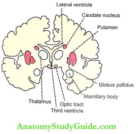
1. Corpus Striatum: It is a mass of gray matter situated at the base of cerebral hemispheres in close relation to the thalamus. The internal capsule incompletely divides the corpus striatum into two parts.
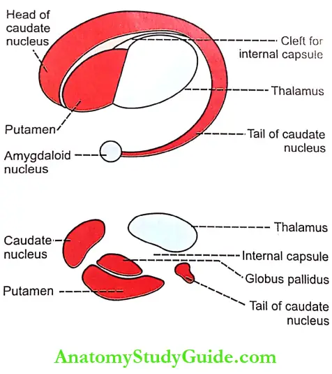
- Caudate Nucleus: It is an elongated arched gray mass, lying medial to internal capsule. Throughout its length, the caudate nucleus is related to lateral ventricle. The caudate nucleus has a head portion and a tail portion. The head is bulged into lateral ventricle and situated rostral to the thalamus. The tail is long and arched. It extends along the dorsolateral surface of the thalamus and ends in the amygdaloid nucleus.
- Lenticular Nucleus: It is a wedge-shaped gray mass, situated lateral to the internal capsule. A vertical plate of white matter called the external medullary lamina, divides the lenticular nucleus into two portions:
- The outer putamen
- The inner globus pallidus.
- Putamen and caudate nucleus are the phylogenetically newer parts of the corpus striatum and these two parts are together called neostriatum or striatum. The globus pallidus is a phylogenetically older part of the corpus striatum. And, it is called pallidum or paleostriatum. The globus pallidus has two parts, an outer part, and an inner part.
2. Substantia Nigra: Substantia nigra is situated below red nucleus. It is made up of large pigmented and small non-pigmented cells. The pigment contains a high quantity of iron.
3. Subthalamic Nucleus Of Luys: This nucleus is situated lateral to red nucleus and dorsal to the substantia nigra.
Connections Of Basal Ganglia
- The afferent and efferent connections of the corpus striatum, substantia nigra, and subthalamic nucleus of Luys are given in Table.
- In addition to afferent and efferent connections, the different components of the corpus striatum of the same side are interconnected by intrinsic fibers.
- Putamen to globus pallidus
- Caudate nucleus to globus pallidus
- Caudate nucleus to putamen.
- The different components of corpus striatum in each side are connected to those of the opposite side by commissural fibers.


Functions Of Basal Ganglia
The basal ganglia form the part of extrapyramidal system, which is concerned with motor activities. The various functions of basal ganglia are:
- Control Of Muscle Tone:
- Basal ganglia control the muscle tone. In fact, the gamma motor neurons of the spinal cord are responsible for the tone of the muscles.
- Basal ganglia exert an inhibitory effect on muscle tone by inhibiting the gamma motor neurons through descending inhibitory reticular system in the brainstem. During the lesion of the basal ganglia, muscle tone increases leading to rigidity.
- Control Of Motor Activity
- Regulation of Voluntary Movements
- The movements during voluntary motor activity are initiated by the cerebral cortex. However, these movements are controlled by basal ganglia, which are in close association with the cerebral cortex. During lesions of basal ganglia, the control mechanism is lost and so the movements become inaccurate and awkward.
- Basal ganglia control motor activities because of the nervous (neuronal) circuits between the basal ganglia and other parts of the brain involved in motor activity. The neuronal circuits arise from three areas of the cerebral cortex:
- Premotor area
- Primary motor area
- Supplementary motor area.
- All these nerve fibers from the cerebral cortex reach the caudate nucleus. From here, the fibers go to putamen. Abmp of the fibers from the cerebral cortex goes directly to the putamen also.
- P-Anmen sends fibers to globus pallidus. The fibers from here run towards the thalamus, subthalamic nucleus of Luys, and substantia nigra. The subthalamic nudes mid substantia nigra are in turn, projected into the thalamus. Now, the fibers from the thalamus are projected back into the primary motor area and the other two motor areas, i.e. premotor area and supplementary motor area.
- Regulation of Conscious Movements
- The fibers between the cerebral cortex and caudate nucleus are concerned with the regulation of conscious movements. This function of basal ganglia is also known as the cognitive control of activity.
- For example, when a stray dog barks at a man, immediately the person, understands the situation, turns away, and starts running.
- Regulation of Subconscious Movements: The cortical fibers reaching the putamen are directly concerned with the regulation of some subconscious movements which take place during trained motor activities, i.e. skilled activities such as writing the learned alphabet, paper cutting, nail hammering, etc.
- Regulation of Voluntary Movements
- Control Of Reflex Muscular Activity
- Some of the reflex muscular activities, particularly visual and labyrinthine reflexes are important in the maintenance of posture. Basal ganglia are responsible for the coordination and integration of impulses for these reflex activities.
- During lesions of basal ganglia, the postural movements, especially the visual and labyrinthine reflexes become abnormal. These abnormal movements are associated with rigidity.
- Rigidity is because of the loss of inhibitory influence from the cerebral cortex on the spinal cord via basal ganglia.
- Control Of Automatic Associated Movements
- Automatic associated movements are the movements in the body, which take place along with some motor activities. Examples are the swing of the arms while walking, and appropriate facial expressions while talking or doing any work. Basal ganglia are responsible for the automatic associated movements.
- The lesion in the basal ganglia causes the absence of these automatically associated movements, resulting in poverty of movements. A face without appropriate expressions while doing any work is called mask like face. A body without associated movements is called a statue-like body.
- Role In Arousal Mechanism: Globus pallidus and red nucleus are involved in arousal mechanism because of their connections with reticular formation. The extensive lesion in globus pallidus causes drowsiness, leading to sleep.
- Role Of Neurotransmitters In The Functions Of Basal Ganglia: The functions of basal ganglia on motor activities are executed by some neurotransmitters released by nerve endings within basal ganglia. Following neurotransmitters are released in basal ganglia.
- Dopamine: It is released by dopaminergic fibers from substantia nigra to corpus striatum (putamen and caudate nucleus – dopaminergic nigro strial fibers). The deficiency of dopamine leads to Parkinsonism.
- Gamma-aminobutyric acid (GABA): It is secreted by intrinsic fibers of corpus striatum and substantia nigra.
- Acetylcholine: It is released by fibers from the cerebral cortex to the caudate nucleus and putamen.
- Substance P and enkephalins: These substances are released by fibers from globus pallidus reaching the substantia nigra.
- Noradrenaline: It is secreted by the fibers between basal ganglia and reticular formation.
- Glutamic acid: It is secreted by the fibers from the subthalamic nucleus to the globus pallidus and substantia nigra.
- Among all these neurotransmitters, dopamine and GABA are inhibitory neurotransmitters. So, the dopaminergic fibers and the fibers releasing GABA are inhibitory fibers. All other neurotransmitters possess excitatory functions.

Applied Physiology Disorders Of Basal Ganglia
- Parkinson’S Disease
- Parkinson’s disease is a slowly progressive degenerative disease of nervous system associated with the destruction of brain cells that produce dopamine. It is named after the discoverer James Parkinson.
- It is also called parkinsonism or paralysis agents. The great boxer Mohammed Ali is affected by Parkinsonism because of the repeated blows he might have received on head resulting in damage of brain cells producing dopamine.
- Causes of Parkinson’s Disease: Parkinson’s disease occurs due to lack of dopamine caused by damage of basal ganglia. It is mostly due to the destruction of the substantia nigra and the nigrostriatal pathway, which has dopaminergic fibers. Damage of basal ganglia usually occurs because of the following causes:
- Viral infection of brain like encephalitis
- Cerebral arteriosclerosis
- Injury to basal ganglia
- Destruction or removal of dopamine in basal ganglia. It occurs mostly due to long-term treatment with antihypertensive drugs like Reserpine. Parkinsonism due to the drugs is known as drug-induced Parkinsonism
- Unknown causes: Parkinsonism can occur because of the destruction of basal ganglia due to some unknown causes. This type of Parkinsonism is called idiopathic Parkinsonism.
- Signs and Symptoms of Parkinson’s Disease: Parkinson’s disease develops very slowly and the early signs and symptoms may be unnoticed for months or even for years. Often the symptoms start with a mild noticeable tremor in just one hand. When the tremor becomes remarkable the disease causes slowing or freezing of movements followed by rigidity. Common signs and symptoms of Parkinson’s disease are
- Tremor: In Parkinson’s disease, the tremor occurs during rest. But it disappears while doing any work. So, it is called static tremor or resting tremor. It is also called drum beating tremor, as the movements are similar to beating a drum. The thumb moves rhythmically over the index and middle fingers, pil is known as pill-rolling movement.
- Slowness of movements: Over time, the movements start slowing down (bradykinesia) and it takes a long time even to perform a simple task. Gradually the patient becomes unable to initiate the voluntary activity (akinesia) or the voluntary movements are reduced (hypokinesia). It is because of the hypertonicity of the muscles.
- The poverty of movements: Poverty of movements is the loss of all automatically associated movements. Because of the absence of the automatic associate movements, the body becomes statue-like. The face becomes mask-like, due to absence of appropriate expressions like blinking and smiling.
- Rigidity: Stiffness of muscles occurs in limbs resulting in rigidity of limbs. The muscular stiffness occurs because of increased muscle tone which is due to the removal of inhibitory influence on gamma motor neurons. It affects both flexor and extensor muscles equally. So, the limbs become more rigid like pillars. The condition is called lead pipe rigidity. In later stages the rigidity extends to neck and trunk.
- Gait: Gait refers to manner of walking. The patient looses the normal gait. Gait in Parkinson’s disease is called festinant gait. The patient walks quickly in short steps by bending forward as if he is going to catch up the center of gravity.
- Speech Problems: Many patients develop speech problems. They may speak very softly or sometimes rapidly. The words are repeated many times. Finally, their speech becomes slurred and they hesitate to speak.
- Emotional changes: The persons affected by Parkinson’s disease are often upset emotionally.
- Dementia: In later stages, some patients develop dementia.
- Treatment for Parkinson’s Disease
- As Parkinson’s disease is due to lack of dopamine caused by damage of dopaminergic fibers, it is treated by dopamine injection.
- Dopamine does not cross the blood-brain barrier. So, another substance called Levodopa (L-dopa) which cross the blood-brain barrier is injected. The L-dopa moves into the brain, and there it is converted dopamine.
- Since L-dopa can be converted into dopamine in the liver, some side effects occur due to excess dopamine content in liver and blood.
- So, along with L-dopa, another substance called carbs dopa is administered. Carbi dopa prevents the conversion of L-dopa into dopamine and, carbi dopa cannot pass through the blood-brain barrier. Thus, L-dopa moves into the brain tissues and is converted into dopamine.
- Some of the symptoms of Parkinson’s disease such as tremor are abolished by surgical destruction of basal ganglia or thalamic nuclei.
- Wilson’S Disease
- Wilson’s disease is an inherited disorder characterized by excess of copper in the body tissues. It is also known as progressive hepatolenticular degeneration. This disease develops due to damage of the lenticular nucleus particularly, putamen.
- In Wilson’s disease, copper is deposited in the liver, brain, kidneys, and eyes. The deposits of copper cause damage of tissues. This makes the affected organs to stop functioning.
- In addition to symptoms of Parkinson’s disease, liver failure and damage to the central nervous system are the most predominant effects of this disorder. Wilson’s disease is fatal if not treated early.
- Chorea: It is an abnormal involuntary movement. Chorea means rapid jerky movements. It mostly involves the limbs. It is due to lesions in caudate nucleus and putamen.
- Athetosis: It is another type of abnormal involuntary movement, which refers to slow rhythmic and twisting movements. It is because of the lesion in the caudate nucleus and putamen.
- Choreoathetosis: It is a condition characterized by aimless involuntary muscular movements. It is due to the combined effects of chorea and athetosis.
- Huntington’s Chorea:
- Huntington’s disease is an inherited progressive neural disorder due to the degeneration of neurons secreting GABA in corpus striatum and substantia nigra. The disease starts mostly in middle age.
- It is characterized by chorea, hypotonia, and dementia. In severe cases bilateral wasting of muscles occurs. It is otherwise called Huntington’s disease, chronic progressive chorea, degenerative chorea or hereditary chorea.
- Hemiballismus:
- It is a disorder characterized by violent involuntary abnormal movements on one side of the body involving mostly the arm. While walking, the arm swings widely.
- These movements are called the flinging movements. These movements are due to the release phenomenon because of the absence of inhibitory influence on movements. Hemiballism occurs due to the degeneration of the subthalamic nucleus of Luys.
- Kernicterus: Ksmiderus is a form of brain damage in infants caused by jaundice. Basal ganglia are the mainly affected parts of brain.
Leave a Reply