Branches Of Vertebral Artery
- Vertebral artery is a branch of 1st part of subclavian artery. It is divided into four parts .
- 1st part: Lies deep in the neck in the vertebral.This part gives no branches.
- 2nd part: Passes in the foramen transversaria of C6-C1 vertebrae. It gives spinal branches that supply
- Meninges, and
- Spinal cord.

- 3rd part: It lies in the suboccipital on the posterior arch of atlas vertebra and gives branches to muscles of suboccipital
- Rectus capitis posterior major
- Rectus capitis posterior minor
- Superior oblique
- Inferior oblique
- 4th part:
- It enters the cranial cavity through foramen magnum.
- It joins with the same artery of opposite side to form basilar artery at the lower border of pons.
- It gives
- Meningeal branches,
- Posterior spinal artery,
- Anterior spinal arteries,
- Posterior inferior cerebellar artery, and
- Medullary branches.
Read And Learn More: Anatomy Important Question And Answers
Table of Contents
Blood-Brain Barrier
- Formed by
- Non-fenestrated capillary endothelium of blood vessels-tight junctions
- Basement membrane of endothelium
- Perivascular foot and cell body of astrocytes
- Network of intercellular space between astrocytes and neurons-20 nm
- Process of cell bodies and neurons
- Blood-brain barrier is absent in the following areas of the brain. It is lined by tanycytes (tanyein—to stretch). They are present around 3rd and 4th ventricles.
- Structures related to 3rd ventricle
- Pineal gland,
- Choroid plexus,
- Subfornical organ,
- Hypothalamus-median eminence,
- Lamina terminalis,
- Posterior lobe of pituitary gland-neurohypophysis,
- Intercolumnar tubercle,
- Supraoptic recess of 3rd ventricle,
- Median eminence,
- Tuberculum cinerium, and
- OVLT (organum vasculosum of lamina terminalis).
- Structures related to 4th ventricle
- Funiculus separans present in the floor of 4th ventricle, and
- Area postrema.
- Astrocytes are concerned with nutrition of the nervous tissue. They are star shaped cells. They form blood-brain barrier.
- Significance of blood-brain barrier:
- It is a physiologic barrier that regulates the passage of various substances from blood to brain.
- It forms stable and balanced ionic composition in the interstitial neuronal environment.
- It protects the cells from any potentially harmful substances.
Blood Supply Of Spinal Cord
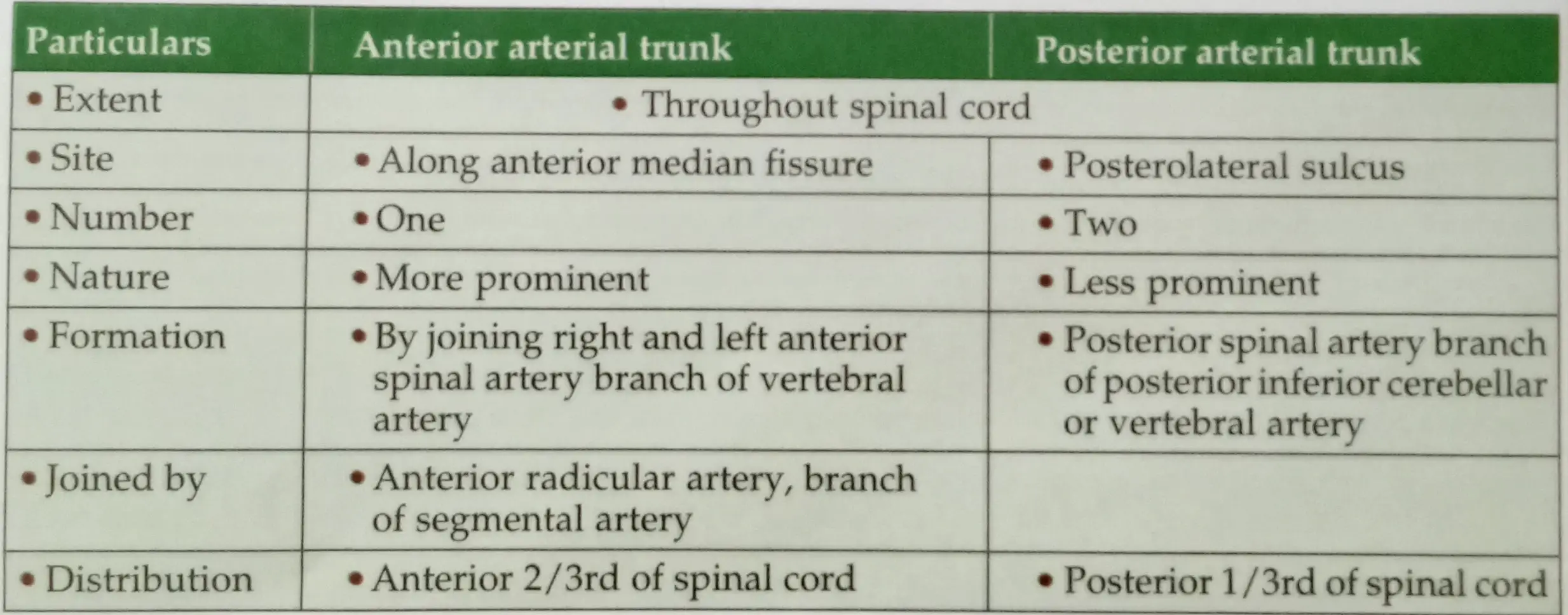
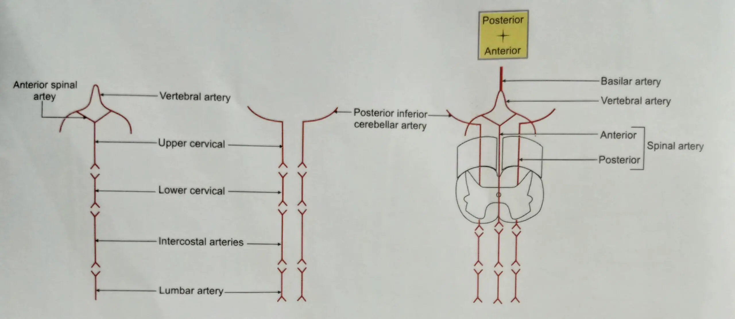
- The blood supply of spinal cord is by
- One anterior spinal artery, and
- Two posterior spinal arteries.

- Segmental spinal branches arise from
- Vertebral artery,
- Ascending cervical artery,
- Deep cervical artery,Cervical segment.
- Intercostal artery-thoracic segment.
- Lumbar artery-lumbar segment.
- Sacral artery-sacral segment.Note: The radicular arteries arise from vertebral, ascending cervical, deep cervical, intercostal, lumbar and sacral arteries. These branches anastomose with one another to form longitudinal vessels. Frequently, one of the radicular arteries is larger than the remainder and is called arteria radicularis magna. It usually arises from one of the intersegmental branches of descending aorta in the lower thoracic or upper lumbar segments.
- Venous drainage: There are about 6 longitudinal venous channels which drain the blood from the spinal cord. They are described as
- Midline venous channels: They are two in number.
- Venous channel present in the anterior median fissure, and
- Venous channel present in the posterior median sulcus.
- Lateral venous channels: They are in two sets.
- One set behind the ventral root of spinal nerve on each side, and
- One set in front of the dorsal root of the spinal nerve.
All above longitudinal veins communicate with the internal vertebral plexus and drain into - Vena cava,
- Azygos vein and into
- Basilar venous plexus.


Branches Of Basilar Artery
- Anterior inferior cerebellar artery (AICA)
- It arises at the lower border of pons.
- It supplies VI, VII and VIIIth cranial nerves.
- Labyrinthine artery:
- Origin: It usually arises from anterior inferior cerebellar artery. There are variations in its origin. It may arise from
- Lower part of basilar artery
- Superior cerebellar artery
- Posterior inferior cerebellar artery
- It accompanies VIIIth nerve and enters the internal auditory meatus to supply the internal ear.
- It is an end artery.
- Origin: It usually arises from anterior inferior cerebellar artery. There are variations in its origin. It may arise from
- Pontine branches
- These are numerous slender branches.
- They pierce the pons both in the medial and lateral parts.
- Superior cerebellar artery
- It arises close to superior border of pons.
- It winds
- Superior border of pons, and
- Middle cerebellar peduncle.
- Function: It supplies
- Pons,
- Middle cerebellar peduncle
it sends many branches to the superior surface of cerebellum.
- Posterior cerebral branches diverge at upper border of pons and supply occipital lobe of cerebrum and midbrain.

SAQ-8 Branches of basilar artery Branches of basilar artery are 5 in number-equal to the numbers of fingers of hand.
- Anterior inferior cerebellar artery
- Superior cerebellar artery
- Pontine arteries are two in number
- Posterior cerebral arteries are terminal branches.
Note: In 80% of cases labyrinthine artery arises from anterior inferior cerebellar artery.
Leave a Reply