OLA-1 What is ‘sternal angle’ and state its clinical importance?
Definition: It is a bony angulation formed at the junction of manubrium and body of sternum.
Sternal Angle Applied anatomy
Table of Contents
- It helps for the counting of ribs,
- It is common site of sternal fracture, is at sternal angle, and
- It is palpable and often visible in young people.
Read And Learn More: Anatomy Important Question And Answers
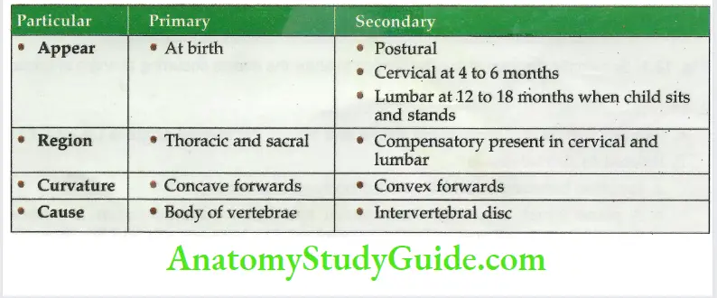
OLA-2 Particular Name the curvatures of vertebral column and classify them.

1. Coronal plane (lateral curve): There is slight lateral curvature in the thoracic region. Its concavity is towards the left. It is possible due to the
- Greater use of the right upper limb, and
- Pressure of the aorta.
The curvatures add to the elasticity of the spine.
The increased number of curves gives a higher resistance to weight.
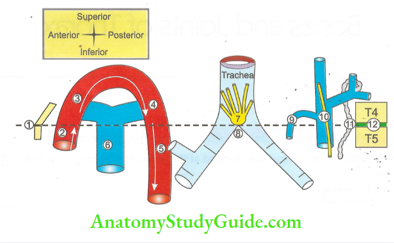
Angle of Louis/sternal Angle
Angle of Louis/sternal Angle Introduction: It is a bony angulation formed at the junction of manubrium sternum and body of sternum.
1. Angle of Louis/sternal Angle Situation: It is situated 5 cm below the suprasternal notch.

- Junction of manubrium sternum and body of sternum.
- Termination of ascending aorta.
- Beginning of arch of sorte.
- Termination of arch of aorta.
- Beginning of descending thoracic aorta
- Bifurcation of pulmonary trunk.
- Deep cardiac plexus.
- Bifurcation of trachea.
- Opening of azygos vein into superior vena cava.
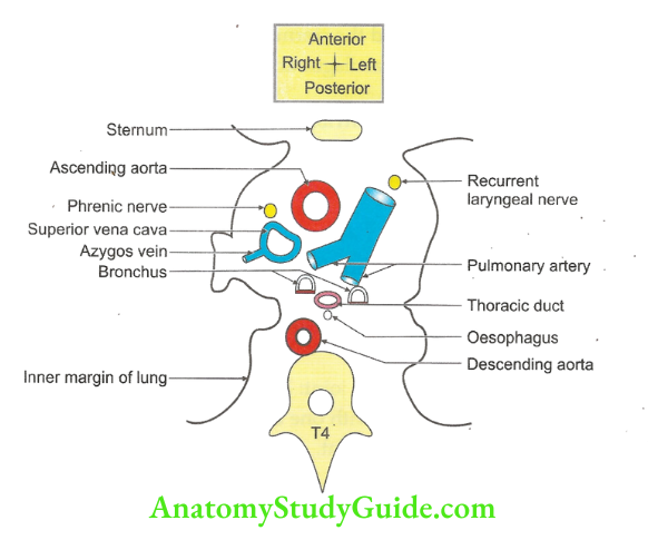
- The plane divides mediastinum into superior and inferior mediastinum.
- Thoracic duct.
- Intervertebral disc between T4 and T5.
2. Events
- Related with skin: Dermatome above and below the sternal angle is C4 and T2.
- Related to skeletal element
- Junction between manubrium and body of sternum.
- A plane which separates the superior and inferior mediastinum. It passes through angle of Louis to the intervertebral disc between 4th and 5th thoracic vertebra,
2nd articular cartilage to the sternum, - 2nd rib lies at this level.
- Related to heart and vessels
- Arterial
- Ascending aorta ends and arch of aorta begins,

- Arch of the aorta ends and descending thoracic aorta begins,
- Ascending aorta pierces the fibrous pericardium.
- Venous
- Superior vena cava pierces fibrous pericardium.
- Azygos vein opens into superior vena cava.
- Pulmonary trunk divides into two pulmonary arteries.
- Nervous: Formation of cardiac plexus.
- Upper limit of the base of heart.
- Related to respiratory system
- In cadaver, trachea divides into two principal bronchi. (In living, trachea divides at T6.NEET Refer; Grey-41st edition)
- Pleural sacs meet at this level.
- Related to lymphatic system: Thoracic duct crosses from right to left.
- Applied anatomy: It is an important landmark for counting the ribs.
Costal Cartilage
1. Costal Cartilage Histologically: It is hyaline cartilage without perichondrium.
2. Costal Cartilage Features
- It represents the unossified anterior parts of the ribs.
- They contribute materially to the elasticity of the thoracic wall.
- There are 10 costal cartilages on each side.
- First 7 cartilages are connected the sternum with the corresponding ribs.
- The 8th, 9th and 10th costal cartilages articulate with one another and form the costal margin.
- The cartilages of the 11th and 12th ribs are small. Their ends are free and lie in the muscles of the abdominal wall.
- The direction of the costal cartilages is variable.
3. Costal Cartilage Joints
- Sternocostal
- 1st sternocostal joint: Synarthrosis (fibrous joint)NEET
- Remaining sternocostal joints: Synovial
- 1st chondrosternal joint: Primary cartilaginous joint
- Interchondral-synovial
- Lateral end forms costochondral joint: Primary cartilaginous joint.
- 5th to 9th cartilages articulate with one another-synovial joints.
- Xiphisternal joint: Symphyseal joint.
4. Costal Cartilage Attachments
- Anterior ends
- 1st costal cartilage
- Articular disc of sternoclavicular joint, II. Sternoclavicular ligament, and
- Subclavius muscle.
- 2nd to 7th costal cartilage: Pectoralis major,
- 2nd to 6th costal cartilage: Sternocostalis,
- 5th, 6th and 7th: Rectus abdominis,
- 7th, 8th and 9th: Internal oblique, and
- 7th to 12th: Transversus abdominis and diaphragm.
Costal Cartilage Superior and inferior borders
- Internal intercostal,
- External intercostalis muscles,
- Anterior intercostal membranes of the corresponding spaces, and
- Musculophrenic artery perforates diaphragm at the level of 9th costal cartilage.
Structures Related To Superior Surface Of 1st Rib
1. Important neurovascular bundle on the superior surface of 1st rib (from anterior to posterior) VAN
- Subclavian vein,
- Subclavian artery (8th part),
- Lower trunk of brachial plexus, and
- Ascending branch of ventral ramus of 1st thoracic nerve.
2. Muscles attached on the superior surface (from anterior to posterior)
- Subclavius,
- Scalenus anterior,
- Serratus anterior, and
- Scalenus posterior.
3. Structures related on anterior border of neck (from posterior to anterior) Sympathetic trunk,
- 1st posterior intercostal Vein
- Superior intercostal Artery, and
- 8th cervical spinal Nerve.

Intervertebral Disc
1. The sum of height of intervertebral discs contributes 1/5th of the length of the vertebral column.
2. Secondary curvatures are postural and are mainly due to the shape of the intervertebral disc.
3. Intervertebral Disc Histology: Fibrocartilaginous structure.
4. Intervertebral Disc Site: Present between the bodies of adjacent vertebrae.
5. Intervertebral Disc Function: Bind the intervertebral disc.
6. Intervertebral Disc Shape: It corresponds to that of the vertebral bodies between which they are placed.
7. Thickness of the disc: It varies from region-to-region and anteroposteriorly in the same disc.
- Cervical and lumbar regions: Discs are thicker in front than behind.
- Thoracic region: Uniform thickness but thinnest in upper thoracic.
- Lumbar region: Thickest.
The intervertebral disc is greater in the cervical and lumbar regions than in the thoracic region.

8. Intervertebral Disc Composition: Each disc is made up of the following two parts.
- Nucleus pulposus: It is the central part of the disc. It is soft. It is gelatinous at birth. It is kept under tension and acts as a hydraulic shock absorber. With increasing age, the elasticity of the disc is much reduced.
- Annulus fibrosus: It forms the peripheral part of the disc. It is made up of a
- Narrower outer zone of collagenous fibres,
- Wider inner zone of fibrocartilage, and
- Arrangement of fibres: They are arranged in the form of incomplete rings. The rings are connected by strong fibrous bands. The outer collagenous fibres blend with the anterior and posterior longitudinal ligaments.
9. Intervertebral Disc Development: The nucleus pulposus is remnant of notochord.
Typical Spinal Nerve
Each typical spinal nerve is formed by dorsal and ventral roots. It divides into ventral and dorsal ramus.
Dorsal ramus goes posteriorly and divides into medial and lateral branches.
1. The medial and lateral branches of dorsal ramus give muscular branches which supply muscles of the back and neck.
2. Lateral branch of the dorsal ramus in the lower half and medial branch of the dorsal ramus in the half are cutaneous. L for L, L = lateral and lower.
3. Ventral rami in cervical, lumbar and sacral region unite together to form plexus.

Leave a Reply