Bones Of The Lower Limb
Attachments to the intertrochanteric line
Table of Contents
The intertrochanteric line provides
1. Attachment to the following ligaments
- Capsular ligament of the hip joint,
- Upper band of the iliofemoral ligament in its upper part, and
- Lower band of iliofemoral ligament in its lower part.
2. Origin to the highest fibres of the following muscles.
- Vastus lateralis from the upper end of the line, and
- Vastus medialis from the lower end of the line.
Read And Learn More: Anatomy Notes And Important Question And Answers
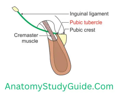
Enumerate the structures attached to Pubic Tubercle
1. Pubic Tubercle Ligament
- Apex of lacunar ligament
- Medial end of the inguinal ligament
- Reflected part of the inguinal ligament
Femur Bone
2. Pubic Tubercle Fascia
- Superficial layer of fascia lata,
- Fascia transversalis.
3. Pubic Tubercle Muscle: Ascending loops of cremaster muscle in male, and
4. Pubic Tubercle Superior crus of saphenous opening.
Greater Sciatic Notch
1. The posterior border of ilium of hip bone is divided by ischial spine into two notches
- Greater sciatic notch—above the spine, and
- Lesser sciatic notch—below the spine.
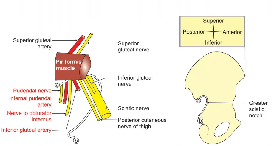
2. The structures passing through greater sciatic notch are divided by the piriformis into
1. Structures above piriformis,
- Superior gluteal vessels—branch/tributary of the posterior division of internal iliac artery, and
- Superior gluteal nerve.
2. Structures below piriformis (from medial to lateral): They are divided into three groups
1. Structures going to the gluteal region
1. Laterally
- Sciatic nerve. It lies at the inferior margin of the greater sciatic notch,
- Nerve to quadratus femoris.
2. Medially: Nerve to obturator internus. It crosses the base of the ischial spine.
3. Intermediate position: Inferior gluteal vessels and nerves.
2. Structures that re-enter into lesser sciatic notch
1. Internal pudendal vessels of smaller terminal branch/tributary of the posterior division of internal iliac vessels, and
2. Pudendal nerve. It lies close to the tip of the ischial spine.
3. Structures going to thigh region: Posterior femoral cutaneous nerve of the thigh.
3. Greater Sciatic Notch Applied anatomy
About greater sciatic notch:
- At the upper border of the greater sciatic notch, the internal iliac artery divides into anterior and posterior divisions.
- The internal iliac vein begins above the greater notch.
- There is a preauricular sulcus above it. It is deep in the female hip bone and more pronounced in the hip bones of multiparous females.
- It is converted into the greater sciatic foramen by the sacrospinous ligament.
- The anterior gluteal line starts from the middle of the upper margin of the greater sciatic notch.
Femur Bone
About lesser sciatic notch:
- The pudendal canal extends from the lesser sciatic notch to the deep perineal pouch.
- It is converted into a lesser sciatic foramen by the sacrotuberous ligament.
- The inferior gemellus muscle arises from the inferior margin of the lesser sciatic notch near the ischial tuberosity.
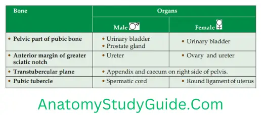
Organs related to hip bone
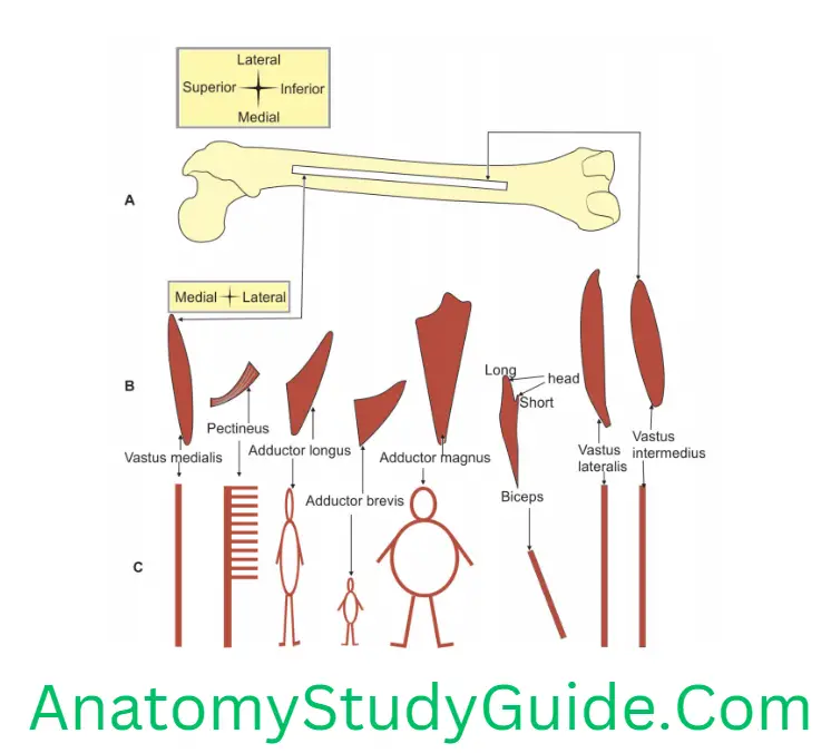
Linea Aspera
(Linea—line, aspera—thick, broad, thickened ridge)
1. Linea Aspera Definition: It is an irregular thick line present on the posterior border of the femur.
2. Linea Aspera Features
1. In the middle 1/3rd of the thigh, it forms the apex of the adductor canal.
2. It gives attachment to intermuscular septa. These septa divide the muscles of the thigh into extensor, adductor and flexor compartments. Following are the muscles attached to linea aspera (from lateral to medial).
- Vastus Intermedius,
- Vastus Lateralis,
- The short head of Biceps femoris
- Adductor Magnus,
- Adductor Brevis,
- Adductor Longus, and
- Vastus Medialis.
3. Linea Aspera Fate of linea aspera
1. It divides into medial and lateral lips at both the ends.
2. Fate of these lips
1. At the upper end.
- The medial lip continues with the spiral line, and
- The lateral lip continues to the gluteal tuberosity.
2. At the lower end
- Medial lip continues as medial supracondylar line, and
- The lateral lip continues as the lateral supracondylar line.
4. Linea Aspera Applied anatomy
- It acts as a buttress to resist compressive forces. Hence, it prevents the anterior bowing of the shaft.
- The nutrient foramen of the femur lies on the linea aspera. It is directed upward.
- The perforating branches of profunda femoris are closely related to the linea aspera.
Adductor tubercle
Introduction: It is the 1st bony prominence felt on the medial side of the thigh as you slide your finger from above downwards.
1. Gives attachment to
- Ischial fibers of adductor magnus muscle, and
- Tibial collateral ligament.
2. Applied anatomy
It forms a bony landmark for surface anatomy.
The epiphyseal line in the femur runs transversely through the adductor tubercle.
Damage of adductor tubercle during surgical intervention. leads to permanent shortening of the lower limb. This is applicable to the bone before ossification.
To palpate the tubercle, flex the knee partly and note the wide, shallow groove that appears posterior to the mass of vastus medialis. The tendon of the adductor magnus can be felt in this groove. It can be traced down to the adductor tubercle.
Femur Bone
Iliac Crest
Iliac Crest Introduction: It is an ‘S-shaped curvature present on the upper border of the ilium.
1. Iliac Crest Curvatures
1. Vertical curvature: It is convex upwards.
2. Iliac Crest Anteroposterior
- It is concave inwards in front and
- Concave outwards behind.
2. It shows two spines at both ends.
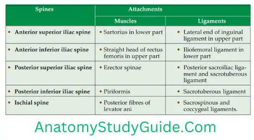
1. Spines
1. Anterior superior iliac spine gives attachment to
- Inguinal ligament, and
- Sartorius.
2. Posterior superior iliac spine gives attachment to the piriformis.
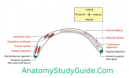
2. Iliac crest has a Ventral segment which is subdivided into
1. Anterior 2/3rd: It gives attachment to the following muscles (from lateral to medial).
- Fascia lata in the whole extent,
- Tensor fasciae latae in front of tubercle,
- External oblique,
- Internal oblique,
- Transversus abdominis, and
- Fascia transversalis.
2. Posterior 1/3rd: It gives attachment to the following muscles (from lateral to medial)
1. Latissimus dorsi,
2. Quadratus lumborum, and
3. The thoracolumbar fascia around quadratus lumborum.
4. Dorsal segment gives attachment to
- Gluteus maximus in outer sloping area, and
- Erector spinae in the inner sloping area.
3. Iliac Crest Ends: It has two ends.
- Anterior end is called the anterior superior iliac spine, and
- Posterior end is called the posterior superior iliac spine.
4. Iliac Crest Bony landmarks
- The highest point of the iliac crest is situated a little behind the midpoint of the crest.
- It lies at the level of the interval between the spines of vertebrae.
5. Iliac Crest Surface anatomy
- Iliac crest can be felt in the living at the lower limit of the flank.
- Anterior superior iliac spine (ASIS) is a prominent landmark. It is easily felt in living individuals.
6. Iliac Crest Applied anatomy
The anterior superior iliac spine is an important surface landmark especially used by tailors for taking measurements.
The Iliac crest is used for bone grafting.
The Iliac crest is used for bone marrow examination.
Tuberosity of the iliac crest is the subcutaneous bone that can be palpated in a fatty patient. It helps to find out the highest point of the iliac crest and anterior superior iliac spine.
Structures attached to spines of the hip bone
Trochanteric Anastomosis
Trochanteric Anastomosis Site: Trochanteric fossa
1. Arteries taking part
1. Ascending branch of the medial
2. Ascending branch of lateral
3. Branches of internal iliac artery
- Descending branch of the superior gluteal artery.
- Inferior gluteal artery,
- Internal pudendal artery
2. Trochanteric Anastomosis Applied Anatomy
It provides a chief source of blood supply to the
Head of the femur, and
Intracapsular part of neck of femur.
Femur Bone
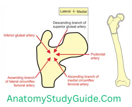
Trochanteric Anastomosis is between branches of the internal iliac and femoral arteries. In case of blockage of one of the arteries, collateral circulation is developed to maintain the blood supply to this region.
Leave a Reply