Classify The Cerebellum As Per Evolution And Function
As per evolution
Table of Contents
- Archicerebellum: Phylogenetically (phylon-tribe, genesis-generation), it is the oldest part of the cerebellum.
- Parts: It is made up of the
- Flocculonodular lobe, and the
- Lingula.
- Connections: It is chiefly vestibular.
- Functions: It controls the
- Axial musculature and the
- Bilateral movements used for locomotion and maintenance of equilibrium.
- Parts: It is made up of the
- Paleocerebellum: It is evolved after archicerebellum.
- Parts: It is made up of the
- Anterior lobe (except lingula),
- Pyramid, and
- Uvula of the inferior vermis.
- Connections are chiefly spinocerebellar.
- Functions: It controls
- Tone,
- Posture, and
- Crude movements of the limbs.
- Parts: It is made up of the
- Neocerebellum is the newest part of the cerebellum to evolve.
- Parts: It is made up of the posterior/middle lobe (the largest part of the cerebellum) except the pyramid and uvula of the inferior vermis.
- Functions: It is primarily concerned with the regulation of fine movements of the body.
Read And Learn More: Anatomy Important Question And Answers
Functionally: The anterior and posterior lobes are organized into three longitudinal zones-lateral, intermediate and vermis.
- Lateral zone: Connected with association areas of the brain and is involved in planning and programming muscular activities.
- Intermediate zone: It is concerned with control of muscles of hands, fingers, feet and toes.
- Vermis: Concerned with control of muscles of trunk, and neck.
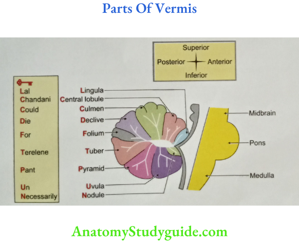
Parts of Vermis of Cerebellum
- Lingula
- Culmen
- Central lobule
- Declive
- Folium
- Tuber
- Pyramid
- Uvula
- Nodule
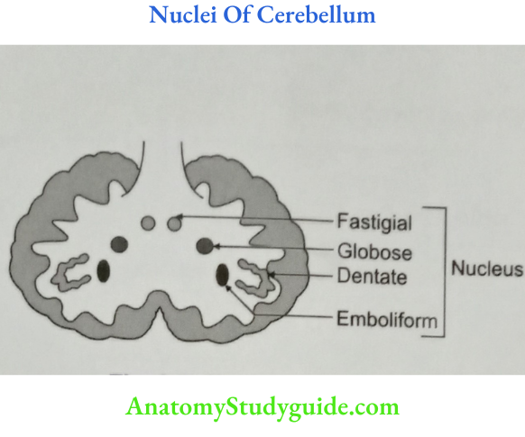
OLA-11 Name the Nuclei of Cerebellum.
They are
- Dentate nucleus,
- Nucleus Emboliformis,
- Nucleus Fastigii, and
- Globose nucleus.
Archicerebellum
- Archicerebellum Evolution: It is the oldest part of cerebellum.
- Archicerebellum Parts
- Parts of vermis
- Lingula
- Nodule.
- Part of hemisphere: Flocculus, sometime paraflocculus.
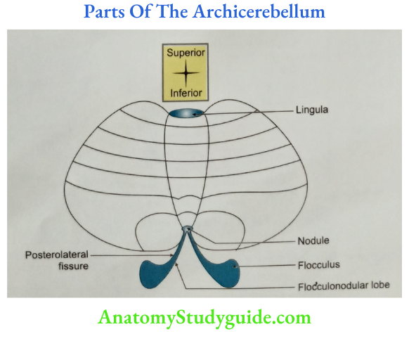
- Parts of vermis
- Archicerebellum Nucleus: Fastigii
- Archicerebellum Connections
- Afferent: Vestibulocerebellar.
- Efferent: Cerebellovestibular.
- Archicerebellum Functions: It controls the
- Axial musculature, and the
- Bilateral movements used for locomotion and maintenance of equilibrium.
- Archicerebellum Applied anatomy: Lesion of the archicerebellum is called archicerebellar syndrome.
- It is affected by tumour, medulloblastoma, particularly in childhood.
- It is characterized by
- Failure of maintenance of equilibrium.
- Failure to walk on a normal base.
- Unable to maintain the upright posture.
- Archicerebellum Morphology: Archicerebellum is separated by rest of the cerebellum by posterolateral fissure. This is the 1st developed part of the cerebellum.

Paleocerebellum
- Paleocerebellum Evolution: It is the older part of cerebellum, evolved next to archicerebellum.
- Paleocerebellum Parts:
- Anterior lobe
- Part of vermis
- Central lobule,
- Culmen.
- Parts of hemisphere
- Ala,
- Quadrangular lobule.
- Part of vermis
- UP of vermis
- Uvula,
- Pyramid.
- Anterior lobe
- Paleocerebellum Nucleus of paleocerebellum: Nucleus emboliformis
- Paleocerebellum Connections
- Paleocerebellum Afferents
- Anterior spinocerebellar,
- Posterior spinocerebellar,
- Cuneocerebellar,
- Par olivocerebellar, and
- Reticulocerebellar.
- Paleocerebellum Efferents: Cerebellospinal.

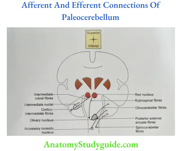
- Paleocerebellum Afferents
- Paleocerebellum Functions: It has a significant role in muscle tone and posture of the limbs.
- Paleocerebellum Applied anatomy: Lesion of the paleocerebellum is due to excessive intake of alcohol or in malnutrition. It is characterized by alteration of gait. Movements of individual legs are much affected. It is associated with increase in tone of extensor group of the muscle.
Neocerebellum
- Neocerebellum Evolution: It is most recently developed during evolution.
- Neocerebellum Parts: Middle lobe excluding UP ( Uvula and Pyramid)
- Parts of vermis
- Declive,
- Folium, and
- Tuber
- Parts of hemisphere are
- Simple lobule,
- Superior semilunar lobule,
- Inferior semilunar lobule,
- Biventral lobule, and
- Tonsil.
- Parts of vermis
- Neocerebellum Nucleus: Dentate nucleus.

- Neocerebellum Connections
- Neocerebellum Afferents
- Cortico-ponto-cerebellar,
- Anterior external arcuate fibres,
- Stria medullaris,
- Olivocerebellar, and
- Trigeminocerebellar.

- Neocerebellum Efferents
- Dentato-rubro-thalamo-cortical,
- Dentato-rubro-spinal.
- Neocerebellum Afferents
- Neocerebellum Functions: It is primarily concerned with the regulation of fine movements of the body.
- Neocerebellum Applied anatomy Lesion of the neocerebellum is called neocerebellar syndrome. It is characterized by
- Neocerebellum Hypotonia: Diminished muscle tone.
- Neocerebellum Asynergia: Loss of muscular coordination.
- Ataxia: Loss of coordination of muscles of trunk, pectoral and pelvic girdle. Dysmetria: Loss of ability to measure the distance for reaching an intended goal.
- Decomposition of movements: Loss of coordination of series of movement. Dysdiadochokinesis: Loss of ability to execute alternate movements in rapid succession, such as pronation and supination of forearm.
- Dysarthria or scanning speech: Loss of coordination of muscles concerned with speech. The speech is slurred, prolonged and explosive.
- Neocerebellum Nystagmus: Involuntary, rhythmical and oscillatory movements of the eyeball.
-
- Intentional tremor is evident during purposeful movements and is diminished or absent with rest.
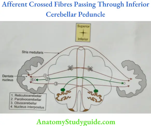
Cerebellar Peduncles


LAQ-18 Inferior Cerebellar Peduncle
Cerebellar Peduncles Introduction: This is the largest collection of white fibres which connect the cerebellum to medulla.
- Cerebellar Peduncles Fibres
- Afferent fibres

- Efferent fibres

- Afferent fibres
- Cerebellar Peduncles Blood Supply
- Superiro Cerebellar Artery
- Posterior inferior cerebellar artery PICA(Branch of vertebral Artery)
- Cerebellar Peduncles Applied Anatomy
- A lesion causes loss of coordination and truncal ataxia,
- Lateral Medullary Syndrome, And
- Medial Medullary Syndrome

Histology of Cerebellum
- The layers of the cerebellum
- The molecular layer consists of dendritic arborizations and thin axons. They run parallel to the surface. There are two types of cells, namely
- Superficial stellate cells, and
- Deep stellate or basket cells.
- A single layer of Purkinje cells.
- The granular layer filled with closely packed granule cells separated by small clear zones called glomeruli.

- The molecular layer consists of dendritic arborizations and thin axons. They run parallel to the surface. There are two types of cells, namely
- Nerve fibres in the cerebellar cortex: Two types of afferent fibres enter the cerebellar cortex. They are
- The climbing fibres originating from the inferior olivary nuclear complex. They end in the dendrites of the Purkinje cells. They excite Purkinje cells.
- The mossy fibres are the other afferent fibres entering the cerebellar cortex. They enter the white matter where they branch repeatedly before entering the granular layer.

- They end as dilated terminals. There is a synapse of granule cell dendrites and the Golgi cell axons. They form a rosette surrounded by glial cells. This is called a cerebellar glomerulus.
- The mossy fibres are excitatory to the granular cells. The Golgi cells are Inhibitory to the granular cells.
- The axons of the Purkinje cells are the only efferent fibres from the cerebellar cortex. They end on the cerebellar nuclei and in the lateral vestibular nuclei.
- All the other neurons in the cortex are inhibitory in nature.
Purkinje Cells
- Purkinje cell layer
- It contains flask-shaped cell bodies of Purkinje cells.
- It is unusual in that it contains only one layer of neurons.
- The multicellular Purkinje cell layer beneath is not uniform. It is subdivided into clusters.
- They form columns that extend rostrocaudally.
- The medial Purkinje cluster cells develop into the future vermis. These Purkinje cells will grow into axons that connect the neurons in the vestibular nuclei and the fastigial nucleus.
- The lateral clusters belong to the future hemispheres. They grow into axons which terminates in the interposed and dentate nuclei.
- Perking cells
- They are characteristic cell of cerebellum.
- They are evenly spaced.
- A dendrite arises from the “neck” of the “flask”. It passes “upwards” into the molecular layer. It divides and subdivides to form an elaborate dendritic tree.
- The branches of this “tree” lie in one plane.
- Their dendritic trees are flattened and orientated perpendicular to the parallel fibres.
- They get stimulated by climbing fibres coming from inferior olivary nucleus.
- The main output of this cell is to cerebellar nuclei and is inhibitory in nature.
- They use gamma aminobutyric acid (GABA) as their neurotransmitter.
- Purkinje cells are the only output neurons of the cortex.
- Large primary dendrites arise from the outer pole of a Purkinje cell.
- The proximal dendritic branches are smooth and are contacted by climbing fibres.
- The distal branches carry a dense array of dendritic spines (spiny branchlets) that receive synapses from the terminals of parallel fibres.
- Inhibitory synapses also receive from
- Basket and
- Stellate cells, and
- Recurrent collaterals of Purkinje cell axons.

- The total number of dendritic spines is 180,000/Purkinje fibre.
- The axon of a Purkinje cell leaves the inner pole of the soma and crosses the granular layer to enter the subjacent white matter.
- The initial axon segment receives axo-axonal synaptic contacts from distal branches of basket cell axons.
- Beyond the initial segment, the axon enlarges, becomes myelinated and gives off collateral branches.
- The main axon ultimately terminates in one of the cerebellar or vestibular nuclei; recurrent axonal collaterals form a sagittal oriented plexus with terminations on neighbouring Purkinje cells and Golgi cells.
- Purkinje Cells Development: In the developing human brain, the Purkinje cells become apparent between 20 and 23 weeks.
- Purkinje Cells Evolution: They are conserved in all vertebrate classes.
Leave a Reply