General Anatomy Cadiovascular System Notes And Important Questions With Answers
Enumerate 4 arteries commonly used for palpating peripheral pulsations
Table of Contents
1. Upper limb—radial artery
2. Lower limb
- Femoral artery
- Dorsalis pedis artery
3. Head, neck and face—superficial temporal artery
Anastomosis
Introduction: It is a precapillary or/and postcapillary communication of vessels. The blood passing through these communications is called collateral circulation.
1. Anastomosis Types: The anastomosis may be of the following types
1. Arterial anastomosis: Anastomosis between the two arteries or branches of the two arteries. It is further divided into
Read And Learn More: Anatomy Notes And Important Question And Answers
1. Actual arterial anastomosis: The main arteries communicate with each other.
In this, the blood spurts through the cut ends on both the sides, e.g.
- Circle of Willis
- Palmar arches
- Labial artery (branch of facial artery).
2. Potential arterial anastomosis: Communication takes place between the terminal arterioles. Such communication is gradually through collateral circulation. Blockage of main artery may fail to compensate the blood, e.g.
- Coronary arteries
- Cortical branch of cerebral arteries.
2. Venous anastomosis: It is the communication between veins or tributaries of veins, e.g. dorsal venous arch of foot and hand.
3. Arteriovenous anastomosis (shunt): It is the communication between an artery and vein. When an organ is active these shunts are closed, and the blood circulates through capillaries. It is divided into simple shunts, e.g. skin of nose, lips and external ear.
4. Specialized, e.g. Skin of digital pads and nail beds. These form a number of small units called glomeruli.
5. Preferential through channels: The blood passes through the capillary network and they form microcirculatory units.
2. Anastomosis Functions: The nutrition of the organ is maintained, in case of blockage of the artery.
End Arteries
End Arteries Introduction: The arteries which do not communicate with neighboring arteries are called end arteries.
1. End Arteries Types: They are of following types
1. Functional end arteries
2. Structural end arteries
1. Functional End Arteries.
- These are not true end arteries.
- Structurally, they communicate,
- But the blood flowing through these communication channels fail to meet the required demand, e.g. coronary arteries.
2. Structural End Arteries
1. There is no structural communication between these arteries.
2. These are true end arteries, e.g.
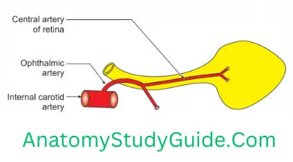
- Central artery of retina,
- Central arteries of the cerebrum,
- Renal arteries, and
- Arteries of spleen.

2. End Arteries Applied Anatomy
Occlusion of an end artery causes serious nutritional disturbances resulting in death of tissue supplied by it, e.g. in case of blockage of right coronary artery, the muscles of the heart undergo ischaemia and results into myocardial infarction.
In case of rupture of central artery of retina, the blood supply of eyeball is lost and person becomes blind.
Bursa
(Bursa—purse)
Bursa Introduction: It is a pocket-like space lined by synovial membrane containing synovial fluid.
1. Bursa Types
- Communicating: Some bursae communicate with joint cavity, e.g. subscapular bursa of shoulder joint.
- Non-communicating: For example, infrapatellar bursa of knee joint.
Bursa Anatomy
2. Bursa Functions
- It reduces the friction between two mobile and tightly opposed surfaces.
- It permits complete freedom of movement within limited range.
3. Bursa Classification: It is classified depending upon the situation
- Subcutaneous: Deep to the skin.
- Subtendinous: Deep to the tendon, e.g. subscapular bursa: It communicates with the shoulder joint. It lies deep to the scapula and is present between the superior and inferior glenohumeral ligaments.
- Submuscular: Deep to the muscle, e.g. semimembranosus bursa: Deep to semimembranosus muscle.
- Subfascial: Deep to the fascia.
Bursa Anatomy
4. Bursa Applied anatomy
Bursitis: Inflammation of bursa is called bursitis, e.g. olecranon bursitis (miner’s elbow, student’s elbow): Inflammation and enlargement of the bursa over the olecranon process of ulna. This bursa lies between olecranon process and the overlying skin.
Clergyman’s knee: It is present below patella and is superficially placed. The person gets pain in movement of knee joint.
Morrant Baker’s cyst: The swelling behind the knee is caused by escape of synovial fluid which lies in space or membrane. It is prominent during extension and disappears during flexion. It is associated with the tendons of semimembranosus or gastrocnemius.
Bursa Anatomy
Housemaid’s knee: The bursa between the skin and anterior surface of patella is called prepatellar bursa and it is inflamed in housemaids.
Weaver’s bottom: The bursa over ischial tuberosity is inflamed in weavers.
Leave a Reply