Cartilage: What It Is, Function And Types
Draw and write a note on articular cartilage
Articular cartilage
Table of Contents
1. Articular cartilage Features
1. The articular surfaces of the bone are not ossified and they remain as articular cartilage.
2. It belongs to hyaline cartilage but differs in the following aspects.
- The perichondrium is absent as it is bathed in synovial fluid.
- It does not ossify with age.
Read And Learn More: Anatomy Notes And Important Question And Answers
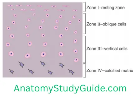
2. Articular cartilage Histology: It shows four zones from superficial to deep surface.
1. Zone 1 (superficial or tangential stratum)
- It contains a few rows of fibrocytes.
- Cells are small and flattened.
- Fibrocytes in the deepest part are parallel to the surface.
- They have a few collagen fibres called lamina splendens.
2. Zone 2 (transitional or intermediate stratum)
- The chondrocytes lie in a random fashion.
- Cells are large and round.
- They may be single or arranged in groups and are obliquely placed.
- They lie within the lacunae.
- EM demonstrates an interterritorial matrix.
- It has obliquely placed collagen fibres.
3. Zone 3 (radiate stratum)
- Contains columns of chondrocytes in lacunae.
- They are arranged radially to the surface.
- EM shows radial collagen fibres in the interterritorial matrix.
4. Zone 4 (calcified stratum)
- They touch the subchondral osseous lamina.
- The cartilage cells die and bone formation starts on the remnants of the matrix.
- It is a type of hyaline cartilage but lacks perichondrium.
- Central regions tend to be thickest on convex osseous surfaces, and thinnest on concave surfaces.
3. Site: It lines most of the articular surfaces of synovial joints except the temporomandibular joint which is lined by fibrocartilage.
4. Articular cartilage Functions of cartilage
- It acts as a cushion and protects the bone.
- It provides smooth surfaces which prevent friction.
5. Articular cartilage Nourishment
- It derives its nourishment from synovial fluid.
- Since the cartilage is avascular tissue, it depends on the surrounding tissue for its nourishment. It has poor healing capacity.
- This also explains why cartilage cannot become very thick, since diffusion of nutrients and gases to the deeper tissue will be insufficient.
6. Articular cartilage Applied anatomy
The thickness of articular cartilage varies from 1 to 7 mm (typically 2 mm) in different joints and decreases from middle to old age. In adults, thickness does not increase in proportion to mechanical loading.
In osteoarthritis, articular cartilage becomes thinner and ultimately breaks. It exposes underlying bone. It is a highly painful condition and leads to a walking disability.
For example,
- Interphalangeal joints of the hand,
- The hip joint, and the Knee joint.
Articular cartilage does not heal if injured, except articular cartilage situated close to synovial membranes.
Draw and write a note on Hyaline Cartilage (Hyalos—glass, transparent)
Introduction: The basic tissue which forms the skeleton of some organs, e.g. larynx.
1. Hyaline Cartilage Types
1. Articular
2. Non-articular
2. Hyaline Cartilage Peculiarities
- It is transparent.
- It contains homogenous intracellular substances.
- It is basophilic, i.e. stains blue with H and E stain.
- The perichondrium may or may not be present.
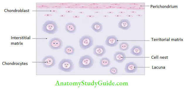
3. Hyaline Cartilage Structures
1. Cells:
- The cartilage cells are called chondrocytes.
- They are present in cell nests.
- They are in groups.
- The cells are kept together by a dense matrix.
- The matrix around the cell nests is stained deeper and is called a lacunar capsule.
- The matrix in between the cell nests is stained pale and is called the interstitial matrix.
2. Ground substance: It contains collagen fibres.
4. Hyaline Cartilage Functions: It resists compressive and tensile forces.
5. Hyaline Cartilage Sites
- Larynx: Thyroid, cricoid, arytenoid cartilage.
- Bronchial part of nasal septum,
- Lateral wall of the nose, and
- Epiphyseal plate
6. Hyaline Cartilage Features of hyaline (glossy) cartilage
- It has a homogeneous, opalescent appearance.
- It is firm and smooth to the touch and shows considerable deformability.
- The size, shape and arrangement of cells vary with sites and age.
- Chondrocytes are flat near the surface perichondrium.
- They occupy the cell nest.
- Each nest has two or more cells.
- Each is surrounded by a basket of fine collagen fibrils.
- The matrices surrounding the cells do not have collagen fibrils. But they are rich in proteoglycans. They exhibit basophilic and metachromatic staining.
Draw and write a note on Fibrocartilage
Fibrocartilage
1. Fibrocartilage Occurrence
It occurs in locations where a tough support or tensile strength is desirable
2. Fibrocartilage Features: It never occurs wholly alone, but merges with neighbouring hyaline cartilage or with fibrous tissue.
3. Fibrocartilage Macroscopic appearance.
- This tissue has an opaque appearance and a firm, fibrous texture.
- A perichondrium is lacking.
4. Fibrocartilage Microscopic structure
1. The cartilage cells are relatively sparse.
2. They occur as single cells, groups and rows.
3. Visible matrix is limited to the near vicinity of cartilage cells.
4. Coarse collagenous fibres are predominant.
5. These fibre bundles usually take a common direction.
5. Fibrocartilage Regenerative ability
- Injuries are not repaired by the cartilage tissue itself. This is because adult cells are imprisoned in a matrix and probably never divide.
- Tissue from the perichondrium and adjacent fascia proliferates and fills in the defect or gap.
- A fracture of a mature cartilage usually becomes united by dense fibrous tissue.
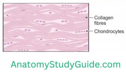
6. Fibrocartilage Histology
1. Cells:
- Large cells (singly, in groups or in rows).
- Single cells are rounded when deeply located.
- The cells are flattened near the surface.
- The cartilage cells are shrunken and vacuolate, owing to loss of water and fat.
B. Matrix:
- It is abundant and basophilic. It stains more deeply in the vicinity of cells.
- The ground substance is largely replaced by collagen fibres.
- Ordinary matrix is seen only near the vicinity of cells.
Draw and write a note on elastic cartilage
1. Elastic Cartilage Features
1. It is highly flexible.
2. It is resistant to degeneration.
3. It has a limited capacity for regeneration.
4. The elastic fibres are profuse.
5. They vary in thickness and quantity in various cartilages.
6. Perichondrium is present. It consists of
- Outer fibrous, and an
- Inner chondrogenic and vascular layer.
2. Elastic Cartilage Histology
1. Elastic fibres
- In H and E stains, they are eosinophilic.
- Elastic fibres are visualised by special staining methods (Orcein stain).
- They appear black with Verhoeff-Van Gieson stain.
- They are more heavily concentrated in the centre of cartilage than near the perichondrium.
- It has numerous branches.
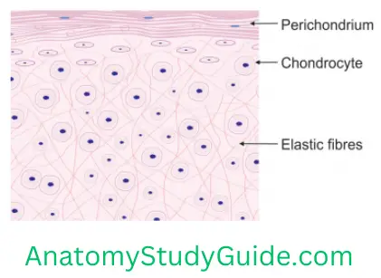
2. Chondrocytes
- Like hyaline cartilage, it contains typical chondrocytes.
- They are either singly or in small groups.
- They are surrounded by a matrix. The matrix in type II collagen fibrils.
3. Elastic Cartilage Matrix
1. It is occupied by very fine yellow elastic fibres.
2. It contains the protein elastin. It does not show periodic banding under the electron microscope.
Examples
- External ear
- External acoustic meatus (lateral part)
- Eustachian tube (medial part)
- Epiglottis
- Corniculate cartilage
- Cuneiform cartilage
3. Elastic Cartilage Applied Anatomy: The traumatic distorted injury of the external ear gets repaired by a cauliflower ear, as seen in participants of some contact sports (wrestlers).
Leave a Reply