Cerebrospinal Fluid Introduction
Cerebrospinal fluid (CSF) is the clear, colorless, and transparent fluid that circulates through the ventricles of the brain, subarachnoid space, and central canal of the spinal cord. It is a part of ECF. The normal amount of CSF is about 150 mL, and it varies between 100 and 200 mL.
Table of Contents
Properties And Composition Of Cerebrospinal Fluid
- Csf Properties:
- Volume: 150 mL
Rate of formation: 0.3mL per minute
Specific gravity: 1.005
Reaction: Alkaline
- Volume: 150 mL
- Csf Composition: The composition of CSF is given in. As CSF is part of ECF, it contains more amount of sodium than potassium. CSF also contains some lymphocytes. The CSF secreted by the ventricle does not contain any cells. The lymphocytes are added when it flows in the spinal cord.
Read And Learn More: Medical Physiology Notes

Formation Of Cerebrospinal Fluid
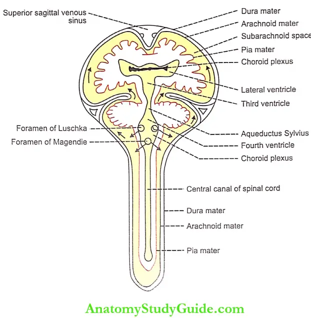
1. Site Of Formation: CSF is formed by the choroid plexuses situated within the ventricles. The choroid plexuses are tuft of capillary projections present inside the ventricles and are covered by pia mater and ependymal covering. A large amount of CSF is formed in the lateral ventricles.
2. Mechanism Of Formation: CSF is formed by the process of secretion. The formation of CSF does not involve ultrafiltration or dialysis. On the other hand, with the expenditure of energy, active transport mechanism is involved in the secretion.
3. Substances Affecting The Formation Of Csf:
- Pilocarpine, ether, and extracts of the pituitary gland stimulate the secretion of CSF by stimulating the choroid plexus
- Injection of isotonic saline also stimulates CSF formation
- Injection of hypotonic saline causes a greater rise in capillary pressure and intracranial pressure and a fall in osmotic pressure leading to an increase in CSF formation
- Hypertonic saline decreases CSF formation and decreases CSF pressure. The increased intracranial pressure is reduced by the injection of 30-35% of sodium chloride or 50% sucrose.
Circulation Of Cerebrospinal Fluid
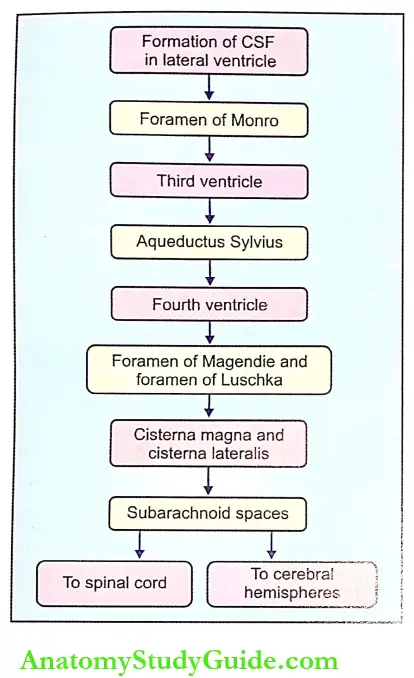
- A major quantity of CSF is formed in the lateral ventricles and passes through the foramen of Monro into the third ventricle. From here, it passes to the fourth ventricle through aqueducts Sylvius. From the fourth ventricle, it enters into the cisterna magna and cisterna lateral through a foramen of Magendie (central opening) and the foramen of Luschka (lateral opening).
- A portion of the cisternal fluid circulates through the spinal subarachnoid space. The greater part of the fluid passes upwards, over the brainstem to the surface of the cerebral hemispheres.
Absorption Of Cerebrospinal Fluid
- It is mostly absorbed by the arachnoid villi into the dural sinuses and spinal veins. A small amount is absorbed along the perineural spaces into cervical lymphatics and into the perivascular spaces.
- The mechanism of absorption is by filtration due to the pressure gradient between hydrostatic pressure in the subarachnoid space fluid and the pressure that exists in the dural sinus blood. The colloidal substances pass slowly and crystalloids are absorbed rapidly.
- Normally, about 500 mL of CSF is formed every day and an equal amount is absorbed.
Pressure Exerted By Cerebrospinal Fluid
The pressure exerted by CSF in man varies in different positions, viz.
Lateral recumbent position = 10-18 cm of H2O
Lying position = 13 cm of H2O
Sitting position = 30 cm of H2O.
Certain events like coughing and crying increase the pressure by decreasing the absorption. Compression of the internal jugular vein also raises the CSF pressure.
Functions Of Cerebrospinal Fluid
1. Protective Function:
- CSF acts as a fluid buffer and protects the brain from shock. Since the specific gravity of the brain and CSF is more or less the same, the brain floats in CSF. When the head receives a blow, CSF acts like a cushion and prevents the movement of the brain against the skull end thereby preventing damage of the brain.
- However, If the head receives a severe blow, the brain move-; ‘forcefully and hits against the skull bone leading to hie u-image of brain tissues. The brain strikes against the skull bone at a point opposite to the point where the blow was applied. So, this type of damage to the brain is known as countercoup injury.
2. Regulation of Cranial Content Volume:
- Regulation of cranial content volume is essential because, if the cranial contents increase in volume, there is an increase in the absorption of substances into the venous sinuses. It happens in cases of cerebral hemorrhage and tumors in the brain.
- When substances are absorbed into the venous sinuses, intracranial pressure is raised. Increased intra-cranial pressure in turn interferes with the cerebral circulation causing asphyxia. It is prevented by greater absorption of CSF to give space for the increasing cranial contents.
3. Medium of Exchange: CSF is the medium through which many substances, particularly the nutritive substances and waste materials are exchanged between blood and brain tissues.
Collection Of Cerebrospinal Fluid
CSF is collected either by cisternal puncture or lumbar puncture. In cisternal puncture, the CSF is collected by passing a needle between the occipital bone and atlas, so that it enters the cisterna magna. In lumbar puncture, the lumbar puncture needle is introduced into the subarachnoid space in the lumbar region, between the third and fourth lumbar spines.
1. Lumbar Puncture:
- The posture of Body for Lumbar Puncture:
- The reclining body is bent forward, so as to flex the vertebral column as far as possible. Then the body is brought near the edge of a table. The highest point of the iliac crest is determined by palpation. A line is drawn on the back of the subject by joining the highest points of iliac crests of both sides.
- Opposite the midplane, this line crosses the 4th lumbar spine. After determining the area of the 4th lumbar spine, the 3rd lumbar spine is palpated. And, the needle is introduced through the soft tissue space between the two spines, so that the needle reaches the subarachnoid space.
- Reasons for selecting this site:
- The spinal cord will not be injured, because, it terminates below the lower border of the 1st lumbar vertebra. The cauda equina may be damaged. Even if it is injured, it is regenerated
- The subarachnoid space is wider in this site. It is because the pia mater is reduced very much.
- Reasons for selecting this site:
- Uses of Lumbar Puncture: Lumbar puncture is used for:
- Collecting CSF for diagnostic purposes
- Injecting drugs (intrathecal injection) for spinal anesthesia, analgesia, and chemotherapy
- Measuring pressure exerted by CSF.
Blood-Brain Barrier
- The blood-brain barrier (BBB) is a neuroprotective structure that prevents the entry of many substances and pathogens into the brain tissues from the blood. It was observed more than 50 years ago, that when trypan blue, the acidic dye was injected into living animals, all the tissues of the body were stained by it except the brain and spinal cord.
- This observation suggested that there was a hypothetical barrier, which prevented the diffusion of trypan blue into the brain tissues from the capillaries. This barrier was named as the blood-brain barrier (BBB). The barrier exists in the capillary membrane of all parts of the brain except in some areas of the hypothalamus.
1. Structure Of Blood-Brain Barrier:
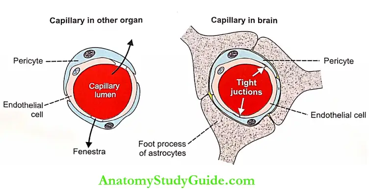
- The tight junctions in the endothelial cells of the brain capillaries are responsible for the BBB mechanism. In capillaries of other organs, the adjacent endothelial cells leave the cleft called fenestra which allows the transcytosis of several substances through the endothelium. However, in the capillaries of the brain, the fenestra are absent because the endothelial cells fuse with each other by tight junctions.
- Tight junctions are formed between the endothelial cells of the capillaries at childhood. At the same time, the cytoplasmic foot processes of astrocytes (neuroglial cells) develop around the capillaries and reinforce the barrier. The astrocytes envelop the vasculature almost completely.
- The pericytes also form the important cellular constituent of BBB. These cells play an important role in the formation and maintenance of tight junctions and the structural stability of the barrier. In brain, pericytes function as macrophages and play an important role in defense.
2. Functions Of Blood-Brain Barrier: BBB acts as both a mechanical barrier and a transport tnacnankms. It prevents potentially harmful chemical substances and permits metabolic and essential maintains into the brain tissues. By preventing injurious materials and organisms, BBB provides a healthy environment for the nerve cells of the brain.
- Substances which can Pass through Blood-Brain Barrier:
- Oxygen
- Carbon dioxide
- Water
- Glucose
- Amino acids
- Electrolytes
- Drugs such as L-dopa, 5-hydroxytryptamine sulfonamides, tetracycline, and many lipid-soluble drugs
- Anesthetic gases such as ether, and nitrous oxide which are lipid soluble
- Other lipid-soluble substances.
- Substances which cannot Pass through Blood-Brain Barrier:
- Injurious chemical agents
- Pathogens such as bacteria
- Drugs such as penicillin and catecholamines. Dopamine also cannot pass through BBS. So, Parkinsonism is treated with L-dopa instead of dopamine.
- Bile pigments. However, since the barrier is not well developed in infants, the bile pigments enter the hum tissues. During jaundice in infants, the bile pigments the brain and causes damage of basal ganglia leading to kernicterus.
Blood – Cerebrospinal Fluid Barrier
It is the barrier between the blood and cerebrospinal fluid that exists at the choroid plexus. The function of this barrier is similar to that of the BBB. It does not allow the movement of many substances from blood to cerebrospinal fluid. It allows the movement of only those substances, which are allowed by BBB.
Applied Physiology Hydrocephalus
- The abnormal accumulation of CSF in the skull associated with enlargement of the head is called hydrocephalus. During the obstruction of any of the foramen through which CSF escapes, the ventricular cavity dilates and this condition is called internal hydrocephalus. It is also known as non-communicating hydrocephalus.
- On the other hand, if the arachnoid villi are blocked, external or communicating hydrocephalus occurs. Hydrocephalus along with increased intracranial pressure causes headache and vomiting. In severe conditions, it leads to atrophy of the brain, mental weakness, and convulsions.
Leave a Reply