Changes During Muscular Contraction Introduction
The muscle contracts when it is stimulated. Contraction of the muscle is a physical or mechanical event. In addition, several other changes occur in the muscle.
Table of Contents
The changes which take place during muscular contraction:
- Electrical changes
- Physical changes
- Histological (molecular) changes
- Chemical changes
- Thermal changes.
Read And Learn More: Medical Physiology Notes
Electrical Changes During Muscular Contraction
- When the muscle is stimulated, electrical changes occur before the mechanical changes. Usually, the electrical events in a muscle (or any living tissue) are measured by using a Cathode Ray Oscilloscope.
- Nowadays, sophisticated electronic equipment like computerized polygraphs are available to record and analyze the electrical activities of any tissue.
1. Resting Membrane Potential:
- Resting membrane potential is the electrical potential difference (voltage) across the cell membrane (between the inside and outside of the cell) under resting conditions.
- It is also called membrane potential, transmembrane potential, transmembrane potential difference or trans-membrane potential gradient.
- When two electrodes are connected to a cathode ray oscilloscope through a suitable amplifier and placed over the surface of the muscle fiber, there is no potential difference, i.e. there is zero potential difference. But, if one of the electrodes is inserted into the interior of the muscle fiber, a potential difference is observed across the sarcolemma (cell membrane).
- There is negativity inside and positivity outside the muscle fiber. This potential difference is constant and is called resting membrane potential. The condition of the muscle during resting membrane potential is called a polarized state. In human skeletal muscle, the resting membrane potential is -90 mV.
2. Action Potential: Action potential is defined as a series of electrical changes that occur in the membrane potential when the muscle or nerve is stimulated. It is also defined as a wave of electrical discharge that travels along the membrane of the cell.
The action potential occurs in two phases:
- Depolarization
- Repolarization.
1. Depolarization: Depolarization is the initial phase of action potential m which the interior of the muscle becomes positive and the exterior becomes negative. That is, the polarized state (resting membrane potential) is abolished resulting in depolarization.
2. Repolarization:
- Repolarization is the phase of action potential when the potential inside the muscle reverses back to the resting membrane potential.
- That is, within a short time after depolarization the interior of the muscle becomes negative and the outside becomes positive. So, the polarized state of the muscle is re-established.
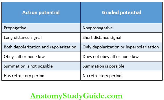
Properties of Action Potential: The Properties of action potential are listed in.
3. Action Potential Curve
- Stimulus Artifact:
- The resting membrane potential is recorded as a straight baseline at -90 mV. When a stimulus is applied, there is a slight irregular deflection of the baseline for a very short period.
- This is called stimulus artifact. The artifact is due to leakage of current from the stimulating electrode to the recording electrode. The stimulus artifact is followed by the latent period.
- Latent Period: This is the period when no change occurs in the electrical potential. It is a very short period with a duration of 0.5-1 milliseconds.
- Firing Level and Depolarization: Depolarization starts after the latent period. Initially, it is very slow. After the initial slow depolarization for about 15 mV (up to -75 mV), the rate of depolarization increases suddenly. The point at which, the depolarization increases suddenly is called the firing level.
- Overshoot: From the firing level, the curve reaches isoelectric potential (zero potential) rapidly and then shoots up (overshoots). beyond the zero potential (isoelectric base) up to +55 mV. It is called overshoot.
- Repolarization: When depolarization is completed (+55 mV), the properties of the action potential are listed in Table repolarization starts. Initially, the repolarization occurs 31-1. rapidly and then it becomes slow.
- Spike Potential: The rapid rise in depolarization and the rapid fall in repolarization are together called spike potential. It lasts for 0.4 milliseconds.
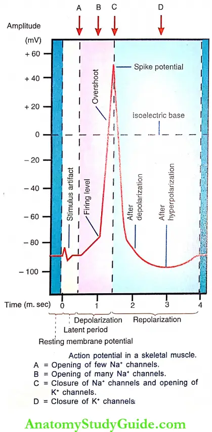
- After Depolarization or Negative after Potential: The rapid fall in repolarization is followed by slow repolarization. It is called after depolarization or negative after potential. Its duration is 2-4 milliseconds.
- After Hyperpolarization or Positive after Potential: After reaching the resting level (-90 mV), it becomes more negative beyond the resting level. This is called after hyperpolarization or positive after potential. This lasts for more than 50 milliseconds. After this, the normal resting membrane potential is restored slowly.
4. Ionic Basis Of Electrical Events:
- Resting Membrane Potential: The development and maintenance of resting membrane potential in a muscle fiber or a neuron are carried out by the movement of ions, which produce ionic imbalance across the cell membrane. This results in the development of more positivity outside and more negativity inside the cell.
The ionic imbalance is produced mainly by two transport mechanisms in the cell membrane:
- Sodium-potassium pump
- Selective -permeability of cell membrane.
1. Sodium-potassium pump
- Sodium and potassium ions are actively transported in opposite directions across the cell membrane by means of an electrogenic pump called sodium-potassium pump. It moves three sodium ions out of the cell and two potassium ions inside the cell by using energy from ATP.
- Since more positive ions (cations) are pumped outside than inside, a net deficit of positive ions occurs inside the cell. It leads to negativity inside and positivity outside the cell. More details of this pump are given.
2. Selective permeability of cell membrane:
- The permeability of the cell membrane depends largely on the transport channels. The transport channels are selective for the movement of some specific ions.
- Their permeability to these ions also varies. Most of the channels are gated channels and the specific ions can move across the membrane only when these gated channels are opened.
Channels for major anions (negatively charged substances) like proteins:
- However, channels for some of the negatively charged large substances such as proteins and negatively charged organic phosphate compounds and sulfate compounds are absent or closed.
- So, such substances remain inside the cell and play a major role in the development and maintenance of resting membrane potential.
Leak channels:
- In addition, the channels for three important ions – sodium, chloride, and potassium also play an important role in maintaining the resting membrane potential. These three ions are unequally distributed across the cell membrane. The Na+ and Cl– are more outside and K+ is more inside.
- Since the CI– channels are mostly closed in resting conditions Cl–ions are retained outside the cell. Thus, only the positive ions, Nat and K/can move across the cell membrane. The Na+ is actively transported (against the concentration gradient) out of the cell and K is actively transported (against the concentration gradient) into the cell.
- However, because of the concentration gradient, Na+ diffuses back into the cell through Na+ leak channels. And, K+ diffuses out of the cell through K+ leak channels. check the negativity. Normally, the negativity (resting membrane potential) inside the muscle fiber is -90 mV, and in nerve fiber, it is -70 mV.
- It is because of the presence of negatively charged proteins, organic phosphates, and sulfates which cannot move out normally. Suppose if the K+ is not present or decreased, the negativity increases beyond -120 mV, which is called hyperpolarization. At this stage, the development of action potential is either delayed or does not occur.
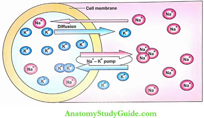
- In resting conditions, almost all the K+ leak channels are opened but most of the Na+ leak channels are closed. Because of this, K+ which were transported actively into the cell, can diffuse back out of the cell in an attempt to maintain the concentration equilibrium.
- But among the Nat which were transported actively out of the cell, only Action Potential a small amount can diffuse back into the cell. That means, in resting conditions, the passive K+ efflux is much greater than the passive Na+ influx. It helps in establishing and maintaining the resting membrane potential.
After the establishment of the resting membrane potential (i.e. inside negativity and outside positivity), the efflux of K+ ions stops in spite of the concentration gradient. The voltage-gated Na+ channels and the voltage-gated K+ channels play important roles in the development of action potential.
It is because of two reasons:
- The positivity outside the cell repels the positive K+ ions and prevents the further efflux of these ions
- The negativity inside the cell attracts the positive K+ ions and prevents further leakage of these ions outside.
Importance of intracellular potassium ions:
- The concentration of K+ inside the cell is about 140 mEq/L. It is almost equal to that of Na* outside. The high concentration of K+ inside the cell is essential to check the negativity. Normally, the negativity (resting membrane potential) inside the muscle fiber is -90 mV, and in the nerve fiber, it is -70 mV.
- It is because of the presence of negatively charged proteins, organic phosphates, and sulfates which cannot move out normally. Suppose if the K+is not present or decreased, the negativity increases beyond -120 mV, which is called hyperpolarization. At this stage, the development of action potential is either delayed or does not occur.
Action Potential:
- The voltage-gated Na+ channels and the voltage-gated K+ channels play important roles in the development of action potential.
- During the onset of depolarization, there is a slow influx of Nat. When depolarization reaches 7-10 mV, the voltage-gated Na+ channels start opening at a faster rate. It is called Na channel activation. When the firing level is reached, the influx of Na+ is very great and it leads to overshoot.
- But the Na+ transport is short-lived. This is because of the rapid inactivation of Na+ channels. Thus, the Na+ channels open and close quickly. The Na+ channels remain in this inactivated state for some time before returning to resting condition. At the same time, the K+ channels start opening. This leads to efflux of K+ out of the cell, causing repolarization.
- Unlike the Na+ channels, the K+ channels remain open for a longer duration. These channels remain open for a few more milliseconds after completion of repolarization. It causes the efflux of more K+ producing more negativity inside. It is the cause of hyperpolarization.
5. Monophasic, Biphasic, And Compound Action Potentials:
- Monophasic Action Potential: Monophasic action potential is the series of electrical changes in a stimulated muscle or nerve fiber by placing one electrode on its surface and the other inside. It is characterized by a positive deflection. The action potential in the muscle discussed above belongs to this category.
- Biphasic Action Potential: Biphasic or diphasic action potential is the series of electrical changes in a stimulated muscle or nerve fiber by placing both the recording electrodes on the surface of the muscle or nerve fiber. It is characterized by a positive deflection followed by an isoelectric pause and a negative deflection.
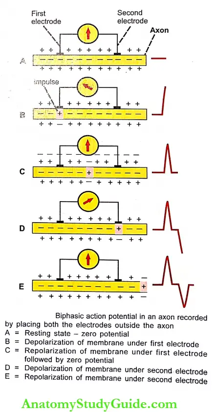
- Recording of Biphasic Action Potential: Biphasic action potential is recorded by extracellular electrodes, i.e. by placing both the recording electrodes surface of a nerve fiber or muscle. Explains the biphasic action potential in an axon. The sequence of events of biphasic action potential:
- In the resting state before stimulation, the potential difference between the two electrodes is zero. So the recording shows a baseline
- When the axon is stimulated at one end, the action potential (impulse) is generated and it travels towards the other end of an axon by passing through the recording electrodes. When the impulse reaches the first electrode, the membrane under this electrode becomes depolarized (outside negative) but the membrane under the second electrode is still in a polarized state (outside positive). By convention, this is graphically recorded as an upward deflection.
- When the impulse crosses and travels away from the first electrode the membrane under this electrode is repolarized. Later when the impulse just travels in between the two electrodes (before reaching the second electrode) the potential difference between both electrodes falls to zero and the baseline is recorded.
- When the impulse reaches the second electrode, the membrane under this electrode is depolarized (outside negative) and a negative deflection is recorded.
- When the impulse travels away from the second electrode the membrane under this gets repolarized. Once again the potential difference between the two electrodes becomes zero and the graph shows the baseline.
- Since this recording shows both positive and negative components it is called biphasic action potential.
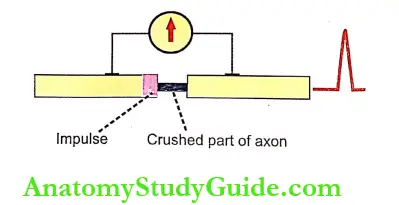
- Effect of crushing or local anesthetics: When a small portion of the axon between the two electrodes is affected by crushing or local anesthetics, the action potential cannot travel through this part of the axon. So, while recording the potential only a single deflection (monophasic) action potential is recorded.
- Compound Action Potential:
- Compound action potential (CAP) is the algebraic summation of all the action potentials produced by all the nerve fibers. Each nerve is made of thousands of axons.
- While stimulating the whole nerve, all the nerve fibers are activated and produce an action potential. The compound action potential is obtained by recording all the action potentials simultaneously.
6. Graded Potential:
- The graded potential is a mild local change in the membrane potential that develops in receptors, synapses or neuro- muscular junctions when stimulated. It is also called graded membrane potential or graded depolarization.
- It is non propagative and characterized by mild depolarization or hyperpolarization. The graded potential is distinct from the action potential and the properties of these two potentials are given in.
- In most of cases, the graded potential is responsible for the generation of the action potential. However, in some cases, the graded potential hyperpolarizes the membrane potential (more negativity than resting membrane potential) and inhibits the generation of action potential.
The graded potentials include:
- Motor end plate potential in the neuromuscular junction
- Electrotonic potential in nerve fibers
- Receptor potential
- Excitatory postsynaptic potential
- Inhibitory postsynaptic potential.
7. Patch Clamp Technique: Patch clamp technique or patch clamping is the method to measure the ion currents across biological membranes. This advanced technique in modern electrophysiology was established by Erwin Neher in 1992. The patch clamp is modified as a voltage clamp to study the ion currents across the membrane of the neuron.
- Procedure: Patch clamp experiments use mostly cultured cells. The cells isolated from the body are placed in dishes containing culture media and kept in an incubator.
- Probing a single cell:
- The dish with tissue culture cells is mounted on a microscope. A micropipette with an opening of about 0.5 μ is also mounted by means of a pipette holder. The pipette is filled with saline solution.
- An electrode is fitted to the pipette and connected to a recording device called a patch clamp amplifier. The micropipette is pressed firmly against the membrane of an intact cell.
- A gentle suction applied to the inside of the pipette forms a tight seal of giga ohms (GO) resistance between the membrane and the pipette.
- This patch (minute part) of the cell membrane under the pipette is studied by means of various approaches called patch clamp configurations.
- Patch Clamp Configurations:
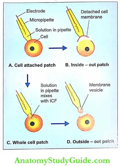
- Cell attached patch: The cell is left intact with its membrane. This allows measurement of current flow through ion channels or channels under the micropipette.
- Inside-out patch:
- From the cell-attached configuration, the pipette is gently pulled away from the cell. It causes the detachment of a small portion of the membrane from the cell. The external surface of the membrane patch faces the pipette solution. But the internal surface of the membrane patch is exposed hence the name inside-out patch.
- The pipette with membrane patch is inserted into a container with a free solution. The concentration of ions can be altered in the free solution. It is used to study the effect of alterations in the ion concentrations on the ion channels.
- Whole-cell patch:
- From the cell-attached configuration, further suction is applied to the inside of the pipette. It causes rupture of the membrane and the pipette solution starts mixing with intracellular fluid.
- When the mixing is complete, the equilibrium is obtained between the pipette solution and the intracellular fluid. A whole cell patch is used to record the current flow through all the ion channels in the cell. The cellular activity also can be studied directly.
- Outside-out patch:
- From the whole-cell configuration, the pipette is gently pulled away from the cell. A portion of the membrane is torn away from the cell. Immediately, the free ends of the torn membrane fuse and reseal forming a membrane vesicle at the tip of the pipette. The pipette solution enters the membrane vesicle and forms the intracellular fluid.
- The vesicle is placed inside a bath solution which forms the extracellular environment. This patch is used to study the effect of changes in the extracellular environment on the ion channels.
- It also helps to study the effects of neurotransmitters and compounds like ozone, G-protein regulators, etc. on the ion channels.
Physical Changes During Muscular Contraction
The physical change, which takes place during muscular contraction, is the change in the length of the muscle fibers or the change in the tension developed in the muscle. Depending upon this, the muscular contraction is classified into two types namely isotonic contraction and isometric contraction.
Histological Changes During Muscular Contraction
1. Actomyosin Complex:
- In the relaxed state of the muscle, the thin actin filaments from the opposite ends of the sarcomere are away from each other leaving a broad ‘H’ zone.
- During the contraction of the muscle, the actin (thin) filaments glide over the myosin (thick) filaments and form an actomyosin complex.
2. Molecular Basis Of Muscular Contraction:
The molecular mechanism is responsible for the formation of an actomyosin complex that results in muscular contraction. It includes three stages:
- Excitation contraction coupling
- Role of troponin and tropomyosin
- Sliding mechanism
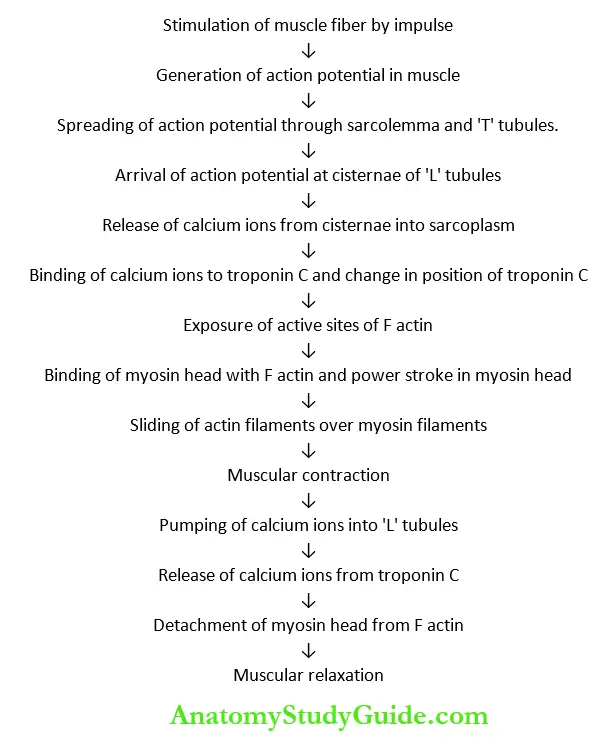
1. Excitation Contraction Coupling: Excitation contraction coupling is the process that occurs in between the excitation and contraction of the muscle. This process involves a series of activities which are responsible for the contraction of the excited muscle.
- Stages of excitation-contraction coupling:
- When the impulse passes through a motor neuron and reaches the neuromuscular junction, acetylcholine is released from the motor endplate. Acetylcholine causes the opening of ligand-gated sodium channels. So, sodium ions enter the neuromuscular junction. It leads to the development of endplate potential.
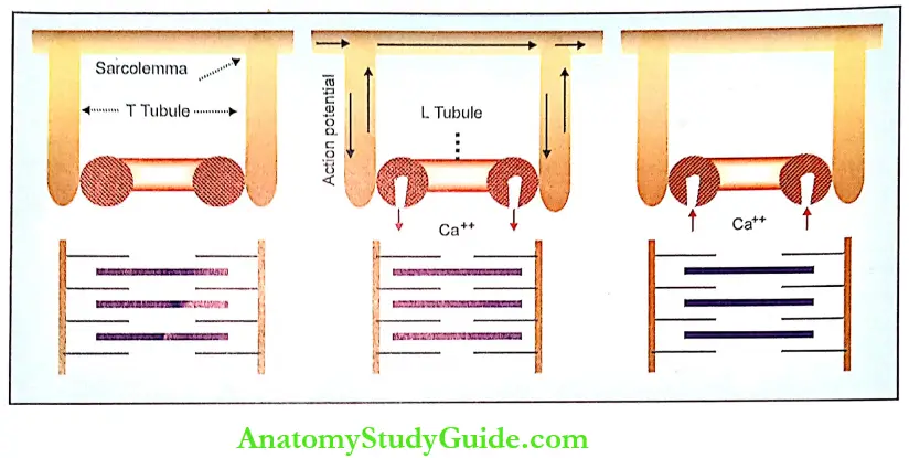
- Endplate potential causes the generation of action potential in the muscle fiber. The action potential spreads over sarcolemma and also into the muscle fiber through the ‘T’ tubules. The ‘T’ tubules are responsible for the rapid spread of action potential into the muscle fiber.
- When the action potential reaches the cisternae of ‘L’ tubules, these cisternae are sarcoplasm move towards the actin filaments to produce the contraction.
- Thus, the calcium ion forms the link or coupling material between the excitation and the contraction of the muscle. Hence, the calcium ions are said to form the basis of excitation-contraction coupling.
2. Role of Troponin and Tropomyosin:
- Normally, the head of myosin molecules has a strong tendency to get attached with the active site of F actin. However, in relaxed conditions, the active site of F actin is covered by tropomyosin. Therefore, the myosin head cannot combine with the actin molecule.
- A large number of calcium ions, which are released from ‘L’ tubules during the excitation of the muscle, bind with troponin C. The loading of troponin C with calcium ions produces some change in the position of the troponin molecule.
- It in turn, pulls tropomyosin molecules away from Factin. Due to the movement of tropomyosin, the active site of F actin is uncovered and immediately the head of myosin gets attached to the actin.
3. Sliding Mechanism and Formation of Actomyosin Complex – Sliding Theory:
- Sliding theory explains how the actin filaments slide over myosin filaments and form the actomyosin complex during muscular contraction. It is also called ratchet theory or walk-along theory.
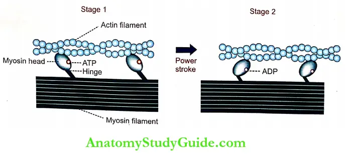
- Each cross-bridge from the myosin filaments has got three components namely, a hinge, an arm, and a head. After binding with the active site of F actin, the myosin head is tilted towards the arm so that the actin filament is dragged along with it. This tilting of the head is called a power stroke.
- After tilting, the head immediately breaks away from the active site and returns to its original position. Now, it combines with a new active site on the actin molecule. And, tilting movement occurs again. Thus, the head of the cross-bridge bends back and forth and pulls the actin filament towards the center of the sarcomere.
- In this way, all the actin filaments of both the ends of sarcomere are pulled. So, the actin filaments of opposite sides overlap and form an actomyosin complex. Formation of the actomyosin complex results in the contraction of the muscle.
- When the muscle shortens further, the actin filaments from opposite ends of the sarcomere approach each other. So, the ‘H’ zone becomes narrow. And, the two ‘Z’ lines come closer with a reduction in the length of the sarcomere. length of the T band decreases. However, the length of the ‘A’ band is not altered. But, the length of the “I” band decreases.
- When the muscular contraction becomes severe, the actin Slaments from opposite ends overlap and, the ‘H’ zone disappears.
Thus, during the contraction of the muscle, the following changes occur in the sarcomere
- The length of all the sarcomeres decreases as the ‘Z’ lines come close to each other
- The length of the ‘I’ band decreases since the actin filaments from opposite side overlap
- The ‘H’ zone either decreases or disappears
- The length of ‘A’ band remains the same.
The summary of the sequence of events during muscular contraction is given in.
Energy for Muscular Contraction:
- The energy for the movement of the myosin head (power stroke) is obtained by the breakdown of adenosine triphosphate (ATP) into adenosine diphosphate (ADP) and inorganic phosphate (Pi).
- The head of the myosin has a site for ATP. Actually, the head itself can act as the enzyme ATPase and catalyze the breakdown of ATP. Even before the onset of contraction, an ATP molecule binds with myosin head.
- When tropomyosin moves to expose the active sites, the head is attached to the active site. Now ATPase cleaves ATP into ADP and Pi, which remains in the head itself. The energy released during this process is utilized for contraction.
- When the head is tilted, the ADP and Pi are released and a new ATP molecule binds with the head. This process is repeated until the muscular contraction is completed.
Relaxation of the Muscle:
- The relaxation of the muscle occurs when the calcium ions are pumped back into the L tubules. When calcium ions enter the L tubules, calcium content in sarcoplasm decreases leading to the release of calcium ions from the troponin.
- It causes detachment of myosin from actin followed by relaxation of the muscle. The detachment of myosin from actin obtains energy from the breakdown of ATP. Thus, the chemical process of muscular relaxation is an active process although the physical process is said to be passive.
Molecular Motors: Along with other proteins and some enzymes, actin and myosin form the molecular motors which are involved in movements.
Chemical Changes During Muscular Contraction
1. Liberation Of Energy: The energy necessary for muscular contraction is liberated during the processes of breakdown and resynthesis of ATP.
Breakdown of ATP:
During muscular contraction, the supply of energy is from the breakdown of ATP. This is broken into ADP and inorganic phosphate (Pi) and energy is liberated.

The energy liberated by the breakdown of ATP is responsible for the following activities during muscular contraction:
- Spread of action potential into the muscle
- Liberation of calcium ions from cisternae of ‘L’ tubules into the sarcoplasm
- Movements of myosin head
- Sliding mechanism.
The energy liberated during ATP breakdown is sufficient for maintaining full contraction of the muscle for a short duration of less than one second.
Resynthesis of ATP: The ADP, which is formed during ATP breakdown, is immediately utilized for the resynthesis of ATP. But, for the resynthesis of ATP, the ADP cannot combine with inorganic phosphate. It should combine with a high-energy phosphate radical. There are two sources from which the high-energy phosphate is obtained namely, creatine phosphate and carbohydrate metabolism.
Resynthesis of ATP from creatine phosphate:
- The immediate supply of high-energy phosphate radicals is from creatine phosphate (CP). Plenty of creatine phosphate is available in resting muscles. In the presence of the enzyme creatine phosphotransferase, high-energy phosphate is released from creatine phosphate. The reaction is called Lohmann’s reaction.
- ADP + CP → ATP + Creatine
- The energy produced in this reaction is sufficient to The creatine should be resynthesized into creatine to maintain muscular contraction only for a few seconds. phosphate and this requires the presence of high-energy phosphate. So, the required amount of high-energy phosphate radicals is provided by the carbohydrate metabolism in the muscle.
Resynthesis of ATP by carbohydrate metabolism:
- Carbohydrate metabolism starts with catabolic actions of glycogen in the muscle. In resting muscle, an adequate amount of glycogen is stored in the sarcoplasm.
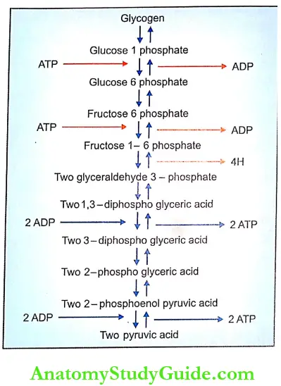
- Each molecule of glycogen undergoes catabolism, to produce ATP. And, the energy liberated during the catabolism of glycogen can cause muscular contraction for a longer period. The first stage of catabolism of glycogen is via glycolysis. It is called glycolytic pathway or Embden- Meyerhof pathway.
Glycolysis:
- Each glycogen molecule is converted into two pyruvic acid molecules. Only a small amount of ATP (2 molecules) is synthesized in this pathway.
- This pathway has 10 steps. And, each step is catalyzed by one or two enzymes as shown in.
During glycolysis, 4 hydrogen atoms are released which are also utilized for the formation of additional molecules of ATP. The formation of ATP by the utilization of hydrogen is explained later. - The further changes in pyruvic acid depend upon the availability of oxygen. In the absence of oxygen, the pyruvic acid is converted into lactic acid that enters the Cori cycle. It is known as anaerobic glycolysis. If oxygen is available, the pyruvic acid enters into Krebs’ cycle. It is known as aerobic glycolysis.
Cori’s Cycle: Lactic acid is transported to the liver where it is converted into glycogen and stored there. If necessary, glycogen breaks into glucose, which is carried by the blood to the muscle. Here, the glucose is converted into glycogen, which enters the Embden-Meyerhof pathway.

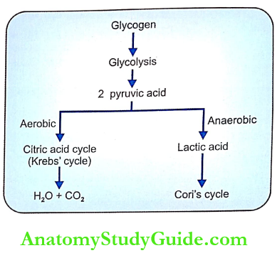
Krebs’ Cycle: It is otherwise known as the tricarboxylic acid cycle (TCA cycle) or citric acid cycle. A greater amount of energy is liberated through this cycle. The pyruvic acid derived from glycolysis is taken into mitochondria where it is converted into acetyl coenzyme A with the release of 4 hydrogen atoms. The acetyl coenzyme A enters the Krebs cycle.
- Krebs’ cycle is a series of reactions by which acetyl coenzyme A is degraded in various steps to form carbon code and hydrogen atoms. All these reactions occur in the matrix of the mitochondrion. During Krebs’ cycle, 2 molecules of ATP and 16 atoms of hydrogen are released. The hydrogen atoms are also utilized for the formation of ATP (see below).
Significance of Hydrogen Atoms Released during Carbohydrate Metabolism:
- Altogether 24 hydrogen atoms are released during glycolysis and Krebs’ cycle:
- 4H : During the breakdown of glycogen into pyruvic acid
- 4H : During the formation of acetyl coenzyme A from pyruvic acid
- 16H: During degradation of acetyl coenzyme A in Krebs’ cycle.
- The hydrogen atoms are released in the form of two pockets into intracellular fluid and it is catalyzed by the enzyme dehydrogenase. Once released, 20 hydrogen atoms combine with NAD (nicotinamide adenine fers the hydrogen atoms to the cytochrome system dinucleotide) which acts as a hydrogen carrier.
- NAD trans- where oxidative phosphorylation takes place. Oxidative phosphorylation is the process during which the ATP molecules are formed by utilizing hydrogen atoms. For every 2 hydrogen atoms 3 molecules of ATP are formed. So, from 20 hydrogen atoms, 30 molecules of ATP are formed.
- The remaining 4 hydrogen atoms enter the oxidative phosphorylation processes directly without combining with NAD. Only 2 ATP molecules are formed for every 2 hydrogen atoms. So, 4 hydrogen atoms give rise to 4 ATP molecules. Thus, 34 ATP molecules are formed from the hydrogen atoms released during glycolysis and Krebs’ cycle.
Summary of Resynthesis of ATP during Carbohydrate Metabolism:
A total of 38 ATP molecules are formed during the breakdown of each glycogen molecule in the muscle as summarized below:
- During glycolysis : 2 molecules of ATP
- During Krebs’ cycle : 2 molecules of ATP
- By utilization of hydrogen : 34 molecules of ATP atoms
- Total : 38 molecules of ATP
2. Changes In Ph During Muscular Contraction: The reaction and the pH of muscle are altered in different stages of muscular contraction.
- In Resting Condition: During resting condition, the reaction of muscle is alkaline with a pH of 7.3.
- During Onset of Contraction: At the beginning of the muscular contraction, the reaction becomes acidic. The acidity is due to the dephosphorylation of ATP into ADP and Pi.
- During Later Part of Contraction: During the later part of contraction, the muscle becomes alkaline. It is due to the resynthesis of ATP from CP.
- At the End of Contraction: At the end of contraction, the muscle becomes once again acidic. This acidity is due to the formation of pyruvic acid and/or lactic acid.
Thermal Changes During Muscular Contraction:
During muscular contraction, heat is produced. Not all the heat is liberated at a time. It is released in different stages:
- Resting heat
- Initial heat
- Recovery heat.
1. Resting Heat: The heat produced in the muscle at rest is called resting heat. It is due to the basal metabolic process in the muscle.
2. Initial Heat: During muscular activity, heat production occurs in three stages:
- Heat of activation
- Heat of shortening
- The heat of relaxation.
- Heat of Activation: It is the heat produced before the actual shortening of the muscle fibers. Most of this heat is produced during the release of calcium ions from ‘L’ tubules. It is also called maintenance heat.
- Heat of Shortening: It is the heat produced during the contraction of the muscle. The heat is produced due to various structural changes in the muscle fiber like movements of cross bridges and myosin heads and breakdown of glycogen.
- Heat of Relaxation: The heat released during the relaxation of the muscle is known as the heat of relaxation. In fact, it is the heat produced during the contraction of the muscle due to the breakdown of ATP molecules. It is released when the muscle lengthens during relaxation.
3. Recovery Heat:
It is the heat produced in the muscle after the end of activities. After the end of muscular activities, some amount of heat is produced due to the chemical processes involved in the resynthesis of chemical substances broken down during contraction.
Leave a Reply