Ciliary Ganglion
Ciliary Ganglion Introduction: It is collection of cell bodies of parasympathetic nerve. It supplies the
sphincter pupillae muscle.
1. Ciliary Ganglion Size: Pinhad.
2. Ciliary Ganglion Content: Cell bodies of multipolar neuron.
3. Ciliary Ganglion Situation: Apex of the orbit in the angle made by optic nerve and lateral rectus “muscle.
4. Ciliary Ganglion Relations
Table of Contents
- Medially: Optic nerve.
- Laterally: Lateral rectus muscle.
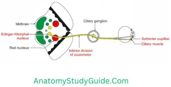
5. Ciliary Ganglion Connections: Three roots
Motor (parasympathetic)
- Preganglionic fibres arise from Edinger-Westphal nucleus > lower division of 3rd nerve > ciliary ganglion > fibres are relayed into ciliary ganglion.
- Postganglionic fibres carried by short ciliary nerve and supply
- Sphincter pupillae, and
- Ciliaris muscle.
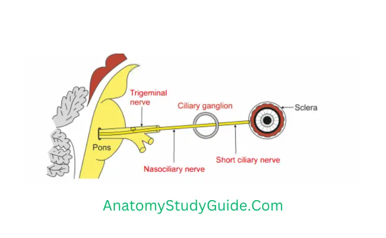
Ciliary Ganglion Sensory: Nasociliary fibres pass through ciliary ganglion without relay.
Ciliary Ganglion Sympathetic fibres pass through the ciliary ganglion without relay.
- Preganglionic fibres arise from spinal nerve and reach to superior cervical sympathetic ganglion.
- Postganglionic fibres arise from the plexus around ophthalmic artery pass through short ciliary nerve and supply dilator pupillae.
6. Ciliary Ganglion Branches:8 toThe10 short ciliary nerves
7. Ciliary Ganglion Peculiarity: postganglionic parasympathetic fibres of ciliary ganglion are myelinated.
8. Ciliary Ganglion Applied anatomy
1. Complete division of oculomotor nerve is manifested as NEEr
- Eyeball is depressed and abducted. “Down and out”. External strabismus: Due
to unopposed action of lateral rectus.
Ophthalmoplegia >Diplopia where false image is higher than true image. - Ptosis: Drooping of eyelid due to involvement of levator palpebrae muscle.
- Sphincter pupillae is not functioning. It results into dilatation of pupil
(mydriasis) - Dilated and fixed pupil.
- Loss of accommodation reflex because of paralysis of three muscles.
Medial rectus-medial convergence of eyeball is lost.
Sphincter pupillae-pupillary constriction is lost.
Ciliaris muscle-thicknss of lens. - Apparent protrusion of eyeball due to flaccid paralysis of most of ocular muscles.
2. Neurosyphilis causes inflammation of posterior cerebral and superior cerebellar arteries. This compresses oculomotor nerve.
3. Weber’s syndrome: Contralateral hemiplegia (upper motor neuron lesion) and ipsilateral paralysis of muscles supplied by oculomotor nerve.
What happens in case of unilateral ocular muscle paralysis?
1. Paralysis of a muscle will cause limitation of movement of the eyeball. One will have double vision when one attempts to use the muscle.
1.When the abducent nerve supplying the lateral rectus is paralyzed, the individual cannot abduct the eyeball on the affected side. The eyeball is fully adducted by the unopposed pull of the medial rectus.
2.In complete 3rd nerve paralysis, the
- Eye cannot be moved upward, downward or inward.
- At rest, the eye looks laterally (external strabismus) because of the activity of the lateral rectus, and
- Downward because of the activity of the superior oblique.
- The patient has double vision (diplopia).
Drooping of the upper eyelid (ptosis) occurs because of paralysis of the levator palpebrae superioris.
The pupil is widely dilated and non reactive to light because of the paralysis of the sphincter pupillae and the unopposed action of the dilator pupillae (supplied by the sympathetic). - Accommodation of the eye is lost.
3. In 4th nerve paralysis, the patient complains of double vision on looking straight downward.
This is because the superior oblique is paralyzed and the eye turns medially as the inferior rectus pulls the eye downward.
4. In 6th nerve paralysis, the patient cannot
- Tum the eyeball laterally.
- When looking straight ahead, the lateral rectus is paralyzed, and
- Unopposed medial rectus pulls the eyeball medially, causing internal strabismus.
Leave a Reply