Classification of Joints
Joints are classified depending upon:
Table of Contents
- Structure,
- Function, and
- Region.
1. Structure of binding material
1. Fibrous joint: Bones are kept together by fibrous tissue. They are sub-classified depending upon nature of fibres
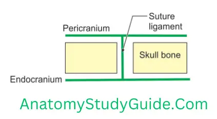
- Suture: The bones are kept together by a thin fibrous tissue or ligaments.
- Syndesmosis: The bones are kept together by fibrous tissue which is in the form of band or interosseous membrane.
- Gomphosis: The bones are kept together by fibrous tissue which is in the form of periodontal membrane.
Types Of Synovial Joints
2. Cartilaginous joint: Bones are kept together by cartilage. They are sub-classified depending upon the type of cartilage
- Primary cartilaginous joints or synchondrosis
- Secondary cartilaginous joints or symphysis
3. Synovial joint: Bones are kept together by synovial fluid.
Read And Learn More: Anatomy Notes And Important Question And Answers
2. Function: They are sub-classified depending upon degree of mobility.
- Synarthroses are fixed joints at which there is no movement. The articular surfaces are joined by tough fibrous tissue. Often the edges of the bones are fixed into one another as in the sutures of the skull.
- Amphiarthroses are joints at which slight movement is possible. A pad of cartilage lies between the bone surfaces, and there are fibrous ligaments to hold the bones and cartilage in place. The cartilages of such joints also act as shock absorbers, e.g. the intervertebral discs between the bodies of the vertebrae, where the cartilage is strengthened by extra collagen fibres.
- Diarthroses or synovial joints are known as freely movable joints. In some joints, the movement is restricted by the shape of the articulating surfaces and by the ligaments which hold the bones together. These ligaments are of elastic connective tissue. The bones forming the synovial joint have articular surfaces. These are covered by articular cartilage. There is fluid, produced by the membrane lining the fibrous capsule. It is called synovial membrane. The fluid produced by synovial membrane is called synovial fluid. It spreads inner surface of the cavity. It acts as a lubricant. It produces free movements.
Types Of Synovial Joints
3. Regional
- Skull type: Immovable.
- Vertebral type: Slightly movable.
- Limb type: Freely movable.
Fibrous Joints
In fibrous joints, the bones are joined by fibrous tissue. These joints are either immovable or permit a slight degree of movement. These can be grouped in the following three subtypes.
1. Sutures: These are peculiar to skull, and are immovable. According to the shape of bony margins, the sutures can be:
- Plane, e.g. internasal suture
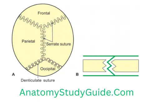
- Serrate, e.g. interparietal suture
- Squamous, e.g. temporoparietal suture
- Denticulate, e.g. lambdoid suture
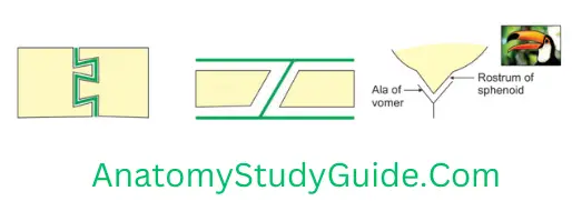
- Schindylesis type, e.g. between rostrum of sphenoid and upper border of vomer.
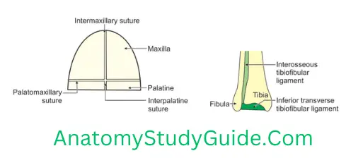
2. Syndesmosis: The bones are connected by the interosseous ligament, e.g. inferior tibiofibular joint.
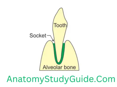
3. Gomphosis (peg and socket joint). Example: Root of the tooth in its bony socket
Suture (Suture—stitch, seam)
Introduction: Joints of skull are connected by fibrous tissue.
1. Functions
- They allow the growth of brain in the cranial cavity.
- They help moulding of head during labour.
2. Movement: No movement.
3. Types: Depending upon articular margin, they are subdivided into:
1. Plane: Margins are straight, e.g.
- Interpalatine
- Intermaxillary
- Palatomaxillary.
2. Serrate(saw): Margins are wavy (saw-like), e.g.
- Sagittal suture
- Coronal suture
Types Of Synovial Joints
3. Denticulate: The articulating margins resemble teeth. The tips are broader than the roots to have effective interlocking, e.g. lambdoid suture (suture between parietal and occipital bone,
4. Squamous: The articulating margins are bevelled, e.g. temporoparietal
5. Schindylesis (splinting a piece of wood): The ridged bone fits into a groove, e.g. rostrum (beak) of sphenoid overlapped by ala (wing) of vomer.
6. Gomphosis (wedge-shaped nail or bolt): Peg and socket type of joint, e.g. tooth and socket.
7. Limbus (border): Borders are mutually ridged or serrated.
4. Age changes
- Ossification of sutural membrane starts at the age of 20 years and is slow.
- Ossification completes at late twenties.
Syndesmoses (Syn—fusion, desmos—band)
Introduction: Bony surfaces are joined together by interosseous membrane or ligament.
1. Characters
- It is a type of fibrous joint.
- The bones are kept together at a distance by interosseous membrane.
- The interosseous membrane persists throughout life.
Types Of Synovial Joints
2. Movement: Slight degree of movement is possible, e.g.
- Inferior tibiofibular joint,
- Middle radioulnar joint, and
- Posterior sacroiliac joint.
Primary Cartilaginous Joint (Synchondrosis)
1. Characters
- The articulating surfaces are covered by a hyaline cartilage.
- They are immovable and strong.
- They are temporary in nature.
- The cartilaginous plate is replaced by bone synostosis.
2. Example:
First chondrosternal joint
Costochondral
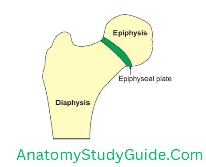
Diaphysis and epiphysis of long bone
Spheno-occipital
Sternoxiphisternal
Secondary Cartilaginous Joint (Symphysis)
Introduction: The articulating surfaces of bones are covered by hyaline cartilage. These hyaline cartilages are separated by fibrocartilage.
1. Characters: The thickness of fibrocartilage is directly related to a range of movements.
2. Functions of secondary cartilaginous joints. They
- Act as a shock absorber.
- Help in flexibility.
- Help in weight transmission.
3. Site: All midline joints of body except
- Symphysis menti (atypical and temporary joint).
- Joint between sternum and xiphoid.
4. Fate: Movements are limited.

5. Duration: Persists throughout life.
6. Example:
Intervertebral joint
Manubriosternal
Pubic symphysis
Sacral joint
Typical Synovial Joint
Introduction: The synovial joints have free movements. There is a cavity between the articulating bones. Hence, they are also called cavitation joints.
1. Characters: Synovial joints are characterized by the following features:
1. Articular cartilage: Articular surfaces are covered by a layer of hyaline cartilage.
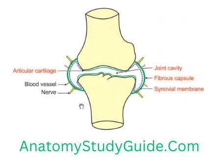
2. Fibrous capsule
- It consists of longitudinal and interlacing bundles of white connective tissue fibres. It is attached to articulating ends of the bones and forms a cuff. It encloses a joint cavity.
- It is pierced by blood vessels and nerves.
- It acts as a watchdog, i.e. it prevents excessive movements and protects the joint from dislocation.
- It has a rich nerve supply, hence it is highly sensitive.
3. Ligaments: It is strengthened by
- Accessory ligaments, and
- Muscles surrounding the joint.
4. Synovial membrane
1. Characters
- It lines the inner surface of the fibrous capsule.
- It is deficient at articular surfaces.
- It secretes hyaluronic acid which is responsible for the viscosity of the synovial fluid.
- The viscosity of the fluid varies with the movements.
- The quantity of the fluid also varies. The knee which is the largest joint contains 0.5 ml.
Types Of Synovial Joints
2. Functions of synovial fluid
- Lubrication, and
- Nutrition.
5. Joint cavity: All synovial joints are enclosed in a joint cavity.
6. Movements: The joint is capable of varying degrees of movement.
2. Functions: Varying degrees of movements.
3. Applied anatomy
Tuberculosis and gonococcal infection affect the synovial joints.
More than one joint may have the same nerve supply, e.g. hip and knee joints supplied by the obturator nerve. Hence, diseases of one joint may cause referred pain to other joints.
Classification of Synovial Joint
Synovial joints are sub-classified depending upon an axis, number of bones, presence of cartilage and the shape of articulating surfaces
1. Axis
- Plane joints: No axis.
- Uniaxial: Movements in only one axis. It is subclassified depending upon the direction of an axis.
1. Hinge variety, if the direction of axis is horizontal.
For example
- Elbow joint
- Knee joint
- Ankle joint
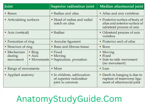
2. Pivot variety, if the direction of axis is vertical.
For example
- Superior and inferior radioulnar joints
- Atlantoaxial joint.
2. Number of bones
1. Simple, if the number of articulating bones is two.
For example
- Shoulder joint
- Hip joint.
2. Compound, if the number of articulating bones is more than two.
For example
- Wrist joint
- Knee joint
3. Presence of cartilage: Complex joint, when joint cavity is divided by an intra-articular disc into upper/lower, medial/lateral compartments it is called complex joint, e.g.
- Temporomandibular joint,
- Acromioclavicular joint, and
- Sternoclavicular joint.
4. Shape of the articulating bones
1. Ball-and-socket or spheroidal joints: The articular surface of one bone is spherical and it fits into the socket of the corresponding bone. Examples are:
- Shoulder joint,
- Hip joint,
- Talonavicular joint, and
- Incudostapedial joint
2. Sellar or saddle joints: The articular surface of one bone is convex and concave and the reciprocating surface is concave-convex. Examples can be recollected by funny Hindi sentence which is followed by body gesture. The sentence is Mera dimag ghutane me nahi hai(esjk fnekx ?kqVuks esa ugh gSA) meaning “My mind is not Kneeling”. The words of the sentence represent as:
Mera represents “sternoclavicular joint”.
Dimag represents “temporomandibular joint and incudomalleolar joint”.
Ghutane me represents the “patellofemoral joint”.
Nahi represents “calcaneocuboid joint”.
Hai represents the “joint of the thumb”
3. Condylar or bicondylar joints :
Examples are:
- Knee joint, and
- Temporomandibular joint.
4. Ellipsoid joint
- Atlanto-occipital joint,
- Wrist joint, and
- Metacarpophalangeal joint.
Pivot Joint (Pivot—a pin on which anything turns)
Introduction: It is a uniaxial (transverse axis) synovial joint.
Leave a Reply