Connective Tissue: Types, Function, Examples, Disorders
Write a note on Dense Regular Connective Tissue
Table of Contents
1. Dense Connective Tissue Contains
- Thicker and densely arranged collagen fibres,
- Fewer cell types
- Fibroblasts are the most abundant. They are located between the dense collagen bundles.
- Less ground substance.
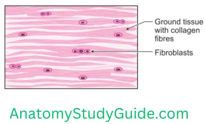
2. Dense Regular Connective Tissue Types: Dense connective tissue is of two types
1. Dense Irregular Connective Tissue
Read And Learn More: Anatomy Notes And Important Question And Answers
1. The collagen fibres are randomly and irregularly arranged.
2. Examples
- Dermis of skin,
- Capsules of various organs, and
- Areas that need strong binding and support.
Nervous Tissue
2. Dense Regular Connective Tissue
1. They contain densely packed collagen fibres.
2. They exhibit a regular and parallel arrangement.
3. Examples
- Tendons, and
- Ligaments.
Write A Note On Adipose Tissue
1. Adipose Tissue Introduction: It is the type of connective tissue.
2. Adipose Tissue Features
- It contains predominantly adipose or fat cells.
- The cells occur singly or in groups.
- They store fat.
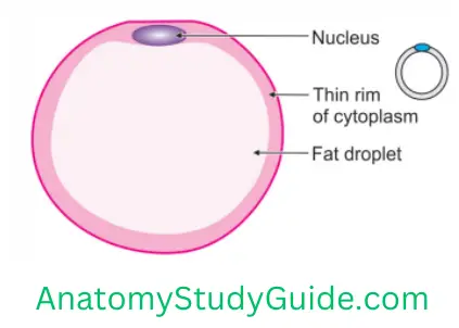
3. Adipose Tissue Sites: They are present in lipid storage organs, e.g. intestinal mesentery.
4. Adipose Tissue Types: There are two types of adipose cells.
1. White adipose tissue
White Adipose Tissue Features
Nervous Tissue
It is more common.
The cells are large and store lipids as a single large droplet.
The lipid stored in the adipose cells are primarily triglycerides (fatty acids and glycerol). They are derived from
- Intestinal lipoproteins
- Very low-density lipoprotein (VLDL) from the liver.
It is
- Distributed throughout the body.
- Highly dependent on gender and age.
- Highly vascular as a result of high metabolic activity.
2. White Adipose Tissue Functions
It serves as an important energy source.
It acts as
- Insulation under the skin.
- Cushion.
- Important endocrine organ.
These cells are the only source of a hormone called leptin.
2. Brown adipose tissue Features
- The cells are smaller.
- It stores lipids as numerous or multiple small droplets.
- It has limited distribution.
- It is present in all mammals.
- It is regulated through the sympathetic nervous system.
- The amount of brown adipose tissue increases as age advances.
2. White Adipose Tissue Sites
- Adrenal gland,
- Great vessels, and
- Neck.
c. White Adipose Tissue Functions: The main function is to supply heat to the body. It is through nonshivering thermogenesis.
What Are The Different Types Of Cells In Connective Tissue? What Are Their Identification Points And Functions?
The main cells of the connective tissue are the
1. Fibroblasts

1. Fibroblasts Identifying points
- A large, rounded vesicular nucleus.
- Prominent nucleus with abundant cytoplasm.
Nervous Tissue
2. Fibroblasts Functions
- Maintain the integrity of connective tissues.
- They secrete and maintain the components of matrix.
2. Fibrocytes
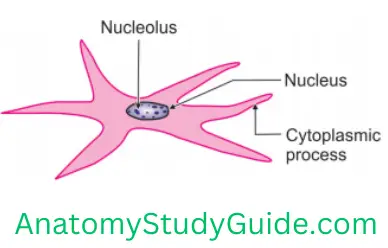
1. Fibrocytes Identifying points
- Spindle-shaped cells
- Flat nucleus
2.Fibrocytes Function: They secrete and maintain the components of the matrix.
3. Fibrocytes White adipose (fat) cells
1.Fibrocytes Identifying points: Large spherical cells with an eccentric nucleus.
2. Fibrocytes Functions: They act as
- Insulators
- Storehouse of energy
- Shock-absorbing cushion.
4. Brown Adipose Cells
1. Identifying points
- They are smaller in size as compared to unilocular.
- They have a centrally placed spherical nucleus.
2. Brown Adipose Cells Functions: The main function is to supply heat to the body through non-shivering thermogenesis.
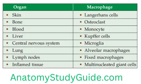
5. Macrophages (Macro—big, phage—to eat)
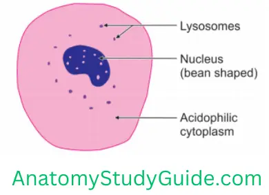
1. Macrophages Identifying points: Large cells with a dark nucleus.
2. Macrophages Functions: Phagocytosis
6. Lymphocytes
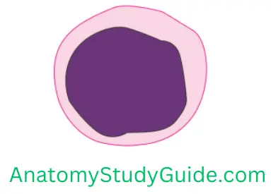
1. Lymphocytes Identifying points
- They have a round nucleus, which is often eccentrically placed.
- In small lymphocytes, a thin rim of cytoplasm is present around the nucleus.
- In large lymphocytes, the rim of the cytoplasm is wider than in small lymphocytes.
2. Lymphocytes Functions
- They are involved in chronic infection.
- They help in an immune mechanism.
Nervous Tissue
7. Plasma cells
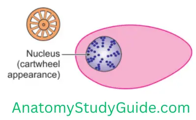
1. Plasma cells Identifying points: Ovoid cells with an eccentrically placed cartwheel nucleus and basophilic cytoplasm.
2. Plasma cells Functions: Involved in antigenic response.
8. Mast cells
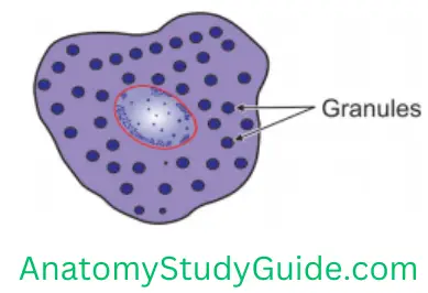
1. Mast cells Identifying points
- Large ovoid cells found along blood vessels.
- Central spherical nucleus.
- Basophilic granules in the cytoplasm.
2. Mast cells Functions
- Release histamine and vasoactive chemicals when exposed to allergens.
- It causes allergic reactions.
9. Neutrophils

1. Neutrophils Identifying points
- These are the most abundant (50–70%). They are present in the blood.
- Their nuclei have 3–5 lobes.
2. Neutrophils Functions
- Engulf and destroy bacteria.
- They provide the first line of defence against infective organisms.
- They phagocytose the microorganisms and destroy them with their enzymes.
10. Reticular cells
1. Reticular cells Identifying points
- Stellate cells with a large nucleus
- Prominent nucleolus.
2. Reticular cells Functions: They produce reticular fibres which support lymphocytes, macrophages and other cells of the lymphoid tissue.
11. Eosinophils
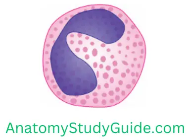
1. Eosinophils Identifying points
- They constitute 1–5% of the leucocytes present in the blood.
- The cells have a bilobed nucleus.
2. Eosinophils Functions
Phagocytize antigen–antibody complexes during allergic reactions.
Describe Plasma Cell
- Area of distribution: Present in serous and mucous membranes of alimentary and lymphoid tissue.
- Characters: Small, rounded or ovoid cells with an eccentric nucleus.
- Nucleus and cytoplasm: Deeply basophilic cytoplasm. Chromatin material in the nucleus is arranged radially and gives a cartwheel appearance.
- Functions: Chief function is the production of antibodies
Leave a Reply