Biology Of Cell Organelles
Cytosol or the cytoplasm is the gel-like ground substance comprising about half the volume of the cell in which the organelles (meaning little organs) of the cells are suspended.
These organelles are the site of major metabolic activities of the cell. As discussed above, most of the organelles are membrane-bound (for example, Mitochondria, endoplasmic reticulum, ribosomes, Golgi apparatus, lysosomes, peroxisomes, and ubiquitin-proteasome system) while the cytoskeleton is without any membrane.
Read And Learn More: General Pathology Notes
In the discussion of organelles that follows, the focus is on their molecular biology and role in major biochemical pathways in cell biology, rather than describing their each and every aspect.
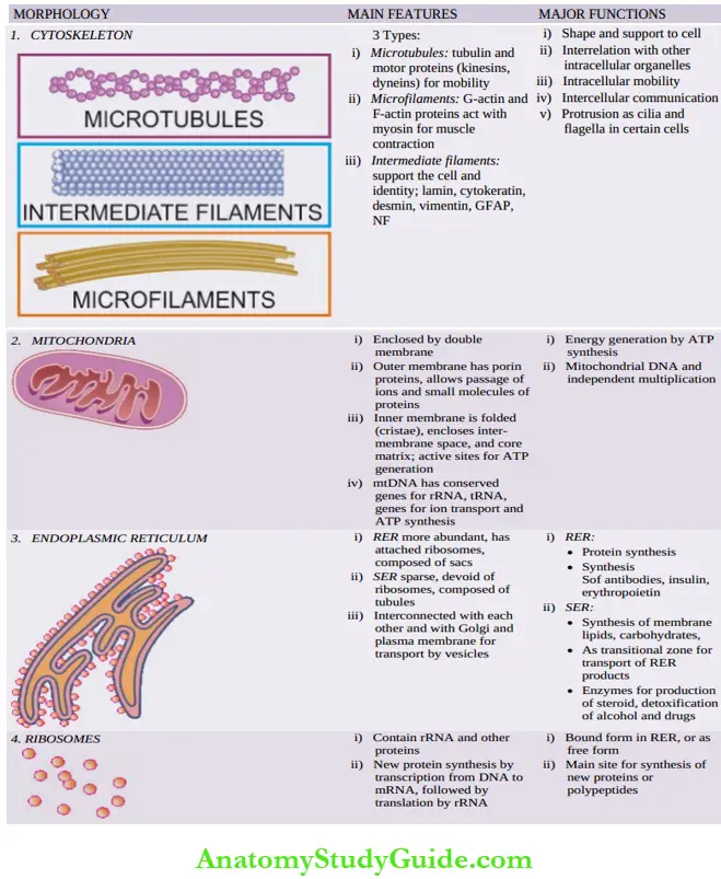
A summary of their main structural composition alongside their illustrated morphologic appearance and a list of their salient functions are presented in
Morphology and functions of major intracellular organelles:


Cytoskeleton: The Cell Scaffolding:
The cytoskeleton is present throughout the cytosol and forms the infrastructure of the cell.
The major general functions of the cytoskeleton are:
- To maintain shape and provide support to the cell.
- To organise interrelationship with other intracellular organelles.
- To provide overall cell mobility (intracellular movement for transport of vesicles, and chromosome movement during cell division).
- To assist in intercellular communication signals.
- To form structures such as cilia and flagella in some cells by protrusions.
Cytoskeleton is composed of three types of cytoskeletal protein fibres:
- Microtubules,
- Microfilaments and
- Intermediate filaments
1. Microtubules:
These are hollow tubular fibrils about 25 nm in diameter and have varying lengths. They are composed of α- and β-tubulin proteins. These fibrils have defined polarity at the two ends i.e. a cationic end (or +) and an anionic end (or -).
The anionic end of the fibril is generally directed towards the microtubule organiser centre or centrosome near the nucleus and is associated with centriole of the chromosomes, while the cationic end of the fibril may elongate or shrink in response to stimuli.
Microtubules contain two types molecular motor proteins powered by ATP, which as the name suggests, are responsible for intracellular ,movements. These are kinesins and dyneins; another motor protein, myosin, is seen in muscle fibres and acts in conjunction with actin in microfilaments.
- Kinesins These proteins move along microtubules and pull the cellular organelles towards the plasma membrane.
- Dyneins These proteins also move along microtubules and pull the intracellular components towards the nucleus.
- Dyneins are also responsible for sliding movement of microtubules as seen in the movement of cilia (e.g. in bronchial epithelium) and flagella (for example Spermatozoa).
2. Microfilaments:
These are contractile actin filaments which are thin, solid rod-like, 5- 9 nm in diameter. They also participate in intracellular organelle movement.
Microfilaments are composed of two types of actin proteins:
- Globular actin (G-actin) proteins are monomeric form and are most abundant protein in the cytosol of the cell.
- Filamentous actin (or F-actin) proteins are polymerised, elongated and double-stranded filaments of G-actin proteins.
- These filaments too have defined polarity as in microtubules and help in maintaining shape of the cell.
- A filamentous motor protein acts in conjunction with actin to make the cell contract which is an ATP-powered process.
3. Intermediate Filaments:
These are rope-like fibrils measuring up to 10 nm in diameter. Intermediate filaments are a form of structural support proteins and provide strength to the respective cells. There are different types of cell-specific intermediate filaments and can be identified by certain techniques (for example Immunohistochemical stains) which helps in assigning the cell of origin to an undifferentiated cancer.
Various types of intermediate filaments are as under:
- Cytokeratin is found in epithelial cells.
- Desmin is found in all types of muscle cells (skeletal, smooth and cardiac muscle).
- Vimentin is found in cells of mesenchymal origin.
- Glial fibrillary acidic protein (GFAP) is present in glial cells around neurons.
- Neurofilaments are seen in axons of neurons of central and peripheral nervous system.
- Lamin is present in nuclear lamina of most cells. Lamins constitute an important component of nuclear membrane; their abnormality may result in diseases such as muscular dystrophies and progeria.

Mitochondria: The Energy Centre
- Mitochondria are unusual organelles in many ways:
- Energy i.e. they are essential for the cell since they produce energy by cellular respiration for various metabolic activities of the cell.
- mtDNA i.e. they have their own small genome (1% of total DNA), having evolutionally evolved in mitochondrial DNA.
- Division i.e. they can divide independently i.e. mitochondrial replication is independent of cell division.
Ultrastructurally, mitochondria are oval or oblong in shape, bounded by a double membrane in which outer membrane fully surrounds while the inner membrane is folded as cristae.The two layers of mitochondrial membranes enclose a small inter-membrane space. The core of mitochondria surrounded by inner membrane contains a mitochondrial matrix
All body cells contain mitochondria in varying numbers; muscle cells, hepatocytes and adipocytes have very large number for giving energy to these cells for performing metabolic activities. Red blood cells, instead, contain haemoglobin to transport oxygen throughout the body for providing energy. Balanced functions of mitochondria are essential for cell survival; its derangements may cause cell death.
Major functions of mitochondria are performed by following mechanisms:
1. Energy Production:
Mitochondria are the power centre for the cell. This is due to structural and biochemical peculiarities of the mitochondrial membranes:
- The outer membrane is studded with protein-based pores, porins, which permit the passage of ions and small molecules of proteins.
- The cristae of inner membrane are also loaded with enzymes; the inter-membrane space is the site for electron transport and synthesis of adenosine triphosphate (ATP).
- The matrix in the mitochondrial core is the site where citric acid cycle enzymes are present.
- Generation of ATP occurs in the mitochondrial matrix, the inner membrane and intermembrane space as follows.
- At the inner membrane, a high-energy electron is passed along an electron transport chain from one to the next, during which energy is released that pumps hydrogen ions (H+) out of the matrix into the inter-membrane space.
- This creates a concentration gradient across the inner membrane that drives H+ back through the inner membrane into the matrix via ATP synthase, which then generates ATP from ADP.
- In the process of ATP generation in which electron chain transport occurs on the inner membrane, the final electron acceptor is O2 which combines with H+ and forms water (H2O).
- Hence, this process of ATP generation is called oxidative phosphorylation.
2. Mitochondrial Dna And Multiplication:
Though evolutionally conserved and quite small, mtDNA is functional. The conserved functions of mtDNA are rRNA genes, tRNA genes, and genes involved in electron transport and ATP synthesis.
Still, the enzymes and proteins required for mitochondrial proteins are synthesised in nuclear genes and transported into the mitochondria.
For multiplication, mitochondria need both mtDNA and nDNA. The process of mitochondrial division is similar to simple asexual multiplication employed by bacteria.
The number of mitochondria in a cell is related to its energy demands; greater in number in cells having high-energy needs.
Endoplasmic Reticulum, Ribosomes And Golgi Apparatus: The Biosynthesis Machinery:
The endomembrane system consists of a set of membrane-bound organelles in the cytosol of the cell; these organelles are interconnected via intracellular membrane transport and they exchange the components with plasma membrane although each organelle performs a specific function.
This system includes endoplasmic reticulum or ER (along with ribosomes), Golgi apparatus, and lysosomes ER and Golgi apparatus are associated with biosynthesis, processing and transport of proteins, lipids and carbohydrates for the cell and are discussed together here.
Lysosomes along with proteasomes, on the other hand, are concerned with their storage, degradation and reutilisation, they are described later.
Endoplasmic Reticulum:
ER is a network of tubules and sacs enclosing an inner space called the lumen. It is spread throughout the cytosol of the cell extending from the cell membrane to the nucleus and forms a continuous connection between the nuclear envelope and plasma membrane via its own lumen.
It is of two types with different structures and functions but both are interconnected for transfer of synthesised contents by special transport vesicles:
- Rough ER (RER): It is so called because it has ribosomes attached to the cytoplasmic surface of its membrane. RER consists of a series of flattened sacs.
- The main functions of RER are:
- Protein synthesis by attached ribosomes by translation of mRNA.
- Synthesised protein may lie in the ER lumen or get integrated into the ER membrane.
- Antibody synthesis in leucocytes.
- Insulin production by islet cells of the pancreas.
- Erythropoietin production by peritubular interstitial fibroblasts in the kidney.


- Smooth ER (SER): It lacks ribosomes and consists of a network of tubules. SER is relatively sparse compared to RER.
- Its main functions are:
- Synthesis of membrane lipids (phospholipids, cholesterol) and carbohydrates.
- As a transitional area for transport of RER products in vesicles to the Golgi.
- Production of enzymes in cells which synthesise steroid hormones (in gonads and adrenal), detoxify certain compounds like alcohol and drugs (in hepatocytes), and assist muscle cells in contraction.
- Since RER is interconnected with SER and the Golgi through intracellular membranes, the products of RER move into SER and are transferred to the Golgi and other destinations within the cell or exported to the exterior by exocytosis.
Ribosomes:
These are small particles where major protein synthesis of the cell takes place. They may exist as ER-bound form (i.e. RER) and as free form in the cytosol. Their number in a cell varies and depends upon the protein-synthesis activity of a cell. They may also be seen in clustered form as polyribosomes.
Ribosomal mass consists of molecules of two subunits: ribosomal RNA (rRNA) and other proteins. Within ribosomes, rRNA molecules direct protein synthesis by combining various amino acids; it is due to this function that rRNA is also called ribozyme.
This process of new protein synthesis or polypeptide synthesis involves following sequential steps of transcription and translation:
- Initiated by transcription of genetic information of cellular DNA to the single-strand of mRNA.
- Next, translation follows in which each mRNA directs complementary DNA synthesis in the same sequence as in the original sequence in the DNA or codon.
- For this, mRNA attracts tRNA molecules.
Thus, a long chain of amino acids is formed resulting in a polypeptide or a new protein.
Golgi Apparatus:
The Golgi apparatus is composed of a complex of membrane-bound flat sacs or compartments (called cisternae) which are continuous with each other and are stacked together. Since the entire complex is continuous via a membrane and is also connected with ER, It has two ends:
- Cis-Golgi network which receives cargo from ER. trans-Golgi network which ships the received cargo to other destinations in the cell.
- The major function of the Golgi is to mediate the manufacture, process, store and transport proteins and lipids, especially proteins synthesised by ER.
In brief, the process of membrane transport through the endomembrane system occurs as follows:
- The protein molecules synthesised in ER exit ER in special transport vesicles and enter cis-Golgi which is often in close proximity of ER.
- These vesicles fuse with Golgi cisternae, releasing their content into the internal portion of Golgi membrane.
- Golgi cisternae contain different protein modification enzymes. As the protein cargo moves through the cisternae of the Golgi, it undergoes post-translational changes: glycosylation (removal of sugar from protein), sulfation (addition of sulphate group), and phosphorylation (addition of phosphate group).
- The Golgi-modified proteins exit through trans-Golgi; the Golgi enzymes direct these proteins to their final destination in the cells i.e. either lysosomes, or storage vesicles, or plasma membrane, or the proteins are extruded from the cell by exocytosis.
Lysosomes, Peroxisomes And Proteasomes: Waste Disposal And Recycling Plants:
Lysosomes, peroxisomes and ubiquitin-proteasome system deal with cellular waste disposal, by catabolism and for reutilising the products of breakdown.
Lysosomes:
These are spherical membrane-enclosed sacs containing a variety of hydrolytic enzymes having acidic pH in their lumen (for example,Proteases, nucleases, lipases, phosphatases, glycosidases etc) which can digest proteins, nucleic acids, lipids and complex sugars.
Lysosomes are formed either by fusion of vesicles which have budded off from trans-Golgi (for intracellular molecules), or by vesicles which bud off from the plasma membrane (for extracellular substances).
Main function of lysosomes is to breakdown macromolecules:
- Into smaller constituents which are then catabolised or recycled as under:
- Lysosomal enzymes are initially synthesised in the ER lumen and then transported through the Golgi where they are tagged with M6P (mannose-6-phosphate) sugar.
- These M6P-modified proteins are transported in vesicles through the Golgi and delivered to the lysosomes for catabolism.
- In receptor-mediated endocytosis and fluid-phase pinocytosis, there is a reverse trafficking of vesicles i.e. a vesicle is formed by pinching off of plasma membrane forming an endosome containing fluid and extracellular molecules which are processed by lysosomal enzymes.
- In heterophagy by phagocytosis, the plasma membrane encloses the extracellular foreign pathogenic materials such as microbes that then merges with lysosome forming phagolysosome, which is then degraded by lysosomal hydrolytic enzymes.
- Autophagy (self-digestion) is a process in which when the cell is starved or is hungry or is senescent, it digests its own organelles, proteins and other cytoplasmic components and recycles metabolites needed to synthesise essential molecules.
- In this process, a membrane-enclosed vesicle is formed containing intracellular worn-out organelles or proteins, which fuse with the lysosome of the cell forming autophagosome which undergoes catabolism by lysosomal enzymes.
Peroxisomes:
Also called microbodies, peroxisomes are small rounded organelles covered by a single layer of membrane. Hundreds of these tiny organelles are present in a cell.
Peroxisomes are formed by biogenesis in which they assemble their membrane and protein and reproduce by dividing. They contain several enzymes which produce hydrogen peroxide as a by-product.
Hydrogen peroxide so generated plays role in the oxidation and decomposition of several organic polymers that include amino acids, fatty acids, and uric acid. Peroxisomes are also involved in synthesis of membrane lipids and bile acids.
Ubiquitin-Proteasome System:
This is a system by which a variety of cellular proteins which suffer from error in biosynthesis or protein misfolding are recognised, targeted and degraded. It consists of two components:
- Proteasome: Proteasome consists of barrel-shaped ATP-dependent protease complex (called 26S proteasome complex) that serves as degradation arm of the ubiquitin-regulated protein catabolism. Multiple proteases comprising proteasomes perform protein unfolding and break down the ubiquitin-tagged misfolded proteins into shorter peptides and digest them.
- Ubiquitin: Ubiquitin is a small regulatory protein (containing 76 amino acids) that is found in almost all human cells (ubiquitous) which helps to regulate the processing of other proteins in the body. The process of bonding of a substrate protein with ubiquitin (i.e. posttranslational modification of protein) involves steps of activation, conjugation and ligation, and is called ubiquitination or ubiquitylation.
Besides proteasome-dependent proteolysis, ubiquitin controls several nonproteolytic cellular activities: in cell cycle regulation, DNA repair, cell growth and signalling etc. Irregularities in ubiquitin is seen is many human diseases from cancer to neurodegenerative disorders.
Biology Of Cell Organelles:
- Cytoskeleton: Cytoskeleton provides shape and support to the cell. Its other major functions are to organise interrelationship with other intracellular organelles, to provide overall cell mobility, intercellular communication and protrude out as cilia and flagella in some cells.
- Cytoskeleton consists of 3 types of protein fibres:
- Microtubules consisting of tubulin and motor proteins (kinesins and dyneins) responsible for movement
- Microfilaments consisting of G-actin and F-actin protein that acts with myosin for muscle contraction, and
- Intermediate filaments in different cells (lamin, cytokeratin, desmin, vimentin, GFAP, neurofilaments).
- Mitochondria: Mitochondria are the source of energy for cell metabolism.
- These are enclosed by double membrane having a space between two layers while the central core contains matrix.
- The outer membrane allows the passage of ions and small molecules. Inner membrane having folds (cristae), inter-membrane space and the core matrix are active sites for generation of
- ATP via a series of biochemical events involving enzymes, proteins and electrons. Mitochondria have a small amount of DNA and they can multiply independent of cell division.
- Endoplasmic reticulum (ER) has two forms: Endoplasmic reticulum rough and smooth ER; both are interconnected through a membrane with each other and with Golgi apparatus.
- RER has attached lysosomes on membrane, and is primarily associated with protein synthesis.
- nSER is sparse and is involved in synthesis of membrane lipids and carbohydrates, as a transitional zone for RER products, and synthesis of detoxifying enzymes in hepatocytes.
- Ribosomes: Ribosomes are the main protein synthesising units of the cell and may exist in bound form in RER or as free form in the cytosol. Synthesis of new proteins or polypeptides occurs by transcription and translation steps involving mRNA and tRNA.
- Golgi apparatus: Golgi apparatus is composed of membrane-bound flat sacs, cisternae, in stacks having a cis-end and trans-end. ER-synthesised proteins enter cis-Golgi as vesicles, undergo modification as they pass through it and exit through trans-Golgi, for delivery to their final intracellular destination or extruded by exocytosis.
- Lysosomes: Lysosomes are spherical membrane-enclosed particles containing hydrolytic enzymes and catabolise proteins. The pathways include M6P-modified pathway, endocytosis, pinocytosis, phagocytosis and autophagy.
- Peroxisomes or microbodies: Peroxisomes or microbodies are multiple small rounded organelles which degrade intracellular proteins and lipids by hydrogen peroxide.
- Ubiquitin-proteasome system: Ubiquitin-proteasome is ubiquitin-directed proteolysis by multiple proteases present in proteasome; it targets misfolded proteins by ubiquitination of the substrate protein
Leave a Reply