Diaphragm
Question – 1: Structures passing through the aortic opening of the diaphragm.
Table of Contents
Answer:
- Descending Thoracic Aorta on the left side (DTA).
- Thoracic duct in the middle.
- Azygos vein on the right side.
Read And Learn More: General Histology Questions and Answers
Referred Pain
It is the area of the skin where the pain is felt by the disease of the organ which is away
Referred pain:

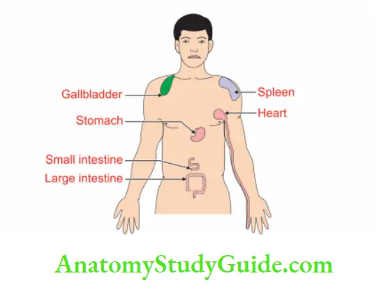
Question – 2: Describe the Diaphragm under the following heads
1. Diaphragm Gross anatomy
2. Diaphragm Openings in the diaphragm
3. Diaphragm Development, and
4. Diaphragm Applied Anatomy.
Answer:
Diaphragm Introduction:
It is the musculotendinous partition which separates the thoracic cavity from the abdominal cavity. It is described as a muscle under origin, insertion, nerve supply and action.
1. Gross Anatomy:
1. Proximal attachments of diaphragm:
- Costal fibres arise from the inner surface of the lower 6 costal cartilages and adjacent parts of the ribs (C).
- Vertebral fibres arise from the upper two lumbar vertebrae by two crura (V).
- Sternal fibres arise from the posterior surface of the xiphoid process (S).
2. Diaphragm Distal attachments: The muscles insert into the trilobed central tendon.
3. Diaphragm Relations:
- Superiorly:
- Pleurae, and
- Pericardium.
- Inferiorly:
- Peritoneum
- Live
- Fundus of stomach
- Spleen
- Kidneys, and
- Suprarenals.
4. Diaphragm Actions: The actions of the diaphragm are
- Inspiration: The Diaphragm is the principal muscle of the inspiration.
- It acts in all expulsive acts, for example sneezing, coughing, laughing, crying, vomiting, micturition, defaecation and parturition.
- It acts as a partition between the thoracic and abdominal cavities.
5. Diaphragm Blood supply:
- Arterial supply:
- Inferior phrenic artery, 1st branch of the abdominal aorta.
- Musculophrenic, a terminal branch of the internal thoracic artery.
- Lower 5 or 6 Posterior intercostal, branches of thoracic aorta.
- Venous drainage:
- The inferior phrenic vein drains into the inferior vena cava.
- Musculophrenic vein drains into internal thoracic vein.
- The posterior intercostal vein drains into the azygos vein.
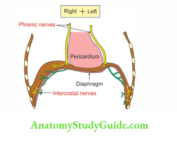
6. Diaphragm Nerve supply: Sensory and motor
- Sensory: It is divided into
- The sensations of the central part are carried by the phrenic nerve (C3, C4 and C5).
- The sensations from the peripheral part are carried by lower intercostal nerves (T6 to T11).
- Motor: Phrenic nerves (C3, C4 and C5)
2. Opening In The Diaphragm:
The major opening in the diaphragm

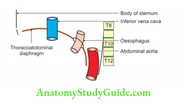
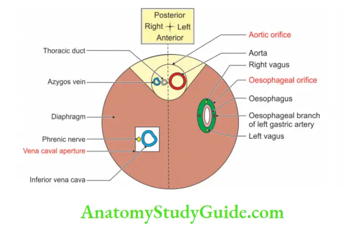
Link Memory:

Linking of the position of letters in words with the position of structures in the opening

Note: All the unpaired Iredlcoloured structures, i.e. arteries are on the left side. All the unpaired |blue colour and red structures, i.e. veins are on the right side.
The minor openings in the diaphragm are as follows:
- The superior epigastric vessels (and some lymphatics) pass between the xiphoid and the costal (7th costal cartilage) origin of the diaphragm.
- This gap is known as Larry’s space or foramen of Morgagni.
- The musculophrenic vessels pierce the diaphragm at the 8th or 9th costal cartilage.
- Several small veins pass through minute apertures in the central tendon.
3. Development
Germ layer: It develops from the mesoderm.
Site: Lateral plate mesoderm.
Sources:
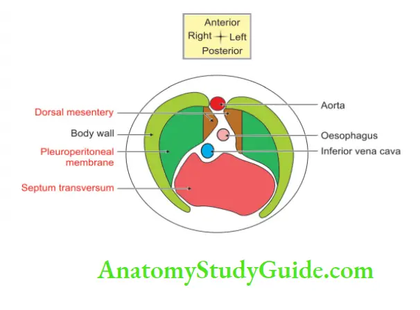
1. Septum transversum forms the principal component of the definitive diaphragm. It forms
- Central tendon
- Anteromedian part
- Vena cava opening, and
- Oesophageal opening.
2. Pleuroperitoneal membranes: It also forms a major contribution to the development of the diaphragm. It gives rise to the circumferential part in the ventrolateral region.
3. Dorsal mesentery of the oesophagus forms
- The posterior part of the diaphragm is between the oesophageal and the aortic openings.
- Crura of the diaphragm
4. Left and right body walls. They form tissue on the periphery of the diaphragm.
Anomalies:
1. Diaphragmatic hernias: It is one of the common anomalies of the diaphragm. The incidence is 1/2000 newborns.
The diaphragmatic hernia may be of the following types depending upon the situation.
- Posterolateral
- Posterior
- Retrosternal, and
- Central.
2. Accessory diaphragm.
3. Congenital eventration (elevation of the dome of the diaphragm) of the diaphragm.
4. Applied Anatomy:
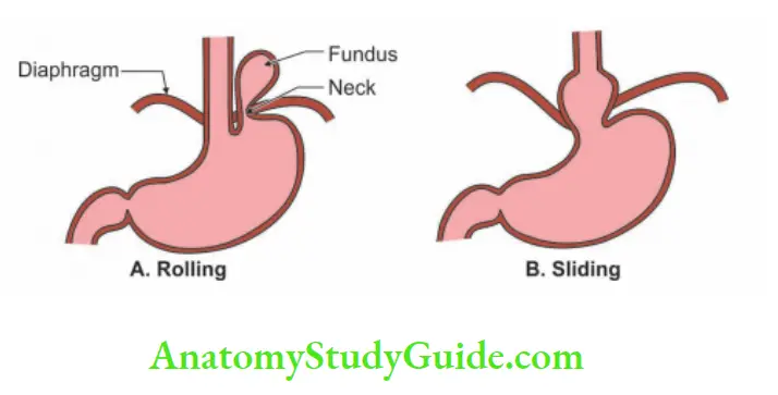
1. Hiatus hernia is relatively common (1%) accounting for 98% of all diaphragmatic hernia.
2. It is HARD to diagnose. Its manifestations are denoted by each letter of the word “HARD”
- Hiccough
- Anaemia
- Regurgitation
- Dysphagia—difficulty in swallowing.
3. The lesion of the phrenic nerve results in the paralysis of the dome of the diaphragm and the affected dome is pushed upwards.
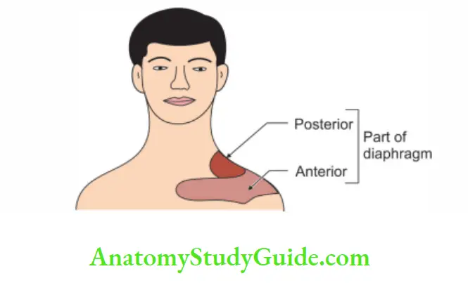
4. Irritation of the diaphragm may cause referred pain in the shoulder.
5. The phrenic nerve, nerve of the diaphragm and supraclavicular nerve supplying the skin of the shoulder have the same root value.
Diaphragmatic Hernia
It is a protrusion of abdominal contents through the thoracoabdominal diaphragm. It is divided into Congenital and acquired.
1. Congenital:
- Retrosternal: It is through the foramen of Morgagni of the space of Larry.
- Posterolateral: Commonest type. It is through the foramen of Bockdalech.
- Posterior hernia: It is due to the failure of the development of the posterior part of the diaphragm.
- Central: It is rare.
- Hiatal hernia
2. Acquired:
- Traumatic
- Hiatal
Leave a Reply