Ear Anatomy Notes And Important Questions With Answers
Pinna (ear)
1. Features
Table of Contents
- The greater part of it is made up of a single crumpled plate of elastic cartilage.
- It is lined on both sides by skin.
- The lowest part of the auricle is soft.
- It is made up of fibrofatty tissue covered by skin.
- This part is called the lobule used for wearing the earrings.
- The rest of the auricle is divided into a number of parts.
Read And Learn More: Head Anatomy Notes And Important Questions With Answers
- These are the helix, antihelix, concha, tragus, and scaphoid fossa.
- The large depression was called the Concha.
- It leads into the external acoustic meatus.
- The external ear has the following muscles.
- Auricularis anterior,
- Auricularis superior, and
- Auricularis posterior.
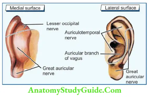
2. Nerve supply

3. Blood supply
- Posterior auricular, 2nd dorsal branch of the external carotid artery, and
- Superficial temporal arteries, one of the small terminal branches of the external carotid artery given at the neck of the mandible.
4. Lymphatic drainage
- Preauricular, and
- Postauricular lymph nodes.
Chorda Tympani Nerve
1. Origin: It is a branch of the facial nerve (VIIth cranial). It arises from the facial nerve about 6 mm above the stylomastoid foramen.
2. Functions
1. It conveys the preganglionic secretomotor fibres to the
- Submandibular,
- Sublingual glands, and
2. Taste fibres from the anterior two-thirds of the tongue except circumvallate papillae.
3. Development: It is a problematic branch of the 1st pharyngeal arch.
4. Course
- It passes through the tympanic membrane.
- It runs between mucous and fibrous layers of the tympanic membrane.
- The course is at the junction of pars flaccida and pars tensa.
- It enters the infratemporal fossa thought the media lend of the erotomanic figure.
anterior ligament of the mallus and anterior tympanic artery accompany it. - It passes downward and forward under the cover of the lateral pterygoid.
- It crosses the medial side of the spine of the sphenoid bone.
- It joins the posterior border of the lingual nerve at an acute angle.
5. Relations
1. In the infratemporal fossa,
- Laterally with
- Middle meningeal artery,
- Auriculotemporal nerve, and
- Inferior alveolar nerve.
2. Medially with
- Tensor palati
- Auditory tube
3. Anteriorly with
- The trunk of mandibular nerve, and
- Otic ganglion.
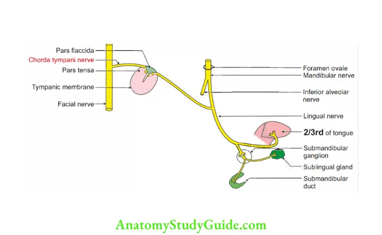
6. Communication branches to the otic ganglion, which probably forms an alternate root of taste sensations from the tongue.
7. Applied anatomy
1. For the operation i the tympanic membrane, the incision is taken posteromedially to avoid the injury to chorda tympani.
2. Damage to the 7th nerve proximal to the origin of the chorda tympani results in
- Loss of taste sensation of anterior two-thirds of the tongue.
- Decrease salivation.
3. A lesion in the region of the spine of the sphenoid may involve chorda tympani and auriculotemporal nerves.
It results in loss of secretion of submandibular, sublingual and parotid glands.
External auditory canal (external auditory meatus)
1. It conducts sound waves from the concha to the tympanic membrane.
1. Shape: S-shaped.
2. It has three parts
- The outer part is directed medially, forward and upwards.
- The middle part is directed medially, backwards and upwards.
- The inner part is directed medially, forward and downwards.
3. It can be straightened for examination by pulling the auricle upwards, backward and slightly laterally.
4. Dimensions
1. Length: 24 mm long.
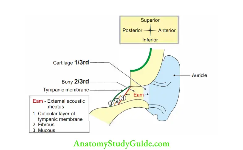
- Medial two-thirds or 16 mm is bony. It is narrower than the cartilaginous part. It is formed by the tympanic plate of the temporal bone which is C-shaped in cross-section.
- Lateral one-third or 8 mm is cartilaginous.
The cartilaginous part is also C-shaped in section, and the gap of the ‘C’ is filled with fibrous tissue.
The lining skin is adherent to the perichondrium and contains hair, sebaceous glands and ceruminous or wax glands.
Ceruminous glands are modified sweat glands.
- Due to the obliquity of the tympanic membrane, the anterior wall and floor are longer than the posterior wall and roof.
- The canal is oval in section. The greatest diameter is vertical at the lateral end and anteroposterior at the medial end.
- The narrowest point, the isthmus lies about 5 mm from the tympanic membrane.
- The posterosuperior part of the palate is deficient. Here the wall of the meatus is formed by a part of the squamous temporal bone.
2. Epithelium lining: The meatus is lined by thin skin, firmly adherent to the periosteum.
2. Blood supply
1. Outer part:
- Superficial temporal and
- Posterior auricular artires. } Branches of the external carotid. artery
2. Inner part: Deep auricular branch of the maxillary artery.
3. Lymphatic drainage
- Preauricular,
- Postauricular, and
- Superficial cervical lymph nodes.
4. Nerve supply
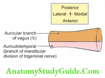
- The skin lining the anterior ½ is supplied by the auriculo temporal nerve branch of the mandibular nerve.
Posterior ½ is supplied by the auricular branch of the vagus.
5. Applied anatomy
- Backward dislocation of the temporomandibular joint destroys the external acoustic meatus.
- Parotid gland disease often causes pain in the external acoustic meatus because of the same innervation by the auriculotemporal nerve.
- Examination of the external acoustic meatus and tympanic membrane begins by u. straightening the meatus.
- In adults, the pinna is grasped and pulled posterosuperiorly (up, out and back).
These movements reduce the curvature of the external acoustic meatus. It makes cartilaginous and bony canals in one line. - The meatus is straightened in infants by pulling the auricle anteroposteriorly (down and back).
Question 1: Describe the external acoustic meatus under the following heads:
1. External features,
2. Relations,
3. Blood supply,
4. Lymphatic drainage,
5. Nerve supply, and
6. Applied anatomy
Answer: Introduction: It is a passage connecting the external ear to the tympanic membrane.
1. External features
1. Parts
- Outer: It forms one-third of the external acoustic meatus. It is 8 mm long.
It is formed by cartilage.
The skin of the cartilaginous part shows many hair follicles and numerous ceruminous glands. It secretes wax. - Inner: It forms two-thirds part and measures 16 mm in length.
It is Bony. (B is the 2nd alphabet and is two-thirds of the external acoustic meatus.) Please associate with the auditory tube.
In the auditory tube, the bony part is one-third and the cartilaginous part is two-thirds.
The medial end of the bony part is smaller in diameter than its lateral part.
It is not developed in newborns and hence, the meatus is much shorter.
2. Attachments
- The medial end is fixed by the fibrous tissue to the circumference of the lateral end of the bony part.
- The lateral end is continuous with the auricular cartilage.
- The diameter is less in the middle. It forms the narrowest region of the meatus.
- Surfaces: The inner surface is lined by skin which is closely adherent to the perichondrium.
- Constrictions: There are two constrictions.
- One at the junction of the bony and cartilaginous parts (at 8 mm depth)
- One at the middle of the meatus (at 12 mm depth).
2. Relations
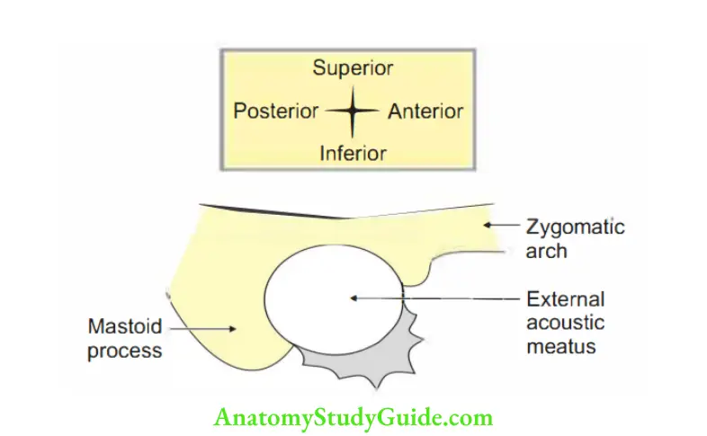
- Anterior: Temporomandibular joint
- Posterior: Mastoid air cells.
- Superior: Middle cranial fossa.
3. Blood supply
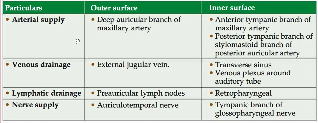
1. Arterial supply
- The anterior auricular artery is the branch of the superficial temporal artery.
- The posterior auricular artery is a branch of the external carotid artery.
- Deep auricular artery, a branch of 1st part of the maxillary artery.
2. Venous drainage
- The anterior auricular vein drains into the superficial temporal vein which drains into the retromandibular vein-external jugular vein.
- The posterior auricular vein drains into the posterior division of the retromandibular vein-external jugular vein.
- The deep auricular vein drains into the maxillary vein-pterygoid venous plexus.
4. Lymphatic drainage
- Parotid group of lymph nodes.
- Mastoid group of lymph nodes.
5. Nerve supply
- The auriculotemporal branch of the mandibular nerve supplies the upper and anterior walls.
- The auricular branch of the vagus supplies the posterior wall and floor.
6. Applied anatomy
- To visualize the tympanic membrane, one has to pull the pinna of the ear upwards, backwards and laterally.
- Since the external acoustic meatus is shorter in newborns, the tympanic membrane is likely to be damaged.
- Infection in the external acoustic meatus is extremely painful due to the tightly adherent skin of the unyielding subcutaneous tissue.
- Removal of the foreign body behind the cartilaginous part may be difficult due to hourglass constriction of the external acoustic meatus.
- Pain from the external acoustic meatus is referred to as teeth because of the same nerve supply.
- There may be cardiac arrest or vomiting while syringing during the removal of foreign bodies because of vagal stimulation.
Tympanic membrane
1. Gross features: It is a thin, translucent partition between the external and the middle ear.
It is an oval in shape, measuring 9 x 10 mm.
It is placed obliquely at an angle of 55° with the floor of the meatus.
It has outer and inner surfaces.
- Outer surface: Covered by thin skin.
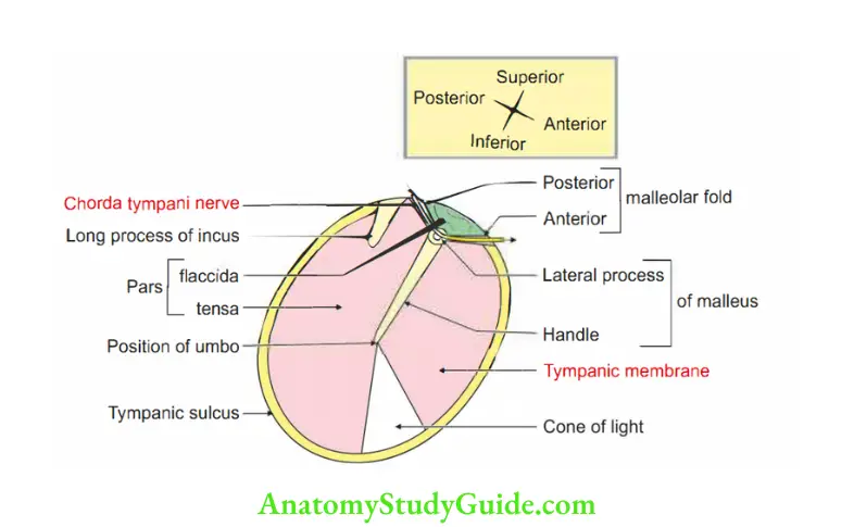
- Inner surface: Provides attachment to the handle of the malleus. The point of maximum convexity lies at the level of the tip, called the umbo.
The membrane is thick at its circumference, which is fixed to the tympanic sulcus.
Superiorly, the sulcus is deficient. Here, the membrane is attached to the tympanic notch.
The greater part of the tympanic membrane is tightly stretched and is called pars tensa.
The part between the two malleolar folds is loose and is called pars flaccid.
The pars flaccida is crossed by the chorda tympani, which passes between the middle fibrous layer and the inner mucous layer.
2. Structure: Composed of three layers.
- Outer cuticular layer.
- The middle fibrous layer is made up of superficial radiating fibres and deep circular fibres. The circular fibres are sparse at the centre and dense at the periphery.
- The inner mucous layer is lined by low-ciliated columnar epithelium.
3. Applied anatomy
- The otoscopic examination reveals the redness, bulging, perforation or retraction of the tympanic membrane.
- The membrane is incised to drain the pus present (acute otitis media) in the middle ear.
The incision is called myringotomy.
It is usually made in the posteroinferior quadrant of the membrane.
In giving an incision, it has to be remembered that the chorda tympani nerve runs downwards and forwards across the inner surface of the membrane.
It is lateral to the long process of incus but medial to the neck of the mandible.

Name the bones in the middle ear
Ear ossicles
1. Malleus
2. Incus, and
3. Stapes
Question 2: Describe the middle ear under the following heads:
1. Gross anatomy,
2. Contents,
3. Blood supply,
4. Nerve supply, and
5. Applied anatomy
Answer: Introduction: Narrow space situated between the external and internal ear present in the petrous part of the temporal bone.
1. Gross anatomy
1. Dimensions: It is biconcave in shape.
- Vertical: 15 mm.
- Anteroposterior: 15 mm.
- Transverse
- Roof: 6 mm.
- Centre: 2 mm.
- Floor: 4 mm.
2. Communication
- Anteriorly: Nasopharynx through auditory tube.
- Posteriorly: Mastoid antrum and air cells through aditus to antrum.
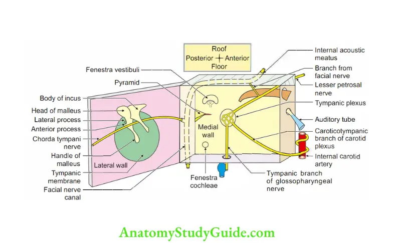
3. Boundaries
1. Roof or tegmental wall: Separates ear from middle cranial fossa. It is formed by thin bone called tegmen tympani which also forms the roof of
- Canal for tensor tympani, and
- Mastoid antrum.
2. Floor or jugular wall
- It is pierced by the inferior canaliculus. It transmits the tympanic branch of the glossopharyngeal nerve.
- Separates the middle ear from the superior bulb of the internal jugular vein.
- Formed by the jugular fossa of the temporal bone.
3. Anterior wall or carotid wall
- It is constricted.
- Consists of three parts:
The upper part forms a canal for tensor tympani.
The middle part forms the opening of the auditory tube.
The lower part forms the posterior wall of the carotid canal. - There is a septum between the canal for tensor tympani and the canal for the auditory tube.
4. Posterior wall or mastoid wall: Represents the following features from above downwards:
- Superiorly: Aditus to mastoid antrum.
- Fossa incudis: Depression for incus.
- Pyramid (projection): The apex of the pyramid presents an opening for the tendon of the stapedius.
- Lateral to pyramid: Posterior canaliculus for chorda tympani.
5. Lateral or membranous wall.
- Separates the middle ear from the external ear.
- Formed mainly by:
Tympanic membrane.
Partly by the squamous part of the temporal bone. - Near the tympanic notch, there are two apertures
Petrotympanic fissure.
Anterior canaliculus for chorda tympani.
6. Medial or labyrinthine wall: It separates the middle ear from the internal ear. It presents the following features :
- Promontory produced by 1st turn of the cochlea.
- Fenestra vestibuli: The oval opening leads to the vestibule of the internal ear.
- Prominence of facial canal.
- Fenestra cochlea ends in the scala tympani and is closed by a secondary tympanic membrane.
- Sinus tympani depression behind promontory.
2. Contents
- Air is the main and important content.
- Bone: Ear ossicles namely malleus, incus and stapes.
- Vessels, which supply and drain the middle ear.
- Muscles: Tensor tympani and stapedius.
- Ligaments of ear ossicle.
- Tympanic cavity proper: Lies opposite to tympanic membrane.
- Epitympanic recess above the tympanic membrane.
3. Blood supply
1. Arterial supply
1. Main and large arteries
- Deep auricular
- A tenor. tympanic. } branch of 1st part of the maxillary artery
- A posterior tympanic branch from the stylomastoid, a branch of the posterior auricular artery.
2. Small and less important arteries
- Superior tympanic (middle meningeal artery).
- Inferior tympanic (ascending pharyngeal).
- Tympanic branch (artery of pterygoid canal, branch of middle meningeal artery).
- Caroticotympanic branch of the internal carotid artery (ICA).
- Petrosal branch of the middle meningeal artery.
2. Venous drainage Veins from the middle ear drain into:
- Superior petrosal sinus.
- Pterygoid plexus of vein.
4. Nerve supply: Tympanic plexus is formed by
- Tympanic branch of glossopharyngeal nerve.
- Caroticotympanic nerve (plexus around ICA).
5. Applied anatomy: Throat infections commonly spread to the middle ear through auditory tubes, which are more common in children.
In children, the tube is wide, small and horizontal.
The pus may be discharged into one of the following
1. Maybe in the external ear following the rupture of the tympanic membrane.
2. The posterior wall of the middle ear is a mastoid process.
It contains mastoid air cells.
They are not true air cells.
They are air-filed pockets.
It is related to the posterior cranial fossa.
The bone may be very thin and in some rare cases, it may be absent.
The infection from the middle ear can go posteriorly into mastoid air cells and may go to the sigmoid air sinus present in the posterior cranial fossa.
It may end up in thrombophlebitis of the sigmoid sinus and there may be severe and serious impairment of blood drainage of the system.
It can go back and produce an infection of the cerebellum.
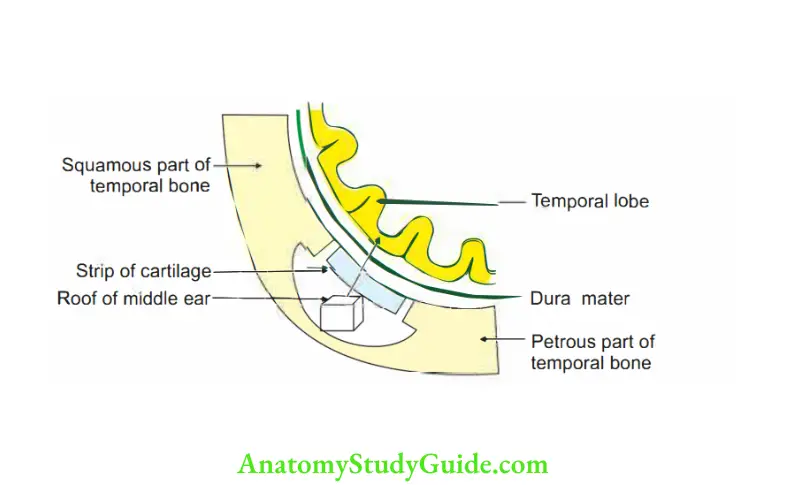
3. Roof: It is very thin and there is a petrosquamous suture. It may not be ossified is weak in children and causes infection to spread to the meninges or temporal lobe of the brain. It results in
- Extradural or subdural abscess.
- Meningitis.
- Temporal lobe abscess or infection.
- May erode the roof and result in meningitis.
- May erode the floor spread downward and cause thrombosis of the internal jugular vein.
4. Posterior wall
- Mastoid air cells are connected to the sigmoid sinus by emissary veins. The infection from the posterior wall can go to the sigmoid sinus and cause thrombosis.
- May cause mastoid abscess.
- Fracture of the middle cranial fossa can cause bleeding through the ear.
Muscles of the tympanic cavity
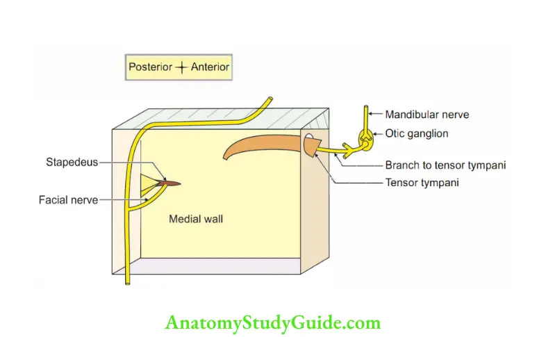
1. Tensor tympani
1. Origin: It lies in the bony canal and opens into the anterior wall of the middle ear. It arises from
- Bony canal
- Cartilaginous part of the auditory tube.
- Inferior surface of the greater wing of the sphenoid bone.
2. Insertion: Into the upper end of the handle of the malleus.
3. Nerv supply: The branch of the branch to the medial pterygoid. The branch to the medial pterygoid is a branch of the mandibular nerve, arising from the trunk.
4. Action: It protects the ear from loud sounds.
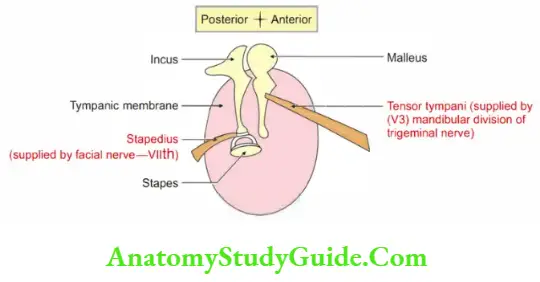
2. Stapedius
- Origin: It is a small muscle which lies in the bony canal. It arises from the wall of the bony canal.
- Insertion: Posterior surface of the neck of stapes.
- Nerve supply: Branch of the facial nerve.
- Action: It protects the ear from loud sounds.
5. Applied anatomy: Paralysis of stapedius muscles gives rise to a condition called hyperacusis in which normal sound appears too loud.
Spiral organ of Corti
Introduction: End organ for hearing.
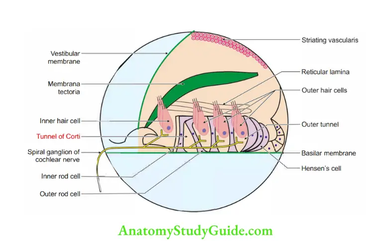
1. Gross: It is located on the basilar membrane of the cochlear duct.
2. Microscopic structure: Consists of the following parts:
- Basilar membrane: Extends from the osseous spiral lamina to the outer cochlear wall. It consists of collagen fibres.
- Rods of Corti: Enclose tunnel of Corti. The details of the rods are as follows

3. Hair cells are essential components of the organ of Corti. It bears stereocilia.
3. Functions
- Detects the movements of endolymph.
- Detects the vibrations of the basilar membrane.
- Transfers vibration into nerve impulses going to the cochlear nerve.
4. The hair cells are divided into inner and outer hair cells.
Cochlea
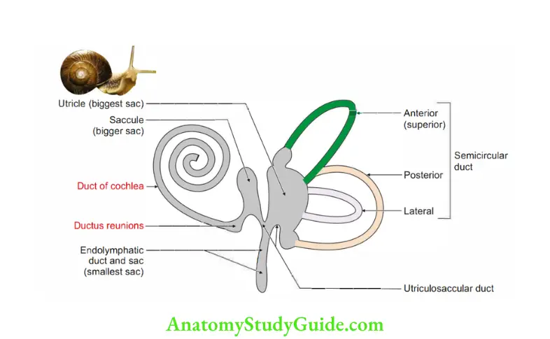
1. Cochlea: Shell of a snail. Shape: conical. 2 and 3/4 turns
2. Location: Anterior to the vestibule.
- Apex is towards the anterosuperior part of the medial wall of the middle ear.
- The base is at the floor of the internal acoustic meatus, perforated by the cochlear nerve.
3. Dimension
- From base to apex 5 mm.
- Width 9 mm (at base).
4. Structure
1. Central bony axis: Modiolus (axis of wheel) with spiral bony canal around. The bony canal is divided into three channels which are spirally arranged.
1. Scala media (cochlear duct): lar, bounded by
- Basilar membrane attached to the osseous spiral lamina.
- Vestibular or Reissner’s membrane.
- The outer wall of the cochlea lies between basilar and vestibular membranes.
- The apex of the cochlea is blind. It contains endolymph.
- The spiral organ of Corti lies on the basilar membrane.

2. Scala vestibuli
- Canal above scala media.
- Communicates with
- Bony vestibule at base;
- With scala tympani at the apex of the cochlea through helicotrema (spiral opening).
3. Scala tympani: Canal below scala media separated from the middle ear cavity by secondary tympanic membrane.
Both scala vestibuli and scala tympani are filled with perilymph.
4. Modiolus: It is broad at the base, narrow at the apex, and contains blood vessels and spiral ganglion.
It gives out osseous spiral lamina which is attached to the basilar membrane.
5. Openings at basal tum of bony cochlea
- Oval window: Occupied by foot plate of stapes.
- Round window: Closed by secondary tympanic membrane.
- Cochlear canaliculus: Communicates scala tympani with subarachnoid space.

Leave a Reply