Esophagus under the following headings:
- Esophagus Introduction
- Esophagus Constriction
- Esophagus Blood supply
- Esophagus Nerve supply
- Esophagus Histological features and
- Esophagus Applied Anatomy.
Answer:
Esophagus Under The Following Headings:-
1. Esophagus Introduction:
- Esophagus is a 10″ (25 cm) long muscular tube that connects the pharynx to the stomach and provides passage for a bolus of food and liquids during the third stage of
deglutition. - Esophagus extends from the lower border of the cricoid cartilage/body of C6 vertebra to the cardiac orifice of the stomach (at the level of the lower border of the T11 vertebra).
Read And Learn More: Anatomy Question And Answers
2. Esophagus Constrictions:
Normally, the esophagus shows four sites of constrictions/narrowings.
From above downward, these are:
- At its commencement (caused by cricopharyngeus muscle) (1)
- Where it is crossed by the aortic arch (2)
- Where it is crossed by left bronchus (3)
- Where it pierces the diaphragm(4)
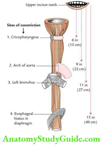
These sites in order from above downward are 6″ (15 cm), 9″ (22 cm), 11″ (27 cm), and 15″ (40 cm) away from the upper incisor teeth.
Note: The narrowest part of the esophagus is at its commencement.
3. Esophagus Blood supply:
- Arterial supply:
- The cervical part of the esophagus, by branches of the inferior thyroid arteries
- The thoracic part of the esophagus, by branches of the thoracic aorta
- Abdominal part of the esophagus, by left gastric and left inferior phrenic arteries
- Venous drainage:
- From the cervical part, by inferior thyroid veins into the superior vena cava
- From the thoracic part, by azygos veins into the superior vena cava
- From the abdominal part:
- By left gastric veins into the portal vein and Hemiazygos vein into the inferior vena cava
4. Esophagus Nerve supply:
- By sympathetic: sympathetic fibers derived from T5–T9 spinal segments, which are sensory and vasomotor
- By parasympathetic: parasympathetic fibers derived from the vagus and recurrent laryngeal nerves, which are sensory, motor, and secretomotor
Trachea Anatomy
5. Esophagus Histological structure:
In the transverse section, the esophageal tube presents an irregular lumen. Its wall from inside out consists of 4 layers, viz. mucosa, submucosa, muscular layer, and adventitia.

- Mucosa:
- iMucosas thrown into folds.
- Epithelium – Stratified squamous of nonkeratinized variety.
- Lamina propria – Thick and contains lymphoid aggregations.
- Muscularis mucosae – Thick prominent and consists of longitudinal smooth muscle fibers.
- Submucosa: It is wide and contains mucous secreting small esophageal glands.
- Muscular layer:
- Muscular layer structure varies in the upper middle and lower 1/3rd.
- In the upper 1/3rd, it is made up of skeletal muscle fibers.
- In the middle 1/3rd, it is made up of both skeletal and smooth muscle fibers.
- In the lower 1/3rd, it is made up of smooth muscle fibers.
- Tunica adventitia: Covers most of the esophagus except the lower part which is covered by serosa.
6. Esophagus Applied anatomy:
- Esophageal Varices:
- Esophageal Varices is a dilation and tortuosity of the anastomotic venous channels between the tributaries of the left gastric vein and hemiazygos vein (portocaval anastomosis) at the lower end of the esophagus, in portal hypertension.
- The rupture of these varices leads to hematemesis (vomiting of frank red color blood).
- Achalasia Cardia:
- Achalasia Cardia is a clinical condition in which there is a failure of relaxation of musculature at the lower end of the esophagus.
- As a result, food accumulates in the esophagus causing regurgitation.
- Note the regurgitation does not include gastric contents; hence, it is not sour-tasting. It occurs due to neuromuscular incoordination of the muscles of the lower end of the esophagus causing loss of peristalsis.
Note:
- Achalasia cardia: Achalasia cardia is the most common esophageal motility disorder with an incidence of 1 in 1,00,000.
- Esophageal carcinoma: It is mostly adenocarcinoma and occurs in the lower 1/3rd of the esophagus.
- Dysphagia: It is a name given to painful/difficult swallowing.
- Achalasia Cardia occurs due to:
- Compression of the esophagus from the outside by the aortic arch aneurysm, etc., or
- By narrowing of lumen due to stricture. It can be diagnosed by barium swallow.
Leave a Reply