Fetal Circulation And Respiration Introduction
- Fetal circulation is different from that of adults because of the presence of the placenta.
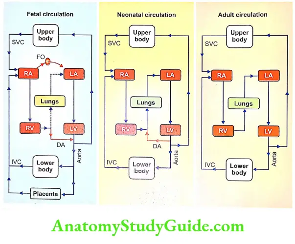
- Since fetal lungs are nonfunctioning, the placenta is responsible for the exchange of gases between fetal blood and the mother’s blood. So the blood from the right ventricle is diverted to the placenta.
Read And Learn More: Medical Physiology Notes
Table of Contents
- The development of the heart is completed in the fourth week of intrauterine life and, it starts beating at the rate of 65 per minute.
- Along with the heart, the blood vessels also develop. The heart rate gradually increases and reaches the maximum rate of about 140 beats per minute just before birth.
- The fetus is connected with the mother through the placenta. Fetal blood passes to the placenta through umbilical vessels and the maternal blood runs through uterine vessels.
- These two sets of blood vessels lie in close proximity in the placenta through which the exchange of substances takes place between the mother’s blood and fetal blood.
- However, there is no direct admixture of maternal and fetal blood.
Blood Vessels In Fetus
- As the fetal lungs are non-functioning, there is no necessity for a large amount of blood to be pumped into the lungs.
- Instead, the fetal heart pumps a large quantity of blood into the placenta for the exchange of substances.
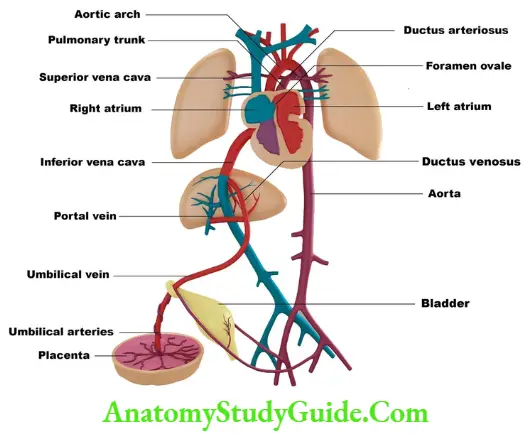
- From the placenta, the umbilical veins collect the blood, which has more oxygen and nutrients.
- The umbilical vein passes through the liver. Some amount of blood is supplied to the liver from the umbilical vein.
- However, a large quantity of blood is diverted from an umbilical vein into the inferior vena cava through ductus venosus. The liver receives blood from the portal vein also.
- In the liver, the oxygenated blood mixes slightly with deoxygenated blood and enters the right atrium via the inferior vena cava.
- From the right atrium, the major portion of the blood is diverted into the left atrium via the foramen ovale. Foramen ovale is an opening in intra-atrial septum.
- Blood from the upper part of the body enters the right atrium through the superior vena cava.
- From the right atrium, blood enters the right ventricle. From here, blood is pumped into the pulmonary artery.
- From the pulmonary artery, blood enters the systemic aorta through ductus arteriosus.
- Only a small quantity of blood is supplied to the fetal lungs. Blood from the left ventricle is pumped into the aorta.
- Fifty percent of blood from the aorta reaches the placenta through the umbilical arteries.

Fetal Lungs
- Pulmonary vascular resistance is the resistance offered to blood flow through the pulmonary vascular bed.
- This resistance is very high in fetuses because of the nonfunctioning of fetal lungs.
- The high resistance in fetal lungs increases the pressure in the blood vessels of the lungs.
- Because of the high pressure, the blood is diverted from the pulmonary artery into the aorta via ductus arteriosus.
Changes In Circulation And Respiration After Birth – Neonatal Circulation And Respiration
1. Changes In Circulation And Respiration First Breath Of The Child
- When the fetus is delivered and the umbilical cord is cut and tied, the lungs start functioning.
- When placental blood flow is cut off, there is sudden hypoxia and hypercapnia.
- Now, the respiratory center is strongly stimulated by these two factors, the respiration starts.
- Initially, there is gasping, which is followed by normal respiration.
2. Changes In Circulation And Respiration Flow Of Blood To Lungs
- Lungs expand during the first breath of the infant. The expansion of the lungs causes an immediate reduction in pulmonary vascular resistance and a sudden fall in pressure in the blood vessels of the lungs.
- Therefore, the blood flow from the pulmonary artery to the lungs increases.

3. Changes In Circulation And Respiration Closure Of Foramen Ovale
- When blood starts flowing through the pulmonary circulation, the oxygenated blood from the lungs returns to the left atrium. It causes an increase in the left atrial pressure.
- Simultaneously, due to the stoppage of blood from the placenta, pressure in the inferior vena cava is decreased.
- It leads to a fall in right atrial pressure. Thus, the pressure in the right atrium is less and the pressure in the left atrium is already high. This causes the closure of the foramen ovale.
- Within a few days after birth, the foramen ovale closes completely and fuses with the atrial wall.
4. Changes In Circulation And Respiration Reversal Of Blood Flow In Ductus Arteriosus
- In a fetus, since pulmonary arterial pressure is very high, the blood passes from the pulmonary artery into the aorta via ductus arterioles.
- However, in neonatal life, since the systemic arterial pressure is more than pulmonary arterial pressure, the blood passes in the opposite direction in ductus arteriosus, i.e.
- from the systemic aorta into the pulmonary aorta. The reversed flow in ductus arteriosus is heard as a continuous murmur in infants.
5. Changes In Circulation And Respiration Closure Of Ductus Venosus
- Due to the contraction of smooth muscle near the junction between the umbilical vein and ductus venosus, the constriction and closure of ductus venosus occur.
- Later, the ductus venosus becomes a fibrous band.
6. Changes In Circulation And Respiration Closure Of Ductus Arteriosus
- The ductus arteriosus starts closing due to narrowing. It closes completely after two days and the adult type of circulation starts. In some rare cases, the ductus arteriosus does not close. It remains intact producing a continuous murmur.
- The condition with intact ductus arteriosus is known as patent ductus arteriosus.
Leave a Reply