Forensic Dentistry Essay Question
Question 1. Enumerate the age determination in forensic dentistry or dental age determination.
Answer:
Table of Contents
- Age estimation for an individual is requested for criminal investigations and civil purposes. Age estimation of unidentified bodies assists in narrowing the age group of the search and thereby focusing on the antemortem data analysis. Dental age estimations are based on age-related findings in teeth.
- Tooth development and eruption pattern are used in children and adolescent groups, whereas tooth morphology and regressive changes like attrition and abrasion are applied for the age estimation of adults and elderly individuals.
Read And Learn More: Oral Medicine and Radiology Question And Answers
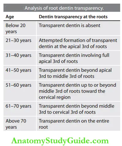
Gustafson’s Method of Age Determination: Sectioning of teeth shows some characteristic signs at advancing ages and these signs are used in age estimation.
- Amount of secondary dentin deposition
- Plane of gingival attachment
- Sclerotic apical dentin—the apical trans-lucency is absent or minimal at 20 years whereas it gradually increases with age and involves half of the root in around 70 years of age.
- Amount of secondary cementum at the apex.
- The extent of resorption at the apex.
By analyzing the root dentin transparency, the age of an individual from 20 years onward can be identified.
Dental Radiographs in Age Determination: Dental radiographs are assessed for the following features:
- Morphology of jaw bones
- Appearance of tooth germs
- Evidence of mineralization in deciduous teeth during intrauterine life
- Beginning of mineralization in permanent teeth
- Amount of crown completion
- Level of root development in both erupted and unerupted teeth
- Amount of root resorption in deciduous teeth
- Presence of open apices in roots
- The dimension of the pulp chamber and root canals—secondary dentin deposition will reduce the pulp cavity size in the advancing age group
- Tooth-to-pulp ratio
- Stage of third molar development.
Age estimation is grouped into three phases based on the above radiographic features:
- Prenatal, neonatal, and postnatal
- Children and adolescents
- Adults.
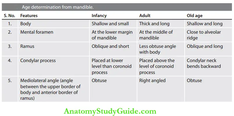
Age Determination from Mandible: Age determination from mandible is possible only for a broad range of groupings like infancy, adult, and older age.
Forensic Dentistry Short Notes
Question 1. Describe comparative dental identification.
Answer:
- Comparative dental identification is the process of comparing the dental findings in postmortem and antemortem dental records to determine similarities and exclude discrepancies.
- The forensic dentist prepares the post¬mortem record by charting and written descriptions of the teeth, supported by the evidence of radiographs, photographs, and dental casts. This record is compared with an available antemortem dental record for personal identification.
- This comparative analysis is helpful in the determination of sex, race, and age by analyzing the teeth, soft and hard tissue anatomical landmarks.
- Forensic odontologists may obtain more information about the deceased based on tobacco habits, occupation, and other causes that bring about characteristic morphological changes in the teeth.
The comparative analysis may lead to four types of results:
- Positive identification: There is adequate matching between antemortem and post-mortem data in essential features without any significant discrepancies to prove that they belong to the same individual.
- Possible identification: There will be sufficient matching between antemortem and postmortem data, but the insufficient details of either the postmortem remains or the antemortem evidence will make it impossible to establish the identity positively.
- Insufficient evidence: The existing details are inadequate to validate the matching.
- Exclusion: The antemortem and postmortem data are obviously mismatched. This finding has as equally significant as positive identification.
Dental identification Advantages:
- Comparative dental analysis is routinely used as a personal identification technique in criminal investigations, calamity, deca¬yed or traumatized bodies, and in situations where visual identification is not possible.
- Dental identifications are rapid, more accurate, and economically productive.
Dental identification Disadvantages:
- Poor quality of antemortem records.
- Inability to trace and obtain the antemortem data.
- The discrepancy between antemortem data and postmortem dental records.
Question 2. Describe the dental records or dental charts or antemortem records in dentistry.
Answer:
- To identify a person, a forensic dentist must have a dental record received from the deceased person’s dentist.
- A dental record is the detailed documentation of dental problems, a comprehensive medical history, oral examination findings, diagnosis, treatment, and follow-up details of a patient.
- The record also includes demographic information, diagnostic and treatment radiographs, study models, clinical photographs, laboratory reports, and drug prescriptions.
- The dental team should be cautious and well-organized in the record-keeping tasks.
- All information in the dental record should be legible, and when new entries are made, it should be certified with a sign and date. The information should be clear and should not contain any short forms or acronyms.
Retention and Storage:
- The dental clinic/hospital should have a record retention policy. Dental records may be stored with a records storage service, preserved on microfilm, or scanned for electronic storage.
- The documentation and maintenance should be accurate. The release of the details for legal purposes if necessary is part of the dentist’s professional responsibility.
Question 3. Describe Gustafson’s technique of age determination.
Answer: This method involves the removal of one or a few teeth from the dead body and preparing longitudinal ground sections. The sections are assessed for the following:
- Level of attrition
- Amount of secondary dentin
- Periodontal attachment
- Secondary cementum deposition
- Evidence of sclerotic or translucent dentin.
All these features accessed from the deceased need to be compared with sections taken from teeth of known age to identify the age of the deceased. By this technique, the age at death may be determined within about 7 years either way from the calendar age.
Question 4. Describe the rugoscopy or palatal rugae in personal identification.
Answer:
- Rugae are the ridges formed by small folds of mucous membranes on the palate. Rugoscopy is the study of rugae patterns and is used in the identification of an individual. There is an average of three to seven ridges for an individual, and the size, shape, and number of these ridges are considered unique to an individual.
- Hence rugoscopy is regarded as a reliable method in postmortem cases. Palatal rugae form an intrinsic and integral pattern for every single individual and can also help in sex determination. The ease of reproducibility and lower level of variation makes palatal rugae a potential tool in forensic odontology.
By determining the length of all rugae, Thomas et al. identified three categories:
- Primary rugae (5-10 mm)
- Secondary rugae (3-5 mm)
- Fragmentary rugae (<3 mm).
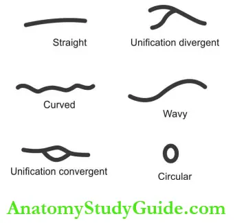
The rugae are also classified into five types based on their shape:
- Straight: Runs from the origin to the ending in a straight line.
- Curvy: Crescent shape ridge with a gentle curve.
- Circular: Continuous ring shape.
- Wavy: Serpentine form.
- Unification pattern: Two rugae that are united at a point.
Limitations of Rugoscopy:
- Identification is not possible without antemortem records. It is essential to keep the scanning record of rugae patterns, preservation of dental casts, and computer records to apply rugoscopy as evidence.
- Worn-out dentures, misalignment of teeth, and palatal lesions can cause alterations in rugae patterns.
Different patterns of rugae are genetically determined and can be used in population differentiation than individual identi¬fication. - Palatal rugae are destroyed in fire accident cases and degeneration processes, hence cannot be used in such incidents.
Question 5. What is coloscopy or lip print?
Answer:
- Lip prints are the anatomical lines present on the lip between the labial mucosa and outer skin. The pattern is characteristic for each and does not change during the life of a person.
- The study about the pattern of lip lines is known as coloscopy and is useful in personal identification.
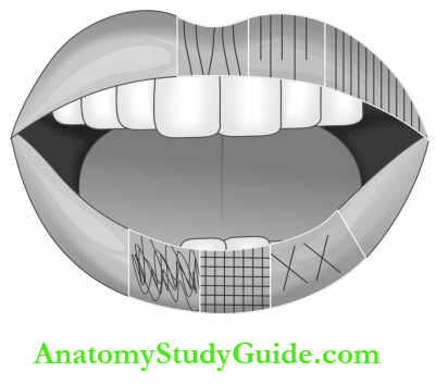
Suzuki and Tsuchihashi, in 1970, devised a classification method of lip prints:
- Type 1: A clear-cut groove running vertically across the lip.
- Type 2: Partial-length groove of type 1.
- Type 3: A branched groove.
- Type 4: An intersected groove.
- Type 5: A reticular pattern.
- Type 6: Other patterns.
Recording the Lip Prints:
- The Magna brush method is frequently used to mark lip prints. The lip impression is made against a glass or smooth, nonporous surface.
- After some time the forensic white powder is sprinkled over the print Magna brush that will reveal the lip print in a definite pattern.
Lip Prints in Personal Identification:
- Lip prints, being unique, can be used in the personal identification process. Lip prints are traced along with tooth marks on food products.
- It is possible to locate lip prints in windows, paintings, doors, plastic bags, and cigarette ends and frequently collected in incidents of murders, rapes, and theft incidents. A record of clear lines permits the personal identification of human beings.
Question 6. Describe the bite marks in personal identification.
Answer:
Bite marks may be seen in nonaccidental injuries to the child and in a variety of crimes. Bite marks were also left in the foodstuffs and other materials in the site of the crime.
- Bite marks on the flesh: Bites are multifaceted injuries produced by crushing pressure from teeth. The typical human bite mark consists of a double arcade of marks corresponding to the upper and lower six anterior teeth and forming an oval or round shape impression. In the center of the oval, a bruise due to tongue pressure or suction will be present.
- Bite marks in foodstuff and other materials: Foods which tend to take the best impression of teeth are apples, cheese, and chocolate. Wooden materials are also used to identify the marks.
Collection of Evidence:
- Recognition of mark as a human bite
- Preservation of evidence in a form suitable for analysis.
Processing Bite Mark Evidence :
- Morgue Procedure:
- Photograph (black and white, color)
- Saliva washings
- Tissue specimens (for microscopic study)
- Impressions as needed.
- Photographs should show the bite mark about the nearby anatomical landmarks, close-up exposure of the entire bite mark as well as the entire arch pattern.
- Saliva washings for enzyme and blood group determination are obtained by swab¬bing the area with 10% cotton threads mois¬tened with distilled water.
Interpretation of Bite Marks:
- Incisors—Rectangles
- Canines—Triangles with some variation
- Premolars—Single or dual triangles, dia¬mond with some variation
- Molars—Rarely leave marks, but when present, reflect the shape of the marking area.
Correlation of Evidence:
- Bite mark evidence is collected and interpreted to connect an individual with a crime during which the bite mark was made. To explore the possibility of particular dentition as a cause for a mark or not, an oral examination and an accurate model of the dentition are needed.
- The evidence collected is then interpreted and a comparison is made with the bite pattern in the tissue. The bite mark in the tissue usually exists as a photograph record. In this comparison, it is necessary to decide whether the dentition in question is the common origin of the bite marks in the tissue.
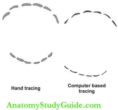
Conclusion: There are three possibilities of bite mark analysis:
- With reasonable dental certainty, the bite marks in the tissue and the exemplars have been left by the same teeth (high degree of possibility for two bite marks has common origin).
- The bite mark in the tissue and exemplars are consistent (middle-degree conclusion).
- The bite mark in the tissue and exemplars are not constant (two bite marks do not have the same origin).
Question 7. What are the various types of bite marks?
Answer:
- Amorous bite mark:
- This mark represents the areas where the tissues are stretched around the neck of the teeth and between the teeth.
- The central area of the bite will be caused by tongue pressure pushing the tissues against the rugae on the palate.
- Moderately aggressive bite marks:
- This mark is usually found on the arms of the victim of attempted rape.
- The bite has been made quickly and with moderate force.
- It often leaves the biting edge of the teeth scraped across the tissue at the central parts of the mark.
- Aggressive bite marks:
- This wound cannot be positively identified as a bite mark.
- Not possible to identify which tooth has caused the bite.
- Tissue scraping will be present.
- Very aggressive bite marks:
- Most aggressive bites will result in tissue tears.
- Usually involved sites are ears, nose, or nipples.
- Very difficult to interpret because the tissue is removed by a combination of biting and tearing and there is only a rare possibility to relate individual parts of the mark to the individual teeth.
Question 8. Describe the DNA analysis or DNA testing in forensic odontology or DNA analysis for personal identification from dental samples.
Answer: Dental source for DNA analysis includes components of tooth and tooth-supporting structures, jaw bones, oral fluids, biopsy specimens, and mucosal swabs.
The steps to be followed:
- The collection method should be legible
- Collected material should be verified for quantity and quality
- Authenticated methodology for DNA extraction and testing
- Analysis of results.
- After the sample collection, it should be preserved adequately and should not be subjected to temperature or moisture changes.
- Teeth: Pulp, dentin, cementum, and bony fragments provide DNA. It is obtained either by horizontal or vertical splitting, crushing/ grinding, endodontic assessment or ultrasonic wash.
- Saliva or mucosal cells: Desquamated epi¬thelial cells act as a source of DNA. Saliva is collected as a whole by using cotton swabs, filter papers, specialized tips or cytobrush.
- Biopsy materials: They provide epithelial and connective tissue cells for DNA extraction. DNA extraction is a three-step procedure that includes cell rupture, protein denaturation and inactivation, and DNA extraction.
The various DNA extraction techniques are:
- Organic method (phenol-chloroform)
- Chelex 100 (rapid technique, minimal chance for contamination, expensive)
- FTA paper (absorbent cellulose paper with chemical substances)
- Isopropyl alcohol method (less expensive).
Question 9. Describe the sex determination in foren¬sic dentistry.
Answer:
In forensic dentistry, the sex determination of an individual is carried out based on the developmental pattern of teeth, eruption sequence, and tooth morphology (size and shape). Three methods are broadly applied to gender determination:
1. Visual method or clinical identification: This method of identification is carried out using gender-differentiating dental records like tooth size and shape.
- Tooth size: Buccolingual (BL) width of all maxillary teeth and mandibular lateral incisors and canines are larger in males than in females. The mesiodistal width of the maxillary and mandibular canine and first molar are larger for men. The size of the mandibular canine and inter-canine distance is greater for males and helps in sex differentiation with 74% accuracy.
- Canine dimorphism: A distal accessory ridge of upper and lower canines (present at the lingual surface between the mesial and distal marginal ridge) is frequently common in males and also more prominent in males than in females.
- The dental index includes the incisor index, mandibular canine index, crown module, and crown index.
- The incisor index is determined by using the formula: Ii = MDI2/MDI1 x 100 where MDI2 = Maximum mesiodistal diameter of the maxillary lateral incisor.
- MDI1 = Maximum mesiodistal diameter of the central incisor. The incisor index is commonly used, and the value is higher for males.
2. Microscopic methods:
- Barr bodies in sex determination:
- The study of X and Y chromosomes in the cells is utilized for sex determination. Buccal smears and tooth pulp are considered as suitable samples for chromosome analysis, and quinacrine mustard stain is specific in the identification of the Y chromosome.
- The pulp tissue is stained with 5% quinacrine hydrochloride and examined under a fluorescent microscope. Cells that have Y chromatin display a blue-violet fluorescent spot and are labeled as positive cells.
3. Advanced methods:
- Polymerase chain reaction (PCR): Dental pulp is subjected to ultrasonication to extract DNA followed by PCR for amplification of X and Y sequences in males and the X sequence in females.
- Sex determination using enamel protein:
- Amelogenin or AMEL is a protein matrix of enamel. The AMEL gene that encodes amelogenin is located on the X chromosome for females and the Y chromosome for males.
- The female has two identical AMEL genes, whereas the man has two different AMEL genes. This pattern of expression is used to find out the sex from DNA samples.
Question 10. Dental jurisprudence.
Answer: Dental jurisprudence is a branch of law that deals with dental practice. It includes legal requirements for patient safety, ethical boundaries, and related matters.
Dental Jurisprudence Risk Management: It describes the series of actions followed by dental practitioners to prevent legal complaints against them.
Legal Responsibilities of a Dental Practitioner
- The dentist should avoid:
- Breach of contract
- Maligning a patient
- Permitting a hazard in the dental clinic
- Technical assault.
- Dental malpractice:
- Malpractices include the following:
- Professional misconduct
- Any unreasonable lack of skill execution:
- Failure to diagnose
- Improper diagnosis
- Failure to inform the diagnosis or treatment of the patient
- Wrong treatment (for example removal of wrong teeth)
- Mortality and morbidity due to negligence.
- Lack of reliability in the performance of professional duties
- Violating the rules and regulations of professional practice.
- Malpractices include the following:
Dentist’s Responsibilities:
- Must be a registered practitioner
- Must execute a reasonable skill, care, and decision
- Must perform any procedure only getting consent from the patient
- Must refer cases to a specialist if the training/skill is insufficient
- Must use standard drugs, materials, and techniques
- Must give adequate instructions and information to the patient
- Must charge reasonably for the offered services
- Must respect the patient’s privacy.
The dentist should obtain written consent from the patient when:
- Administering new drugs
- Carry out trial procedures or clinical trial
- Presenting or publishing the patient’s photograph
- Administering general anesthesia
- Providing treatment for children in a public program
- The duration of treatment is more than 1 year.
Question 11. Describe the recent advances in forensic dentistry.
Answer:
Forensic Dentistry Role of DNA:
- The use of DNA as a biological material for personal identification is a recent advance in forensic dentistry. Teeth are an excellent source of DNA material for deceased individuals because of their protected nature in the oral cavity.
- DNA extracted from the teeth of an unidentified individual is compared with a suspected known antemortem sample or to a parent or sibling.
Forensic dentistry Genomic DNA:
- Present in the cell nucleus and can be used as a source of DNA for forensic examination.
- Subjecting this DNA sample for PCR analysis will provide DNA profile that can be compared with known antemortem records.
Forensic dentistry Mitochondrial DNA: Cells contain a high number of mitochondria and mitochondrial DNA (mDNA). mDNA is maternally inherited.
For the forensic analysis, DNA from the teeth is obtained by either:
- Cryogenic grinding
- Restriction fragment length polymerization.
Question 12. Enumerate the medicolegal importance of teeth.
Answer:
- Teeth help to identify both living and deceased in the following:
- Age estimation
- Blood group detection
- DNA sample extraction for comparison.
- Habit detection:
- Blackish brown or reddish stains—Habit of betel nut chewing
- Dark brown stains on the inner aspect of incisors—Habit of smoking.
- Racial identification :
- In Chinese, Inuit, and Mongoloid, mandibular 1st premolar has three cusps
- In Mongoloids, mandibular molars have three roots.
- Professional identification: Notched incisors suggest being the profession of tailor or cobbler.
- Identification of toxins on examination of teeth and gingiva:
- Sulfuric acid causes a chalky white appearance of teeth
- Nitric acid causes a yellow stain
- Cocaine produces black stains on teeth
- The lead poison causes a bluish line along marginal gingiva
- Copper sulfate causes bluish-green discoloration at the junction of the tooth and gingiva.
Question 13. Enumerate the work of forensic odontologists.
Answer:
- Identification of unknown bodies from dental records
- Bite marks identification on the victim’s body
- Tracing of bite marks from the materials, such as foodstuffs
- Match the identified bite marks with the teeth of the suspect and submit this proof as an experts witness for the legal purpose
- Age estimation of skeletal remains
- Sex determination by microscopic examination of teeth.
Forensic Dentistry Multiple-Choice Questions
Question 1. The Demirjian system is used for dental age identification.
- Sex determination
- Dental age identification
- Race identification
- Ethnic group identification
Answer: 2. Dental age identification
Question 2. Whitehouse standards are useful for.
- Dental age identification
- Bone age identification
- Personal identification
- Comparative identification
Answer: 2. Bone age identification
Question 3. Boyde’s method of age determination is based on microscopic observation of.
- Root development
- Root resorption
- Dentin deposition
- Cementum deposition
Answer: 3. Dentin deposition
Question 4. The biochemical method of age determination uses the.
- Collagen in pulp
- Collagen in dentin
- Undifferentiated cells in the apex
- DNA extraction from gingiva
[Note: Biochemical method of age determination uses the collagen (ascorbic acid) in dentin].
Answer: 2. Collagen in dentin
Question 5. The half-formed roots of the wisdom tooth denote the age of the individual as around.
- 21 years
- 13 years
- 30 years
- 17 years
Answer: 4. 17 years
Question 6. The angle of the mandible is sharp in males and round in females.
- Sharp in females and round in males
- Obtuse in males and females
- Sharp in males and round in females
- Oblique in males and females
Answer: 3. Sharp in males and round in females
Question 7. Barr bodies in the nucleus are the indication for the.
- Male sex
- Female sex
- Jacob syndrome
- Jacobsen syndrome
[Note: The absence of Barr bodies in the nucleus denotes male sex, in males with Klinefelter’s syndrome (genotype 47XXY), one Barr body is present].
Answer: 2. Female sex
Question 8. Strangulation or victims of drowning may lead to.
- Black discoloration of teeth
- Greenstick fracture of the mandible
- Tongue protrusion
- Pink discoloration of teeth
(Note: Strangulation or victims of drowning may lead to pink discoloration of teeth after 1.5-2 weeks).
Answer: 4. Pink discoloration of teeth
Question 9. In WINID tooth codes, the primary codes denote.
- Modifiers
- Supernumerary teeth
- Restored surfaces
- Prosthetic replacement
Answer: 3. Restored surfaces
Question 10. In WINID tooth codes, the secondary codes are.
- Modifiers
- Supernumerary teeth
- Restored surfaces
- Prosthetic replacement
(Note: In WINID tooth codes, the primary codes denote restored surfaces and the presence or absence of a tooth. The secondary codes are modifiers, allowing for extra information but not for best match comparison).
Answer: 1. Modifiers
Question 11. Dorian’s method of bite mark analysis is applied when the bite is insufficiently defined to be a.
- Animal bite
- Human bite
- Teeth mark
- Postmortem evidence
(Note: Dorian method of bite mark analysis involves removal of bitten tissue from assault victim for microscopic examination especially when the bite is insufficiently defined to be a human bite).
Answer: 2. Human bite
Question 12. The study of biting marks made by teeth is known as.
- Tooth sculpture
- Dental sculpture
- Odontoscopy
- Endoscopy
Answer: 3. Odontoscopy
Question 13. In a population study, the intergroup marker is.
- Molar
- Canine
- Premolar
- Central incisor
Answer: 4. Central incisor
Question 14. The study of the oral health status of an ancient population from the fossil or skeletal remnants is known as.
- Podiatry
- Paleontology
- Trace evidence analysis
- Gerodontology
Answer: 2. Paleodontology
Forensic Dentistry Viva Voce
Question 1. What is odontography?
Answer:
- Identification of a deceased person using dental records and dentition from a legal perspective is known as odontography.
- The dentition helps in the identification of individuals where other recognized methods like face recognition, moles, thumb or other fingerprints. They play a crucial role in the facial reconstruction process for identification.
Question 2. What is a shovel tooth or blade shape tooth?
Answer:
- Upper and lower incisors are called as shovel-shaped teeth if they manifest a pronounced mesial and distal marginal ridge with a deep pit (palatal fossa) on the palatal surface (blade shape).
- This pattern is helpful in population analysis and is present in 100% of Mongoloid origin population’s central incisors.
Forensic Radiology Essay Questions
Question 1. Describe the intraoral radiographic techniques in forensic dentistry.
(or)
Briefly explain the radiographic techniques to obtain the intraoral image for a deceased.
Answer:
Intraoral Radiographic Sources (for Intact Jaws): To take intraoral radiographs of the dead body (morgue) with the entire jaw, the possibility of gaining access to place the film in the proper position is a difficult task. Mechanical mouth props are required if the jaws are in rigor mortis. To place the film in position, hemostats, taps, and gauze are used.
Intraoral Radiographic Sources (for Jaws Resected):
- If the body is skeletonized, and jaws can be resected, conventional radiographic techniques can be used. The specimen should be wrapped in multiple layers of plastic wrap.
- The radiographic film is then taped to the specimen and exposed. If the procedure can be delayed, then the sample should be stored in 10% formalin solution.
- The postmortem radiograph should duplicate the antemortem film as closely as possible. If antemortem radiographs are not available then, conventional periapical and bitewing films should be obtained.
- Exposure time should be reduced by one-third for resected jaws and up to one-half for skeletonized jaws.
Question 2. Describe the role of dental radiology in personal identification.
Answer:
- For identification purposes, properly exposed, mounted, and labeled dental radiographs (both antemortem and postmortem) are as good as fingerprints.
- In the case of “direct identification” the dentists compare the postmortem details with antemortem records and produce the evidence.
- In the case of “unknown identification”‘ no presumption of identification exists, and the dentist needs to provide a detailed description of the dentition and surrounding structures for comparison of a missing person.
- Personal identification of human remains is possible only when a specific dental finding on the corpse is matched with the individual’s data obtained during his life. The technique used for getting antemortem and postmortem radiographs should be as similar as possible to prevent matching difficulties.
Two requirements are essential for positive radiographic identification in forensic den¬tistry:
- The detail must be unique
- The detail should be stable for a long time.
- Usually, one to four similarities and the absence of disparity are taken for positive identification. Both extraoral-and intraoral radiographs, various anatomical landmarks, physical characteristics and disease status are used for identification purposes if antemortem data are available.
- Missing and unerupted teeth, fillings, caries, root canal fillings, and periapical and periodontal pathology are the common features useful for identification. The type of prosthetic restorations, the placement of retention pins and posts, and the placement and type of implants can also be evaluated.
- Accurate assessment of tooth development and age determination in a child and juvenile is also possible with dental radiographs.
- Bone trabeculation patterns, anatomical landmarks like mental foramen and mandibular canal, and nutrient canals are also served for identification.
Question 3. What is panoramic radiography in forensic identification?
Answer:
- The panoramic radiographic image reveals a wide range of anatomical, pathological, and therapeutic details in both jaws and related structures on a single film. Hence the comparative analysis of antemortem and postmortem panoramic radiographs will reveal sufficient details in the oral and maxillofacial complex.
- In postmortem cases, dissection is not required for using the panoramic technique. The head portion of the body is introduced to the X-ray scanning area. A machine that can accommodate a patient in a prone position is preferred.
- In a cadaver, the tube head travels 240° under the patient’s head during exposure, and the radiograph is examined for forensic purposes. Processing should be done with care by adopting technical recommendations to get an image of adequate quality and details.
Forensic Radiology Short Notes
Question 1. What is virtopsy?
Answer:
- “Virtopsy” refers to virtual autopsy—An imaging technique used to provide objective and permanent records of postmortem evidence. It is a blend of computed tomography (CT) and magnetic resonance imaging (MRI) to obtain a three-dimensional view.
- The benefit of this method is the avoidance of any sort of damage to the body by puncturing or resection and to “freeze” findings during the investigation. It is also possible to revisit the image on later periods either for submitting to court or for other purposes.
Question 2. What is the role of dental radiology in age estimation?
Answer:
- Frontal sinus in age estimation: Frontal sinus can be detected radiographically above the age of 6 years. It increases in size rapidly during puberty and stops increasing by 20 years of age.
- The formation and eruption stages of dentitions identified on the panoramic radiographs can be used for age estimation in children.
- In adults, pulp chamber volume is reduced by the increased secondary dentin deposition and is proportional to the age of an individual. This can be identified through dental radiographs and considered for age estimation.
- The chronology of third molar eruption also helps in age estimation
Forensic Dentistry Highlights
- Personal identification is a part of criminology investigation to identify the crimes and unknown deceased individuals. For establi¬shing the details of the unidentified victims, dental findings can be used alone or in combination with other evidence. The art and science of applying dentistry in the detection or investigation of an unknown person are evolved as a new branch named forensic odontology/dentistry.
- Keiser-Neilson in 1970 defined forensic odontology as a branch of forensic medicine that deals with the proper handling and examination of dental evidence and the proper evaluation and presentation of the dental findings to support the law and justice.
- Identification is made by comparing the known dental findings/records of an individual (antemortem data) with the identified characteristics of an unknown body (postmortem data). This chapter deals with the methods, interpretation, and reporting system of dental and oral findings for personal identification.
Leave a Reply