Heart-Lung Preparation Introduction
Heart-lung preparation is an experimental setup devised by Starling.
Table of Contents
It is used to demonstrate the effects of various factors on the activities of the heart, particularly heart rate and cardiac output.
This preparation is also used to record the cardiac function curves.
Read And Learn More: Medical Physiology Notes
Procedure
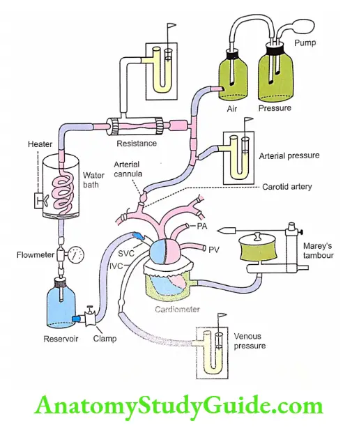
Heart-lung preparation is usually done in dogs. After giving anesthesia to the dog, the neck is opened and a tracheal cannula is inserted into the trachea.
The trachea cannula is connected to a respiratory pump so that, the respiration in the animal is controlled artificially to avoid any disturbance during the experimental procedure.
Then chest is opened and an arterial cannula is inserted into one of the branches of the aorta.
All the other branches from the arch of the aorta and descending aorta are ligated.
The arterial cannula is connected to two instruments:
- Mercury manometer to measure the arterial blood pressure
- Air bottle, which provides elasticity artificially (as in the case of arterial wall).
Thus, the blood ejected from the left ventricle passes into an air bottle through the arterial cannula and rubber tubes.
From the air bottle, the blood is diverted through a tube that provides artificial resistance.
The air bottle is also connected to a pressure bottle. The pressure bottle is attached to a pressure pump.
The pump is used to maintain the pressure within the setup.
The artificial resistance is offered by applying pressure surrounding the resistance tube. The resistance tube is also connected to a manometer.
After passing through the resistance tube, the blood is allowed to flow through a warming glass coil which is kept inside a water bath with a heater.
The temperature of a water bath is controlled, so that, the temperature of could be maintained.
The warming coil is connected to a venous reservoir through a flowmeter.
The flowmeter determines the amount of blood flow (cardiac output). The venous reservoir is connected to the superior vena cava by a rubber tube.
A screw-type clamp is fitted to the rubber tube.
This clamp is used to adjust the amount of blood returning to the heart (venous return).
A thermometer is also fitted to the tube to note the temperature of the blood.
A third mercury manometer is connected to the inferior vena cava. It is used to determine the venous pressure.
A cardiometer is fitted to the ventricle. This audiometer is connected to a recording device like Marey’s tambour or polygraph to record the ventricular volume changes.
The pulmonary circulation is kept intact for continuous oxygenation of blood.
Uses Of Heart-Lung Preparation
Thus, in this set up, the heart works as an isolated organ. So, the effects of various factors can be demonstrated on the activities of heart, like heart rate, ventricular volume, and cardiac output.

For example,
- When the venous return decreases, stroke volume decreases.
- When the venous return increases, the stroke volume increases.
- When the resistance increases, the cardiac output decreases.
- When the resistance decreases, the cardiac output increases.
The heart-lung preparation is also used to record two types of cardiac function curves:
- The cardiac output curves
- The venous return curves.
Though the cardiac function curves are obtained in experiments using animals, these curves represent the functions of the ventricles in the human heart also.
Leave a Reply