Histology of Blood Vessels
Draw and describe the Muscular Artery
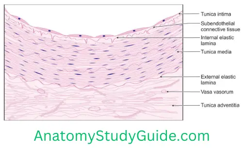
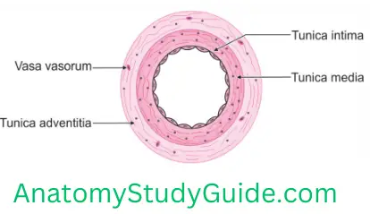
1. Muscular Artery Synonymous: It is the medium size artery (distributing artery).
Table of Contents
2. Muscular Artery Layers: It consists of three layers
1. Tunica intima
- It shows folded appearance.
- The internal elastic lamina is prominent and wavy.
2. Tunica media: It shows plenty of smooth muscle cells, arranged concentrically.
3. Tunica adventitia
Read And Learn More: Anatomy Notes And Important Question And Answers
- It is as thick as media.
- The diameter of the lumina is less than thickness of the wall
3. Functions: It regulates the flow of blood in different regions.
4. Examples: Most of the arteries of body distal to subclavian artery.
- Branches of common carotid,
- Thoracic, and
- Abdominal aorta.
Draw And Describe The Elastic Artery
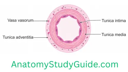
1. Elastic Artery Synonymous: It is a large size artery called elastic artery.
2. Elastic Artery Layers: It consists of three layers
1. Elastic Artery Tunica intima
- It is the innermost coat and lined by simple squamous epithelium.
- It contains elastic fibres.
- The subendothelial layer is prominent and contains elastic fibres
- The internal elastic lamina is present but indistinguishable.
2. Elastic Artery Tunica media
1. It is a middle and thickest coat.
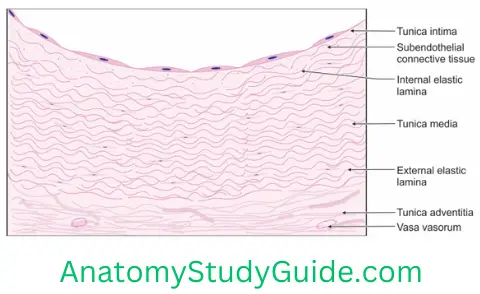
2. It consists of a concentric layer of fenestrated elastic fibres.
3. It also contains a little amount of smooth muscle and collagen fibres.
3. Elastic Artery Tunica adventitia
- It is thin and contains elastic fibres.
- The fibres merge with the external elastic lamina.
3. Lumina: It is irregular. The diameter of the lumina is more than the thickness of wall.
4. Elastic Artery Functions: It conducts blood from heart to the different parts of body.
5. Elastic Artery Examples: Aorta, large arteries of head and neck.
Draw and Describe Vein
1. Vein Histology
1. It exhibits three layers or tunica.
- The tunica intima shows endothelium and subendothelial connective tissue.
- The muscular layer is much thinner and less prominent. The smooth muscles intermix with connective tissue fibres.
- The tunica adventitia is the thickest and best-developed layer of the three layers.
2. Vein Features
1. Vein Wall
- 1. It is thin as compared to arteries of similar size.
- 2. Thickness is not correlated exactly with the size of the vein and varies in different regions. For example, wall is thicker in veins of the leg than it is in veins of a similar size in the arm.
2. Vein Muscle
1. Amount of muscle is considerably less than in arteries.
2. The collagen and elastic fibres predominate.
3. In most veins of the limbs, the muscles are arranged circularly.
4. Longitudinal muscles are present in the following veins
- Iliac,
- Brachiocephalic,
- Portal,
- Renal,
- Superior, and
- Inferior venae cavae.
5. Muscles are absent in
- Maternal placental veins,
- Dural venous sinuses,
- Pial and retinal veins,
- Veins of trabecular bone, and
- Venous spaces of erectile tissue;
These veins consist of endothelium supported by variable amounts of connective tissue.
6. Distinction between the media and adventitial layers is often difficult.
7. Discrete internal elastic lamina is absent.
8. Collapse of the veins is prevented
- Connective tissue fasciae and
- Surrounding tissues.
9. Vein Valves
- Veins have valves which convey blood against gravity.
- They assist venous blood flow toward the heart by preventing backflow.
- When blood flows toward the heart, pressure in the veins forces the valves to open.
- As the blood begins to flow backwards, the valve flaps close to the lumen. Thus, it prevents the backflow of blood.
- They are found in small veins and at the junctions of tributaries.
- The valves are semilunar (cusps) and attached by their convex edges to the venous wall.
- Valves are absent in the veins of the thorax and abdomen.
3. Classification of veins: The veins are arbitrarily classified as
- Small,
- Medium, and
- Large.
4. Vein Functions
1. The structure of the venous walls allows
- Flexibility, and
- Accommodation of a large amount of blood. As a result, veins contain most of the blood present in the body.
2. Pressure within the venous system does not normally exceed 5 mm Hg.
3. Pressure decreases as the veins grow larger.
4. It maintains cardiac output by effectively mobilizing the blood.
5. A sudden fall in blood volume (e.g. following a haemorrhage) is compensated by
- Elastic recoil of the vein, and
- Reflex constriction of vein.
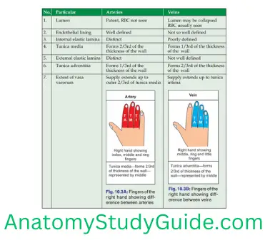
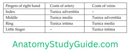
The differences between arteries and veins are tabulated in Table.
Leave a Reply