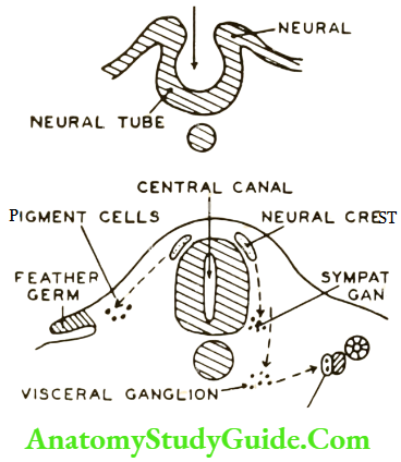
• Skip to main content
  • Skip to secondary menu
  • Skip to primary sidebar
  • Skip to footer
  • Anatomy
    • Anatomy Question And Answers
    • Face Anatomy
    • Neck Anatomy
    • Head Anatomy
    • Oral Anatomy
    • Lower Limb
    • Upper Limb
  • Endodontics
    • Paediatric Dentistry
  • General Histology
    • Oral Histology
    • Genetics
  • Pediatric Clinical Methods
  • Complete Dentures
    • Pharmacology for Dentistry
  • Medical Physiology
    • Body Fluids
    • Muscle Physiology
    • Digestive System
    • Renal Physiology
    • Endocrinology
    • Nervous System
    • Respiratory System
    • Cardiovascular System
    • Reproductive System
    • Oral Physiology
  • General Medicine
  • General Pathology
    • Systemic Pathology
    • Oral Pathology
    • Neoplasia
    • Homeostasis
    • Infectious Diseases
    • Infammation
    • Amyloidosis Notes
  • Periodontology
  • General Surgery
    • Basic Principles Of Surgery
    • General Surgery

Anatomy Study Guide

Anatomy Study Guide

  • About Us
  • Contact Us
  • Privacy Policy
  • Terms of Use
  • Disclaimer
  • Sitemap
Home » Human Nervous System – Structure, Function & Parts

Human Nervous System – Structure, Function & Parts

October 17, 2023 by Marksparks arkansas Leave a Comment

Nervous System

Neural Crest

Introduction: It is ectoderm present at the junction of the neural tube and rest of the ectoderm.

Table of Contents

  • Nervous System
  • Neural Crest

Nervous System - Neural crest

Read And Learn More: General Histology Question And Answers

Neural derivatives:

1. Sensory neurons of the cranial ganglia V, VII, VIII, X, and XI.

2. Sensory neurons of the spinal dorsal root ganglia and their peripheral sensory receptors.

3. Satellite cells in all sensory ganglia.

4. Sympathetic ganglia and plexuses: Neurons and satellite cells.

5. Parasympathetic ganglia and plexuses: Neurons and satellite cells.

6. Enteric plexuses: Neurons and glial cells.

7. Schwann cells of all the peripheral nerves.

8. Medulla of the adrenal gland, chromaffin cells.

9. Carotid body type I cells (type II, satellite type cells).

10. Calcitonin: Producing (C cells)

11. Melanocytes.

12. Odontoblast.

13. Leptomeninges.

14. Head mesoderm, etc.

Filed Under: General Histology

Reader Interactions

Leave a Reply Cancel reply

Your email address will not be published. Required fields are marked *

Primary Sidebar

Recent Posts

  • Esophagus Anatomy
  • Lacrimal Apparatus: Anatomy, Parts & Function
  • Scalp Temple And Face Question and Answers
  • Orbicularis Oculi Muscle Anatomy
  • Extraocular Muscles Anatomy
  • Ciliary Ganglion Anatomy
  • Femoral sheath Anatomy
  • Femoral Artery – Location and Anatomy
  • Adductor Canal: Anatomy And Function
  • Ankle Joint: Anatomy, Bones, Ligaments And Movements
  • Risk Factors For Breast Cancer
  • Cervical Tuberculous Lymphadenitis Notes
  • Carbuncles: Causes, Symptoms, and Treatments
  • Sinuses And Fistulas Notes
  • Cellulitis: Treatments, Causes, Symptoms
  • Pyogenic Liver Abscess: Causes, Symptoms, and Diagnosis
  • Acid Base Balance Multiple Choice Questions
  • General Surgery Multiple Choice Questions
  • Hypertrophic Scarring Keloids Multiple Choice Questions
  • Surgical Site Infection Multiple Choice Questions
  • Facebook
  • Pinterest
  • Tumblr
  • Twitter

Footer

Anatomy Study Guide

AnatomyStudyGuide.com is a student-centric educational online service that offers high-quality test papers and study resources to students studying for Medical Exams or attempting to get admission to different universities.

Recent

  • Esophagus Anatomy
  • Lacrimal Apparatus: Anatomy, Parts & Function
  • Scalp Temple And Face Question and Answers
  • Orbicularis Oculi Muscle Anatomy
  • Extraocular Muscles Anatomy

Search

Copyright © 2026 · Magazine Pro on Genesis Framework · WordPress · Log in