.Describe the Inguinal Canal under the following headings:
- Inguinal Canal Introduction
- Inguinal Canal Boundaries
- Inguinal Canal Contents
- Inguinal Canal Defense/protective mechanisms
- Inguinal Canal Development and
- Inguinal Canal Applied anatomy.
Answer:
1. Inguinal Canal Introduction:
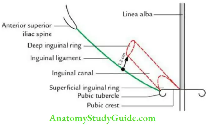
- It is an oblique intermuscular slit-like passage in the lower part of the anterior abdominal wall for the transmission of spermatic cord in the male
- It is situated above the medial half of the inguinal ligament.
- It is about 4 cm in length.
- It begins at the deep inguinal ring and terminates at the superficial inguinal ring.
- It is directed downward, forward, and medially.
- It is larger in males than in females.
Read And Learn More: Anatomy Question And Answers
2. Inguinal canal Boundaries:
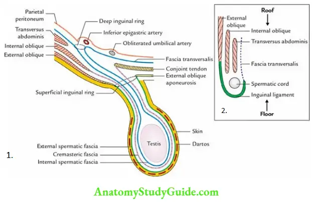
- Anterior wall: It is formed by the external oblique aponeurosis in its whole extent and by the lower fleshy fibers of the internal oblique in its lateral 1/3rd.
- Posterior wall: It is formed by fascia transversalis in its whole extent, a conjoint tendon in its medial half, reflected part of the inguinal ligament in its medial fourth, and interfoveolar ligament in its lateral 1/3rd.
- Floor: It is formed by the upper grooved surface of the inguinal ligament and at the medial end by the lacunar ligament.
- Roof: It is formed by the lower arched fibers of the internal oblique and transversus abdominis muscles.
- Inlet and outlet: The inlet is formed by the deep inguinal ring and the outlet by the superficial inguinal ring.
- Deep inguinal ring: It is an oval aperture in the fascia transversalis, half an inch (1.25 cm) above the mid inguinal point and just lateral to the inferior epigastric artery.
- Superficial inguinal ring: It is a small/oblique triangular aperture in the aponeurosis of the external oblique muscle, above and lateral to the pubic tubercle. The medial and lateral margins of the superficial inguinal ring are called crura.
The lateral crus is attached to the pubic tubercle and the medial crus is to the front of the pubic symphysis. The base of the superficial inguinal ring is formed by the pubic crest.
3. Contents/structures passing through the inguinal canal:
- Spermatic cord in males and the round ligament of the uterus in females
- Ilioinguinal nerve
4. Defense/protective mechanisms of the inguinal canal:
These are the mechanisms that prevent the abdominal contents from entering the inguinal canal, thus preventing inguinal hernias from occurring.
These are as follows:
- Flap-Valve Mechanism: The increased intra-abdominal pressure approximates the anterior and posterior walls and obliterates the inguinal canal.
- Slit-Valve Mechanism: The contraction of the external oblique approximates two crura of the superficial inguinal ring.
- Shutter Mechanism: The contraction of the arching fibers of the internal oblique approximates the roof with the floor of the inguinal canal like a shutter.
- Ball-Valve Mechanism: When the cremaster muscle contracts, it draws upward the spermatic cord to plug the superficial inguinal ring.
- Posterior and Anterior Guards:
- Posterior guard: The superficial inguinal ring is guarded posteriorly by the conjoint tendon and reflected part of the inguinal ligament.
- Anterior guard: The deep inguinal ring is guarded anteriorly by the fibers of the internal oblique muscle.
5. Inguinal canal Development:
Developmentally, the inguinal canal is formed by the formation of processus vaginalis and the descent of the testis through the anterior abdominal wall.
6. Inguinal canal Applied anatomy:
Inguinal hernias:
The inguinal canal is the site of the inguinal hernia because it is the region of potential weakness in the lower part of the anterior abdominal wall. Therefore, when intraabdominal pressure increases, the abdominal contents especially the intestinal loop surrounded by the peritoneal sac are pushed into the inguinal canal, leading to a clinical condition called an inguinal hernia.
If the abdominal contents are pushed into the inguinal canal through a deep inguinal ring, it is called an indirect inguinal hernia. On the other hand, if abdominal contents are pushed into the inguinal canal directly through the posterior wall of the inguinal canal, it is termed a direct inguinal hernia.
Indirect inguinal hernias: These are subdivided into 3 types
- Vaginal, i.e. complete because processus vaginalis remain patent along the entire extent and hernia reaches the base of the scrotum.
- Congenital, i.e. present since birth and
- Bubonocele, i.e. incomplete and does not protrude through the superficial inguinal, ring. It produces a bulge only in the groin.
Direct Inguinal Hernias:
These are subclassified into two types:
- Medial direct inguinal hernia if abdominal viscus is pushed through the medial part of the Hesselbach’s triangle, i.e. medial to obliterated umbilical artery; and
- Lateral direct inguinal hernia if the abdominal viscus is pushed through the lateral part of the Hesselbach’s triangle, i.e. lateral to the obliterated umbilical artery.
Leave a Reply