Joints Of The Lower Limb
Question 1. Describe the hip joint under the following headings:
- Hip Joint Classification,
- Hip Joint Ligaments,
- Hip Joint Relations,
- Hip Joint Movements and muscles producing them and
- Hip Joint Applied anatomy.
Answer:
Hip joint:
Hip joint Classification:
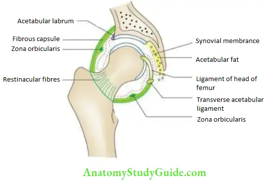
It is a synovial joint of ball and socket variety. It is formed between the rounded head of the femur and the cup-shaped acetabulum of the hip bone. The acetabulum is deepened at its margins by a fibrocartilaginous rim – the labrum acetabular.
Hip joint Ligaments:
Hip joint Capsular ligament:
- Above, it is attached to the acetabular margin and transverse acetabular ligament. Below, it is attached to the femur on the intertrochanteric line anteriorly and on the femoral neck about 1 cm above the intertrochanteric crest posteriorly.
- The capsular fibers are reflected from their lower attachment upward on the femoral neck to form the retinacula.
Hip joint Iliofemoral ligament (Bigelow’s ligament):
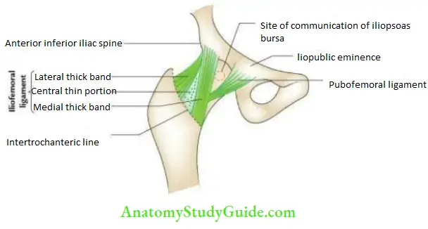
It is a strong inverted ‘Y’–shaped ligament. The apex/stem of ‘Y’ is attached to the anterior inferior iliac spine, while its medial and lateral limbs are attached to the intertrochanteric line.
- Pubofemoral ligament: It is triangular in shape and attached above the iliopubic junction and below the anteroinferior part of the capsule, adjacent to the intertrochanteric line.
- Ischiofemoral ligament: It is attached above to the ischium, and below some fibers are attached to the base of the greater trochanter, but the majority of fibers spiral and blend with the capsule around the femoral neck to form zona orbicularis.
- Ligament of the head femur (round ligament/ligamentum teres): It is a flat and triangular ligament with an apex attached to the fovea capitis of the femoral head and its base to the transverse acetabular ligament.
Hip joint Note:
The ligamentous stability of the hip joint is provided by the following three ligaments:
- The iliofemoral ligament restricts hyperextension of the hip to prevent the backward fall while standing.
- The pubofemoral ligament supports the joint anteromedially.
- The iliofemoral ligament supports the joint posteriorly.
Hip joint Relations:
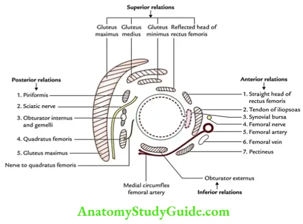
The relations of the hip joint are given in the box below and shown.

Hip joint Movements and muscles:
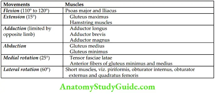
These are given in the box below:
Hip joint Applied anatomy:
Hip joint Dislocation:
Acquired dislocation mostly occurs posteriorly and often injures the sciatic nerve. (Note that congenital dislocation is most common in hip joints.)
Hip joint Fracture of neck of femur:
It commonly occurs between 40 and 60 years of age, especially in females.
Hip joint Referred pain:
In diseases of the hip, pain is referred to as the knee.
Question 2. Give the anatomical basis of avascular necrosis of the head of the femur following the fracture neck of the femur.
Answer:
Avascular necrosis:
Avascular necrosis of the head of the femur occurs if the fracture line is intracapsular because the retinacula which carries retinacular vessels, the main arterial supply to the head is torn.
Question 3. Describe the knee joint under the following headings:
- Knee Joint Classification,
- Knee Joint Ligaments and menisci,
- Knee Joint Relations,
- Knee Joint Movements and muscles producing them and
- Knee Joint Applied anatomy.
Answer:
knee joint:
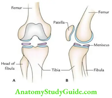
Knee Joint is one of the strongest and most important joints of the body. It consists of 3 bones: the femur, tibia, and patella.
knee joint Classification:
knee joint Anatomically:
knee joint It is a compound synovial joint with the following components:
- Condylar joint (modified hinge joint), between medial and lateral condyles of femur and tibia.
- Saddle joint, between femur and patella.
knee joint Physiologically:
It is a modified hinge joint that permits flexion and extension and a small degree of medial and lateral rotation.
knee joint Ligaments
knee joint Capsular ligament:
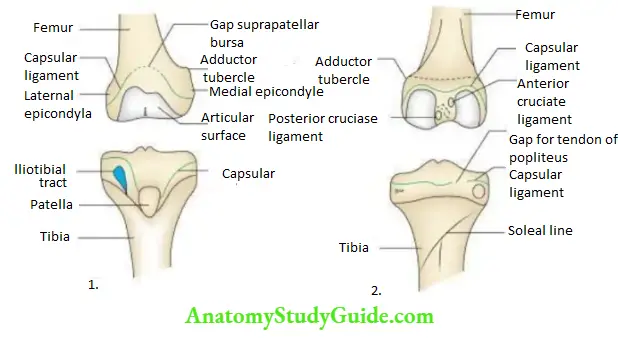
It is attached to the margins of articular surfaces except anteriorly where it is deficient and supplemented by the extensor apparatus of the knee joint consisting of the tendon of quadriceps, patella, and ligamentum patellae. Posterolaterally, it prevents an opening for the passage of the tendon of the popliteus.
knee joint Medial (tibial) collateral ligament:
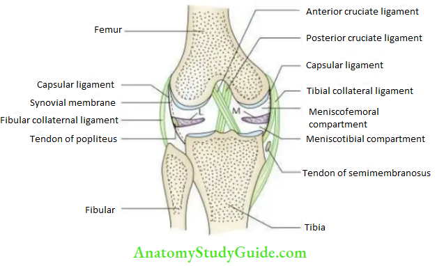
It consists of superficial and deep parts. The superficial part is attached above to the epicondyle of the femur and below the upper part of the medial border of the tibia. The deep part is firmly attached to the medial meniscus.
knee joint Lateral (fibular) collateral ligament:
It is attached above the epicondyle of the femur and below the head of the fibula. It lies away from the meniscus.
knee joint Cruciate ligaments (anterior and posterior):
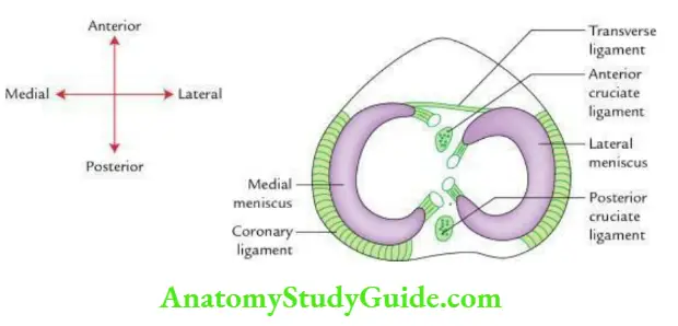
They are intracapsular and intrasynovial. The anterior cruciate ligament extends from the anterior part of the intercondylar area of the tibia to the medial side of the lateral femoral condyle. It prevents hyperextension and resists forward movement of the tibia on the femur.
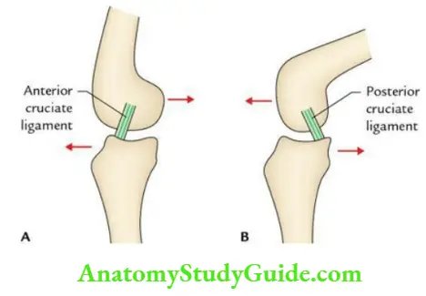
The posterior cruciate ligament extends from the posterior part of the intercondylar area of the tibia to the lateral side of the medial femoral condyle. It becomes taut in hyperflexion and resists posterior displacement of the tibia on the femur.
knee joint Menisci (semilunar cartilages):
These are semilunar fibrocartilaginous plates that lie on the articular surfaces of the superior surface of the tibia. The medial meniscus is larger and ‘C’–shaped, while the lateral meniscus is relatively smaller and ‘O’–shaped. They are attached to the tibial intercondylar area by their horns (anterior and posterior) and peripherally by coronary ligaments.
knee joint Relations:
These are given in the box below:

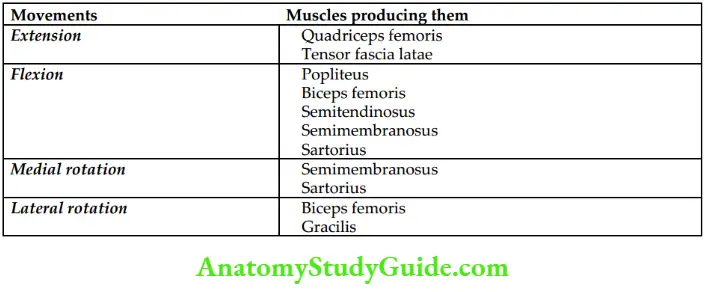
knee joint Movements:
These are given in the box below:
knee joint Applied anatomy:
knee joint Osteoarthritis:
Being a weight-bearing joint, osteoarthritis (degenerative changes in articular cartilages and surfaces) is most common in the knee joint.
knee joint Meniscal tear in young adults:
- The most common cause of meniscal tear in young adults is traumatic injury.
- The meniscal tear occurs when the knee joint is twisted awkwardly (i.e. rotated internally or externally) in a flexed position, with the foot sitting on the ground.
- This commonly occurs in football players. The lateral meniscus often escapes injury by being attached to the popliteal tendon, which moves it away from being caught. Thus medial meniscus is commonly torn.
knee joint An unhappy triad of the knee joint:
- A combination of injuries involving
- tibial collateral ligament,
- medial meniscus, and
- anterior cruciate ligament is termed an unhappy triad of the knee joint.
Question 4. Write a short note on locking and unlocking the knee joint.
Answer:
knee joint:
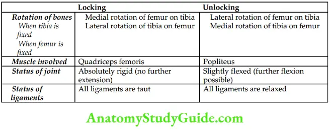
- The full extension of the knee is called the locking of the knee joint. It occurs due to the medial rotation of the femur on the fixed tibia or lateral rotation of the tibia on the fixed femur in the terminal phase of the extension.
- The initial flexion of a locked knee is called the unlocking of the knee joint. It occurs due to lateral rotation of the femur on the tibia. The purpose of locking and unlocking the knee is to provide stable movements at the knee joint.
knee joint Anatomical basis of locking and unlocking:
- Articular surfaces of the tibia and femur are not proportionate and incongruent.
- During the terminal phase of the knee extension, the small articular surface of the tibia is used by the femur. Now to accommodate this unused articular surface of the femur on the tibia, the femur or tibia rotates to have a stable movement at the knee.
knee joint Differences between locking and unlocking of the knee joint:
These are given in the box below:
Question 5. Enumerate bursae around the knee joint.
Answer:
knee joint:
knee joint There are approximately 12 bursae around the knee joint:
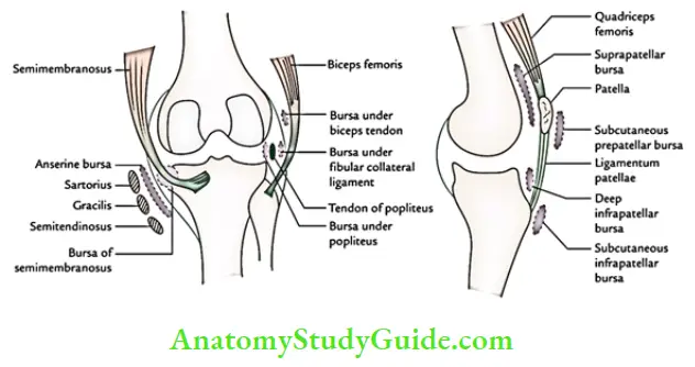
knee joint Anterior bursae:
- Subcutaneous prepatellar bursa
- Suprapatellar bursa
- Infrapatellar bursae:
-
- subcutaneous and
- deep
knee joint Lateral bursae:
- Between the fibular collateral ligament and tendon of the biceps femoris
- Between the fibular collateral ligament and tendon of the popliteus
- Between tendon of popliteus and lateral femoral condyle
- Between tendons of semimembranosus and semitendinosus
knee joint Medial bursae:
- Anserine bursa, between the tendons of sartorius, gracilis, and semitendinosus
- Between semimembranosus and medial tibial condyle
knee joint Posterior bursae:
- Deep to the lateral head of the gastrocnemius
- Deep to the medial head of gastrocnemius (Brodie’s bursa)
knee joint Note:
- Inflammation of the prepatellar subcutaneous bursa leads to Housemaid’s knee,
- inflammation of the subcutaneous infrapatellar bursa leads to Clergyman’s knee, and
- inflammation of the bursa deep to the tendon of the semimembranosus leads to Baker’s cyst.
Question 6. Describe briefly arterial anastomoses around the knee.
Answer:
Briefly arterial anastomoses around the knee:
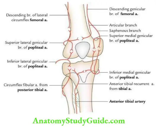
The arterial anastomoses around the knee are formed by genicular branches of the following arteries:
- Femoral artery
- Popliteal artery
- Anterior tibial artery
- Posterior tibial artery
Question 7. Describe the ankle joint under the following headings:
- Classification,
- Ligaments,
- Relations,
- Movements and
- Applied anatomy.
Answer:
Ankle joint:
Ankle Joint Classification:
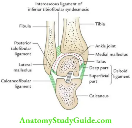
- It is the synovial joint of the modified hinge variety.
- It is formed between the lower ends of the tibia and fibula, and talus.
Ankle joint Ligaments:
Ankle joint Capsular ligament:
It encloses the articular surfaces. It is lax anteriorly to permit uninhibited hinged movements.
Ankle joint Medial collateral (deltoid) ligament:
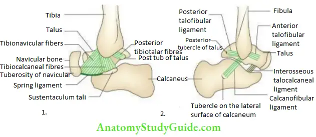
It is a strong triangular ligament and consists of superficial and deep parts. The deep part is a vertical band extending between the medial malleolus and talus. The superficial part is fan–shaped. It is attached above to the medial malleolus and below (from front to back) the tuberosity of the navicular, spring ligament, sustentaculum tali, and posterior tubercle of the talus.
Ankle joint Lateral collateral ligament:
It consists of 3 bands/components: anterior talofibular, posterior talofibular, and calcaneofibular ligament. The anterior talofibular ligament is attached to the neck of the talus, the posterior talofibular ligament to the lateral tubercle of the talus, and the calcaneofibular ligament to the lateral surface of the calcaneus.
Ankle Joint Relations:
Ankle joint Anterior:
Ankle joint From medial to lateral:
- Tendon of tibialis anterior
- Extensor hallucis longus
- Anterior tibial vessels
- Deep peroneal nerve
- Extensor digitorum longus
- Peroneus Tertius
Ankle joint Mnemonic: The Himalayas are never dry places.
Ankle joint Posterior:
Ankle joint (Behind tibial malleolus) From anterior to posterior:
- Tendon of the tibialis posterior
- Flexor digitorum longus
- Posterior tibial artery
- Tibial nerve
- Tendon of flexor hallucis longus.
Ankle joint Mnemonic: The Doctors are not here.
Ankle joint Movements and muscles producing them
Ankle joint These are given in the box below:

Ankle joint Applied anatomy
Ankle joint Ankle sprains:
It occurs due to stretching or tearing of the anterior talofibular (most common) and calcaneofibular ligaments following excessive eversion of the plantar flexed foot. Clinically, it presents pain, swelling, and loss of movement.
Ankle joint Pott’s fracture:
- It includes avulsion of the deltoid ligament (first degree); avulsion of the deltoid ligament and fracture of the medial malleolus (second degree); and avulsion of deltoid ligament, fracture medial malleolus, and fracture lateral malleolus (third degree).
- In severe cases of third-degree Pott’s fracture, there is a fracture of the posterior lip of the tibial facet (also called third malleolus). The Pott’s fracture occurs when the foot is caught in the rabbit hole and everted forcibly.
Question 8. Give a brief account of talocalcaneonavicular, subtalar, and midtarsal joints.
Answer:
Midtarsal joints:
Midtarsal jointsTalocalcaneonavicular joints:
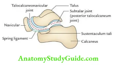
It is a compound synovial joint of ball and socket variety (roughly). It is formed between the head of the talus above (ball) and the calcaneum, navicular, and spring ligament below (socket). It may be divided into two components: posterior and anterior subtalar joints.
Midtarsal joints Subtalar joint:
It is the posterior subtalar joint between the body of the talus and the middle third of the superior surface of the calcaneum.
Midtarsal joints Midtarsal joint:
It consists of calcaneocuboid and talonavicular joints. Both of them are plane-type of synovial joints.
Question 9. Describe inversion and eversion in brief.
Answer:
Brief:
When the foot is off the ground.
- Inversion is the movement in which the medial border of the foot is raised so that the sole faces medially.
- Eversion is the movement in which the lateral border of the foot is raised so that the sole faces laterally.
Brief Characteristic features
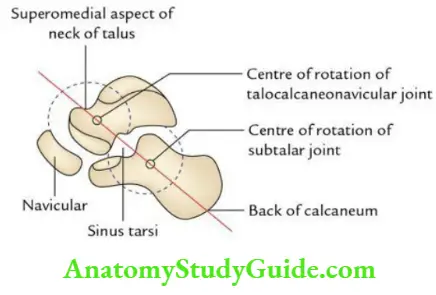
- These movements take place at the talocalcaneonavicular (mainly) and midtarsal joints.
- These movements take place around an oblique axis, which passes forward, upward, and medially from the back of the calcaneum through the sinus tarsi to the superomedial aspect of the neck of the talus.
- The inversion is akin to supination and eversion is akin to pronation of the forearm.
- The range of motion is more in inversion than that in eversion.
- Inversion is produced by the tibialis anterior and tibialis posterior, while eversion is produced by the peroneus longus and peroneus brevis.
Leave a Reply