Kidney Ureter And Suprarenal Glands
Question 1. Describe the kidney under the following headings:
- Kidney Introduction
- Kidney External features
- Kidney Coverings/capsules
- Kidney Relations
- Kidney Arterial supply
- Kidney Venous drainage and
- Kidney Applied anatomy.
Answer:
1. Kidney Introduction:
- The kidneys are paired excretory organs.
- They are located on the posterior abdominal wall one on each side of the vertebral column, behind the peritoneum in the lumbar region.
- Each kidney is about 11 cm long, 6 cm broad, and 3 cm thick.
- Each kidney weighs about 150 gm in males and 135 gm in females.
- Each kidney extends from upper border of T12 to the center of L3 vertebra.
- The right kidney is slightly lower than the left kidney (due to the presence of the liver on the right side).
- The lower poles of the kidneys lie about 2.5 cm above the iliac crest.
2. Kidney External features:
- Two poles:
- The upper pole is broad and in close contact with the suprarenal gland.
- The lower pole is pointed.
- Two surfaces:
- The anterior surface is slightly irregular and partly covered with peritoneum.
- The posterior surface is flat and entirely nonperitoneal.
- Two borders:
- The medial border is concave and shows a depression in its middle called hilum.
- The lateral border is convex.
- One hilum: Three structures enter/leave the hilum. From anterior to posterior, these are:
- Renal vein
- Renal artery
- Renal pelvis
- Accessory renal artery (in 30% of cases)
3. Kidney Relations:
1. Anterior relations: These are given in the box below and shown in
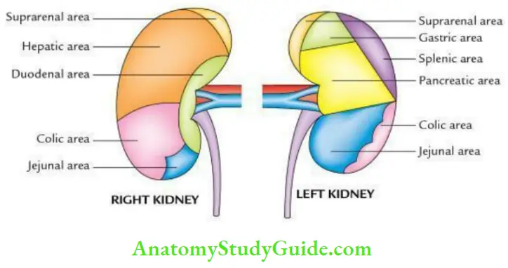

Note: In the right kidney, hepatic and intestinal surfaces are covered by the peritoneum. While in the left kidney gastric splenic and jejunal surfaces are covered by the peritoneum.
2. Posterior relations:
The posterior relations of both kidneys are:
- Four muscles:
- Diaphragm
- Psoas major
- Quadratus lumborum
- Transversus abdominis
- Three nerves:
- Subcostal
- Iliohypogastric
- Ilioinguinal
- One set of vessels: Subcostal vessels
- One or two ribs:
- 12th rib in case of the right kidney
- 11th and 12th ribs in case of left kidney
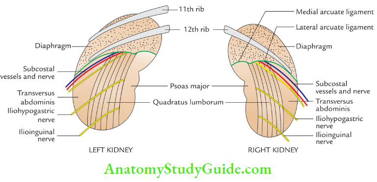
4. Kidney Capsules/covering: There are 4 coverings of the kidney. From within outward these are:
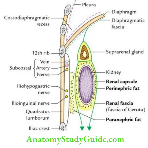
Fibrous capsule (true capsule):
- It is a thin membrane, which closely invests the kidney and lines the renal sinus.
- Normally, it can be easily stripped off from the kidney, but in certain diseases, it becomes adherent to the kidney and cannot be stripped off.
Perirenal/perinephric fat:
- It is a layer of adipose tissue lying outside the fibrous capsule.
- It is thickest at the borders and fills up the extra space in the renal sinus.
Renal fascia/fascia of zero (false capsule):
- It has anterior and posterior layers.
- The anterior layer is called the fascia of Toldt, and the posterior layer is called the fascia of Zuckerkandl.
Read And Learn More: Anatomy Question And Answers
Pararenal/para nephric fat:
It is a variable amount of fat lying outside the renal fascia especially posteriorly. It fills up the paravertebral gutter and acts as a cushion for the kidney.
The fate of renal fascia superiorly:
The two layers first enclose the suprarenal gland in a separate compartment and then fuse with each other to continue with the diaphragmatic fascia.
- Inferiorly, anterior and posterior layers remain separate and enclose the ureter.
- Then, the anterior layer is lost in the extraperitoneal tissues of the iliac fossa, while the posterior layer blends with the fascia iliaca.
- Laterally, two layers merge with fascia transversalis.
- Medially, the anterior layer passes in front of renal vessels and merges with the connective tissue surrounding the aorta and inferior vena cava, while the posterior layer covers the fascia covering the quadratus lumborum and psoas major.
5. Kidney Arterial supply:
- Each kidney is supplied with a renal artery, which arises from the abdominal aorta at the level of the intervertebral disc between the L1 and L2 vertebrae.
- Each artery divides into anterior and posterior branches near the hilum which in turn divides into segmental arteries to supply renal segments viz. apical, upper, lower, middle, and posterior.
6. Kidney Venous drainage:
- Each kidney is drained by the renal vein, which opens into the inferior vena cava.
- The left renal vein is longer than the right renal vein and lies superficial to the abdominal aorta.
7. Kidney Applied anatomy:
Renal angle:
- It is an angle between the lower border of the 12th rib and outer border
of the erector spinal muscle. The renal pain is usually felt here as a dull ache. - The pressure applied here may elicit tenderness in the kidney lesion (Murphy’s punch sign).
- The oblique incision in kidney exposure commences here. The danger of opening the pleural cavity should be kept in mind while exposing the kidney from behind.
- \When the 12th rib is too short to be felt or absent. The 11th rib is mistaken for the 12th and the chances of opening the pleural cavity are increased immensely.
Bimanual palpation:
- Kidney can be palpated bimanually with one hand placed in front of the anterior abdominal wall and the other hand behind the flank.
- When enlarged, the lower pole of the kidney can be palpated on deep inspiration.
Floating kidney/nephroptosis:
- Normally, the kidney more or less remains in place because it lies in the abundant fat within the renal fascia.
- But in chronic debilitating disease, this fat disappears and the kidney becomes hypermobile and can move up and down within the renal fascia, but not from side to side.
- This may cause kinking of ureter and urinary obstruction.
Question 2. Briefly describe thoracolumbar fascia.
Answer:
Thoracolumbar fascia:
- It is diamond shaped deep fascia on the back of the trunk which is specially thickened in the lumbar region.
- It encloses paraspinal muscles, viz. erector spinae on the posterolateral aspects of the vertebral column.
In the lumbar region, it divides into 3 layers, viz. posterior, middle, and anterior:
- Posterior layer: Posterior layer is attached to the tips of the spines of the lumbar vertebrae.
- Middle layer: Middle layer is attached to the tips of transverse processes of lumbar vertebrae.
- Anterior layer: Anterior layer is attached to a ridge on the anterior aspect of the transverse processes of lumbar vertebrae.
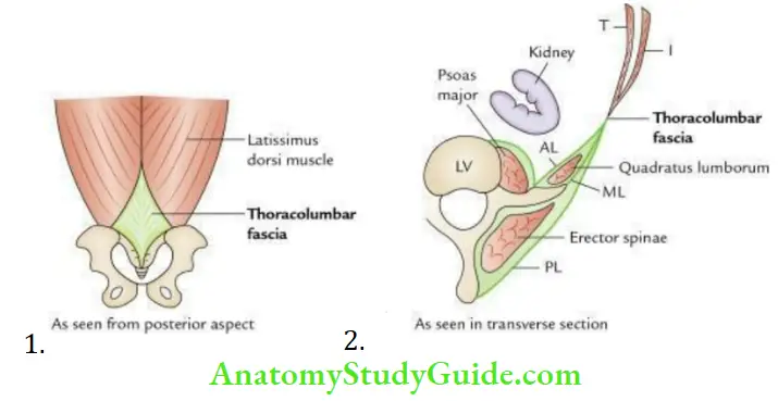
Thoracolumbar fascia Functions:
- Provides stability to the vertebral column, especially in the lumbar region.
- Provides attachments to large muscles namely, latissimus dorsi, lateral muscles of the abdomen, gluteus maximus, etc., and allows them to act on the vertebral column.
Thoracolumbar fascia Applied anatomy:
- Excessive strain of thoracolumbar fascia leads to low back pain.
- Knowledge of its layers and muscles enclosed between them helps the surgeon in exposing the kidney from behind.
Question 3. Discuss the histological features of the kidney.
Answer:
The histological section through the kidney presents an outer cortex and an inner medulla.
Kidney Cortex:
The cortex presents the following features:
- Presence of darkly, stained, small, dense, rounded structures arranged in parallel rows at a right angle to the capsule of the kidney. These are renal corpuscles/glomeruli.
- Pale stained lines run vertically towards the medulla, the medullary rays.
- Presence of many sections of proximal convoluted tubules and some sections of distal convoluted tubules.
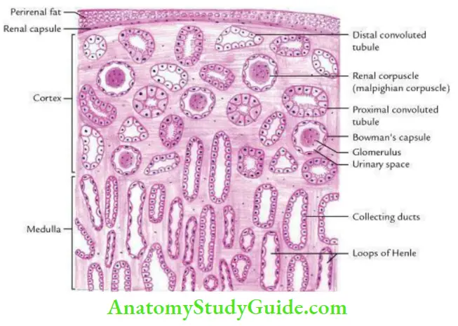
The differences between the sections of proximal and distal convoluted tubules are given in Table.
Differences Between the Sections of Proximal and Distal Convoluted Tubules:

Kidney Medulla:
The medulla presents the following features:
- Numerous light-staining tubular structures run vertically, the collecting ducts.
- Sections of the loop of Henle.
- Capillaries and connective tissues.
Question 4. Describe the development of the kidney and associated common congenital anomalies in brief.
Answer:
Kidney Development:
- Kidney develops from intermediate mesoderm in the pelvis during the 5th week of IUL.
- It ascends in the lumbar region and becomes functional in the 9th week of IUL.
Kidney Sources:
- The secretory part develops from metanephric blastema.
- The collecting part develops from the ureteric bud, which arises from the mesonephric duct.

The components of the secretory and collecting parts of the kidney are given in the box below:

Congenital anomalies:
1. Renal agenesis (absence of kidney): It is unilateral and occurs in 1/2400 births.
2. Horseshoe kidney: In this type usually inferior poles of two kidneys are fused to each other across the midline by an isthmus to form a U shape, like a horseshoe nail. The urethra of both kidneys passes in front of connecting isthmus.
3. Polycystic kidney: In this type, numerous cysts filled with urine are present within the kidney.
It occurs due to:
- Failure of connection between excretory and collecting components (old view).
- Abnormal dilatation of different parts of uriniferous tubules especially loops of the Henle (recent view).
- For details see Textbook of Clinical Embryology by Vishram Singh.
Question 5. Describe the Ureter under the following headings:
Answer:
- Ureter Introduction
- Ureter Course
- Ureter Constrictions
- Ureter Arterial supply
- Ureter Nerve supply
- Ureter Development and
- Ureter Applied anatomy.
Answer:
1. Ureter Introduction:
The ureter is a narrow (3 mm in diameter) thick-walled muscular tube that conveys urine from the kidney to the urinary bladder. It is 25 cm (10”) long, of which 5” lies in the abdomen and 5” in the pelvis.
2. Ureter Course:
- It begins within the renal sinus as a funnel-shaped dilatation termed renal pelvis. The renal pelvis gradually narrows and descends along the medial margin of the kidney.
- At the lower pole of the kidney, it becomes the ureter proper.
- Ureter proper passes downward and lies on tips of transverse processes of lumbar vertebrae and psoas major.
- It crosses the pelvic brim (in front of the termination of a common iliac artery) to enter true pelvis.
- In the pelvis at first, it passes in front of the ischial spine and then runs forward and medially to enter urinary bladder.

3. Ureter Constrictions:
The ureter presents 5 normal anatomical constrictions at the following sites:
- Pelviureteric junction
- Pelvic brim
- The juxtaposition of vas deferens or broad ligament
- Uretero-vesical junction (i.e., the point where it pierces the wall of the urinary bladder)
- Ureteric orifice, i.e., opening into the urinary bladder
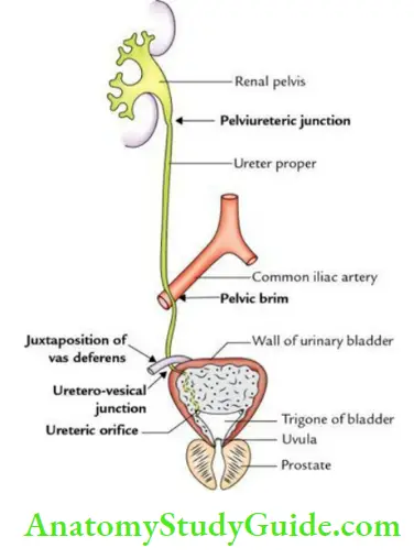
Note: Most of the anatomy textbooks describe only 3 constrictions (pelvic ureteric junction, pelvic brim, and ureterovesical junction).
4. Ureter Arterial supply:
The ureter is supplied by the following three sets of arteries:
- The upper part is by the branches from the renal artery.
- The middle part, by the branches from the abdominal aorta.
- Pelvic part, by the branches from vesical (superior and inferior) middle rectal/uterine arteries.
- Nerve supply
By the sympathetic (T10–L1) and parasympathetic (S2, S3, and S4) fibers through renal, aortic, and hypogastric plexuses.
5. Ureter Development: The ureter develops during the 5th to 9th weeks of IUL from the ureteric bud.
6. Ureter Applied Anatomy:
- Ureteric colic (also called renal colic):
- It is a severe spasmodic pain arising from the ureter when the ureteric stone is lodged in its lumen. Pain typically starts in the loin and radiates down to the groin (scrotum/labium majora) and the inner side of the upper part of the thigh.
- Note pain is referred to the cutaneous areas supplied by T10 to L1 spinal segments, which also supply the ureter.
- Impaction of ureteric stone:
- Ureteric stone is likely to be impacted at one of the sites of normal anatomical constrictions.
- Ureteric stones lodged in the lower part of the ureter in females can be palpated per vaginum because of the close relationship of the ureter to the lateral fornix of the vagina.
Question 6. Give histological features of the ureter
Answer:
The histological section of the ureter presents a star-shaped lumen and a thick wall. The thick wall consists of 3 coats.
From outward, these are the Mucous coat, Muscular coat, and Fibrous coat.

Mucous coat:
- The mucous membrane is lined by transitional epithelium.
- Lamina propria is wide and made up of fibroblastic tissue, which is thrown into 5–6 longitudinal folds giving the lumen a star-shaped appearance.
Muscular coat:
It is made up of smooth muscle fibers.
- In upper 2/3rd, it consists of 2 layers:
- The outer layer of circular muscle fibers
- The inner layer of longitudinal muscle fibers
- In lower 1/3rd, it consists of 3 layers:
- The outer layer of longitudinal muscle fibers
- An intermediate layer of circular muscle fibers
- The inner layer of longitudinal muscle fibers
Adventitia (fibrous coat): It is made up of loose connective tissue containing many elastic fibers, blood vessels, lymphatics, and nerves.
Note: There is no submucous coat in the ureter.
Question 7. Describe the suprarenal gland in brief
- External features
- Arterial supply and
- Venous drainage.
Answer:
1. Suprarenal gland External features:
- The suprarenal glands are a pair of endocrine glands situated over the upper pole of the kidneys.
- It consists of an outer cortex and an inner medulla. The cortex secretes a number of steroid hormones and the medulla secretes adrenalin and noradrenalin.
- Each gland weighs about 5 gm.
- The right gland measures about 4 × 4 × 1 cm.
- The left gland measures about 5 × 3 × 1 cm.
- The right gland is triangular/pyramidal in shape, while the left gland is semilunar in shape.
2. Suprarenal gland Arterial supply
- Each suprarenal gland is supplied by 3 arteries:
- Superior suprarenal artery, a branch of the inferior phrenic artery.
- Middle suprarenal artery, a direct branch from the abdominal aorta.
- The inferior suprarenal artery, is a branch of the renal artery.

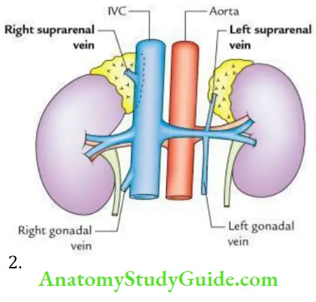
3. Suprarenal gland Venous drainage:
- Each gland is drained by one vein.
- The vein from the right gland drains into IVC, while the vein from the left gland drains into the left renal vein.
Question 8. Give differences between the right and left suprarenal glands.
Answer:
The differences between the right and left suprarenal glands are given in the box below:

Question 9. Give histological features of the suprarenal/adrenal gland.
Answer:
The histological section of the suprarenal gland presents the cortex (outer 2/3rd) and medulla
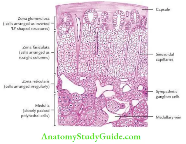
Suprarenal/adrenal gland Cortex:
The cortex from superficial to deep consists of 3 zones.
- Zona glomerulosa: Zona glomerulosa forms 15% of the thickness. It is made up of inverted U-shaped arches or acini of cells with intervening sinusoids. The cells are columnar with dark-staining spherical nuclei.
- Zona fasciculata: Zona fasciculata forms 75% of the thickness. It consists of vertical columns, usually 2– 3 cell thick which are separated by sinusoids. The cells are cuboidal or polygonal, giving a foamy or spongy appearance hence also called spongiocytes.
- Zona reticularis: Zona reticularis forms 10% of the thickness. It consists of an irregular network of branching cords and clumps of cells separated by wide sinusoids. The cells are much smaller than the cells in zona fasciculata.
Suprarenal/adrenal gland Medulla:
- The medulla consists of closely packed clumps of polyhedral chromaffin cells separated by wide sinusoids.
- The prominent medullary vein is characteristically located in the center of the medulla.
Note:
- Zona glomerulosa secretes mineralocorticoids, zona fasciculata secretes glucocorticoids, and zona reticularis secretes androgen hormones.
- Medulla secretes epinephrine (adrenalin) and norepinephrine (noradrenaline).
Leave a Reply