Lymphatic System
Lymphatic system is a dosed system of lymph channels or lymph vessels through which lymph flows. It is a oneway system and allows the lymph to flow from tissue spaces towards the blood.
Table of Contents
Organization Of Lymphatic System
- The lymphatic system arises from tissue spaces as a meshwork of delicate vessels. These vessels are called lymph capillaries.
- The lymph capillaries start from tissue spaces as enlarged blind ended terminals called capillary bulbs.
- These bulbs contain valves, which allow flow of lymph in only one direction. There are some muscle fibers around these bulbs. These muscle fibers cause contraction of bulbs so that, lymph is pushed through the vessels.
Read And Learn More: Medical Physiology Notes
- The lymph capillaries are lined by endothelial cells. The capillaries unite to form large lymphatic vessels.
- The lymphatic vessels become larger and larger because of the joining of many tributaries along their course.
- The structure of lymph capillaries is slightly different from that of blood capillaries.
- The lymph capillaries are more porous and the cells lie overlapping on one another. It allows the fluid to move into the lymph capillaries and not in the opposite direction.
Drainage Of Lymphatic System: The larger lymph vessels ultimately form right lymphatic duct and thoracic duct. The right lymphatic duct opens into right subclavian vein and the thoracic duct opens into left subclavian vein. Thoracic duct drains the lymph from more than 2/3rd of the tissue spaces in the body.
The situation of lymph vessels: Lymph vessels are situated in the following regions
- Deeper Sayers of skin
- Subcutaneous tissues
- Diaphragm
- Wall of abdominal cavity
- Omentum
- Linings of respiratory tract except alveoli
- Linings of the digestive tract
- Linings of urinary tract
- Linings of genital tract
- Liver
- Heart.
The lymph vessels are not present in the following structures:
- Superficial layers of skin
- Central nervous system
- Cornea
- Bones
- Alveoli of lungs.
Lymph Nodes: Lymph nodes are small glandular structures located in the course of lymph vessels. The lymph nodes are also called lymph glands or lymphatic nodes.
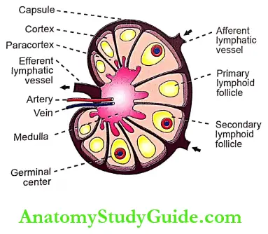
- Structure of Lymph Node: Each lymph node constitutes masses of lymphatic tissue covered by a dense connective tissue capsule. The structures are arranged in three layers cortex, paracortex, and medulla.
- Cortex:
- Cortex of lymph node consists of primary and secondary lymphoid follicles. The primary follicle develops first. When some antigens enter the body and reach the lymph nodes, the cells of primary follicle proliferate.
- The active proliferation of the cells occurs in a particular area of the follicle called the germinal center.
- After the proliferation of cells, the primary follicles become the secondary follicle.
- Cortex also contains some B lymphocytes, which are usually aggregated into the primary follicles. Macrophages are also found in the cortex.
- Paracortex: It is in between cortex and medulla. Paracortex contains T lymphocytes.
- Medulla: Medulla contains B and T lymphocytes and macrophages. Blood vessels of lymph node pass through medulla.
- Cortex:
- Lymphatic Vessels to Lymph Node
- The lymph node receives lymph by one or two lymphatic vessels called afferent vessels. Afferent vessels divide into small channels.
- Lymph passes through afferent vessels and small channels and reaches the cortex. It circulates through the cortex, paracortex, and medulla of the lymph node. From the medulla, the lymph leaves the node via one or two efferent vessels.
- Distribution of Lymph Nodes: The lymph nodes are present along the course of lymphatic vessels in the elbow, axilla, knee, and groin. The lymph nodes are also present in certain points in the abdomen, thorax, and neck where many lymph vessels join.

Formation Of Lymph
- Lymph is formed from interstitial fluid, due to the permeability of lymph capillaries. When blood passes via blood capillaries in the tissues, 9/10th of fluid passes into venous end of capillaries from arterial end. And, the remaining 1/1 Oth ot the fluid passes into lymph capillaries, which have more permeability than blood capillaries.
- So, when lymph passes through lymph capillaries the composition of lymph is more or less similar to that of interstitial fluid including protein content.
- Proteins present in the interstitial fluid cannot enter the blood capillaries because of their larger size. So, these proteins enter lymph vessels, which are permeable to large particles also.
Addition Of Proteins And Fats: The tissue fluid in liver and gastrointestinal tract contains more protein and lipid substances. So, proteins and lipids enter the lymph vessels of liver and gastrointestinal tract in large quantities. Thus, lymph in larger vessels has more proteins and lipids.
Concentration Of Lymph: When the lymph passes through the lymph nodes, the lymph is concentrated because of the following reasons:
- Water and electrolytes are absorbed from lymph into the lymph node
- Proteins and lipids are not absorbed
- Bacteria are phagocytosed by the macrophages of the lymph node.
Rate Of Lymph Flow: About 120 ml of lymph flows into blood per hour. Out of this, about 100 ml/hour flows through the thoracic duct and 20 ml/ hour flows through the right lymphatic duct.
- Factors Increasing the Flow of Lymph: The flow of lymph is promoted by the increase in
- Interstitial fluid pressure
- Blood capillary pressure
- Surface area of lymph capillary by means of dilatation
- Permeability of lymph capillaries
- Functional activities of tissues.
Composition Of Lymph
Usually, lymph is a clear and colorless fluid. It is formed by 96% water and 4% solids. Some blood cells are also present in lymph.
Functions Of Lymph
- The important function of lymph is to return the proteins from tissue spaces into blood
- Lymph flow plays an important role in the redistribution of fluid in the body
- Through the lymph, the bacteria, toxins, and other foreign bodies are removed from tissues
- Lymph flow is responsible for the maintenance of the structural and functional integrity of the tissue. Obstruction to lymph flow affects various tissues, particularly the myocardium, nephrons, and hepatic cells.
- Lymph flow serves as an important route for intestinal absorption. This is the reason for the milky appearance of lymph after a fatty meal
- It plays an important role in immunity by the transport of lymphocytes.

Functions Of Lymph Nodes
- Lymph nodes serve as filters that filter bacteria and toxic substances from the lymph. The functions of the lymph nodes are:
- When lymph passes through the lymph nodes, it is filtered, i.e. the water and electrolytes are removed. But, the proteins and lipids are retained in the lymph
- The bacteria and other toxic substances are destroyed by macrophages of lymph nodes. Because of this, lymph nodes are called defense barriers.
Applied Physiology Swelling Of Lymph Nodes
- During infection or any other processes in a particular region of the body, the activities of the lymph nodes in that region increase. This causes swelling of the lymph nodes. Sometimes, the swollen lymph nodes cause pain.
- The most common cause of swollen lymph nodes is infection. The lymph nodes situated near an infected area, swell immediately. When the body recovers from infection, the lymph nodes restore their original size gradually in one or two weeks.
- Causes for swelling of lymph nodes in different regions of the body are:
- Skin infection of arm causes swelling of lymph nodes in the armpit
- Tonsillitis or throat infection causes swelling of lymph nodes in neck
- Infection of genital organs or leg results in swelling of lymph nodes in groin
- Viral infections such as glandular fever which affect the whole body cause swelling of lymph nodes in various parts of the body
- Cancer in a particular region may spread into the nearby lymph nodes causing the swelling.
Examples:
- Throat cancer may spread into lymph nodes in neck
- Lung cancer may spread into lymph nodes in chest
- Breast cancer may spread into lymph nodes in armpit
- Intestinal cancer may spread into lymph nodes in
abdomen - Lymphomas (cancer of the lymphatic system) and antigen cause swelling of lymph nodes in many
Leave a Reply