Making Primary Impression of Maxillary and Mandibular Edentulous Foundations Introduction
Correctly made primary impression is base for fabrication of good quality, well-fitting complete denture. Primary impression should be made with great care as it is important to fabricate custom trays which are needed for making the final impression. Various materials can be used for making a primary impression depending upon the condition of the denture foundation area. In the present chapter, we will learn to make impressions of edentulous maxillary and mandibular arches with impression compound and alginate impression material.
Table of Contents
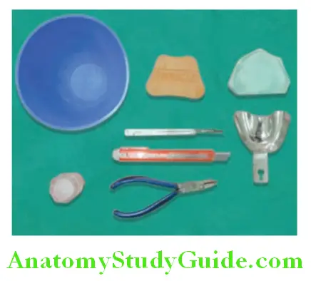
Instruments And Materials
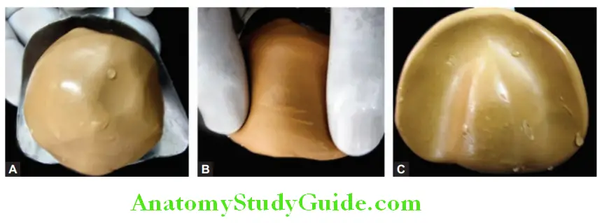
- Maxillary edentulous cast
- Large clean rubber bowl
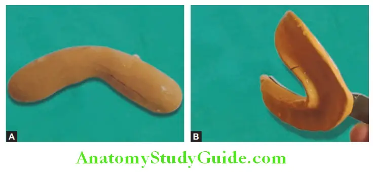
- Maxillary and mandibular edentulous nonperforated stock metal trays
Read and Learn More: Preclinical Prosthodontics Notes
- BP blade and handle
- Pliers
- Vaseline
- Impression compound.
Making Primary Impression
Step 1:
Take edentulous casts of maxillary and mandibular arch. Select proper edentulous
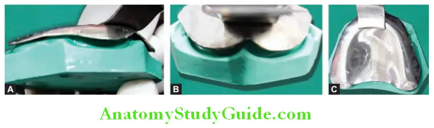
nonperforated maxillary and mandibular trays such that they cover denture bearing area and limiting area. Ensure that there is space of 5–6 mm between the tray and cast to load impression material. Posteriorly it should cover the hamular notch and posterior palatal seal area. The mandibular tray should cover the retromolar pads posteriorly. Sometimes you may need to modify tray flanges with pliers.
Step 2:
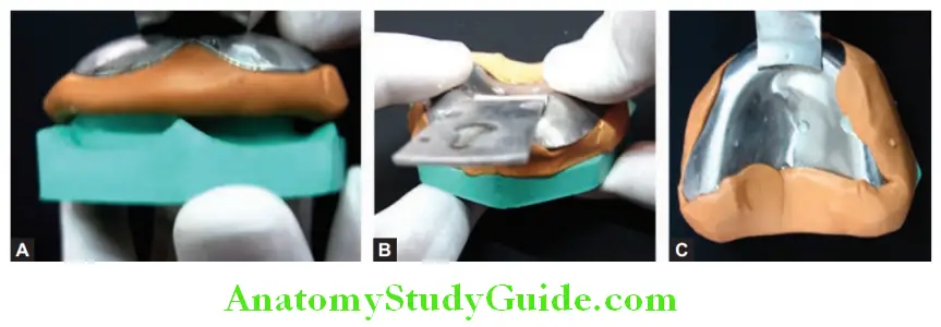
Lightly grease the model/cast with vaseline to prevent adherence of impression material with it or wet the cast in slurry water for a few minutes.
Step 3:
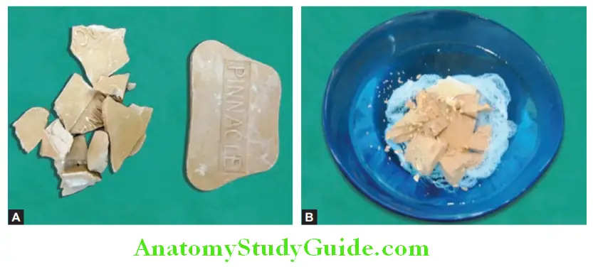
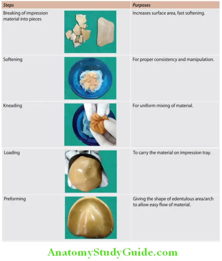
Take water at a temperature of 55–60°C in a clean rubber bowl. Break the impression compound into pieces place it into hot water in a bowl and allow it to soften.
Note: Put a cloth or small handkerchief at the bottom of the rubber bowl containing hot water. This will prevent the adhesion of the impression compound with a rubber bowl.
Step 4:
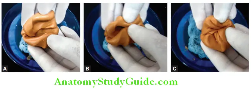
After softening it should be kneaded for uniform mixing.
Step 5:
The kneaded softened compound is loaded on the impression tray in the form of a pellet and roughly shaped like an arch. For mandibular arch impression, the impression compound is rolled in a rope shape and loaded on the tray for performing.
Step 6:
Soften the impression material surface by Glazing and tempering by dipping it in hot water. (Avoid over-glazing, it may traumatize the mucosa in the oral cavity).
Step 7:
Now, the loaded tray is placed over the cast soaked in slurry water, taking the anterior notch as a reference with the labial frenum on the cast. Apply proper pressure to flow the impression compound and record the impression.
Note: Fold excess material on the back side of the impression tray, this will prevent dislodgement of the impression from the tray.
Step 8:
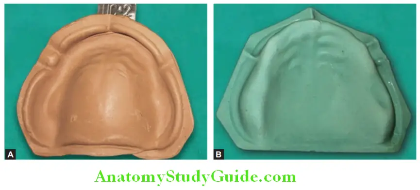
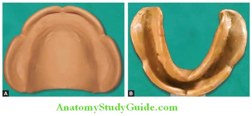
The impression compound is allowed to set (harden). The tray with impression compound is removed from the cast and the impression for its perfection. Inspect it for gross defects, wrinkles, non-recorded areas, etc. Repeat the impression if needed.
Step 9:
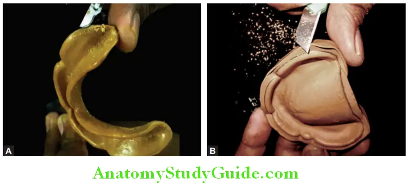
Mark the periphery of the impression with a pencil and remove excess material with a BP blade or paper cutter. Maxillary and mandibular edentulous impressions.
Note: Do not try to remove excess material in bulk, slowly remove it otherwise it may break the impression. Peeling it in a small area and B is the correct method.
Steps In Manipulation Of Impression Compound/Impression Material
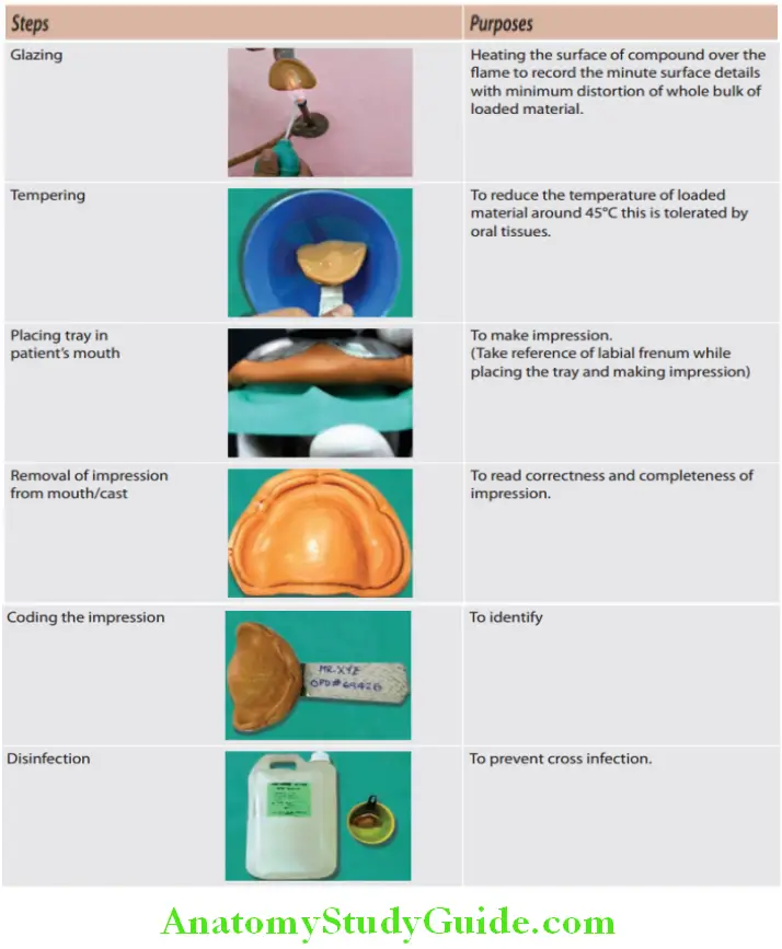
Leave a Reply