Mediastinum (partition)
Mediastinum Introduction: Thoracic mediastinum is the space between right and left pleural sacs and limited on each side by mediastinal pleura.
1. Mediastinum Extent: It extends vertically from thoracic inlet to diaphragm.
2. Mediastinum Division: The mediastinum is divided by an imaginary horizontal plane extending from sternal angle to lower border of 4th thoracic vertebra.
Read And Learn More: Anatomy Important Question And Answers
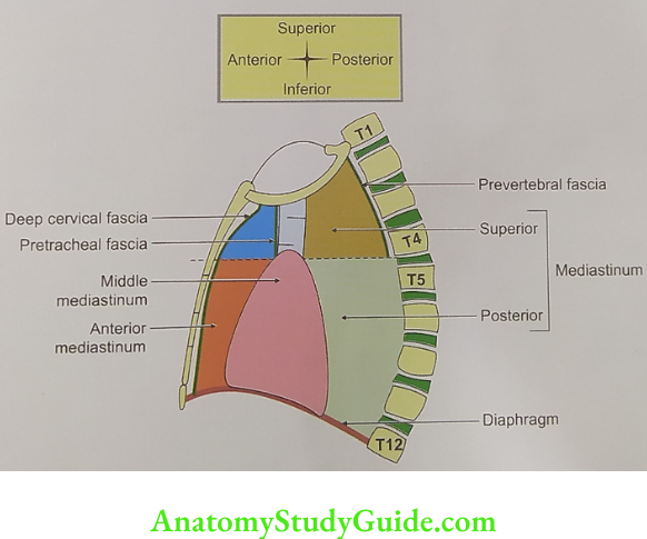
- Superior mediastinum
- Inferior mediastinum: It is subdivided by pericardium and heart into
- Anterior mediastinum,
- Above: Thoracic inlet
- Below: Horizontal plane extending from sternal angle to lower border of 4th thoracic vertebra.
- On each side: Mediastinal pleura.
Mediastinum Contents
Retrosternal structures
- Superior vena cava
- Veins opening into superior vena cava
- Right and left brachiocephalic vein, and
- Left superior intercostal vein.
- Thymus gland
- Muscles: Sternothyroid, sternohyoid.
Mediastinum Anatomy
Table of Contents
Intermediate structures (aorta)
Arch of aorta and its branches from right to left.
- Brachiocephalic trunk,
- Left common carotid artery, and
- Left subclavian artery.
Nerve
- Phrenic nerve,
- Vagus nerve, and
- Cardiac plexus.
Prevertebral structures (trachea and oesophagus) Note: The direction of arrow indicates the direction of the structures.
- Trachea,↓
- Oesophagus,↓
- Left recurrent laryngeal nerve,
- Thoracic duct, ↑
- Muscle-longus colli, and ↑
- Paratracheal and tracheobronchial lymph node. →
Mediastinum Applied anatomy
- Abscess (caries of cervical vertebra) or bleeding behind prevertebral fascia enters superior mediastinum.
- Obstruction to superior vena cava gives rise to engorgement of veins in the upper half of the body.
- Pressure over trachea causes dyspnoea, and cough.
- Pressure on the oesophagus causes dysphagia.
- Pressure on the left recurrent laryngeal nerve gives rise to hoarseness of voice.
- Pressure over sympathetic chain causes Horner’s syndrome.
- Horner’s syndrome: It is due to involvement of the sympathetic nerve, which is contributed by T1 segment of the spinal cord. There is injury to the root of brachial plexus.
Mediastinum Anatomy
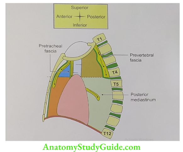
(1) Anterior to pretracheal fascia leads to anterior mediastinum;
(2) Posterior to pretracheal fascia and anterior to prevertebral fascia leads to posterior mediastinum;
(3) Posterior to prevertebral fascia leads to superior mediastinum
Anterior Mediastinum
Anterior Mediastinum Introduction: It is a potential space present in the anterior part of inferior mediastinum.
1. Anterior Mediastinum Boundaries
- Superiorly: Imaginary line extending from sternal angle to lower border of 4th thoracic vertebra.
- Inferiorly: The diaphragm
- Anteriorly: Posterior surface of body of sternum.
- Posteriorly: Fibrous pericardium.
2. Anterior Mediastinum Contents
- Thymus gland is the principal content of the anterior mediastinum.
- Mediastinal artery, a branch of internal thoracic artery.
- Ligaments: Superior and inferior sternopericardial
- Retrosternal lymph node
- Loose areolar tissue.
Mediastinum Anatomy
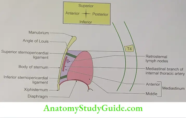
3. Anterior Mediastinum Applied anatomy: Abscess or bleeding or growth in front of the pretracheal fascia of the superior mediastinum enters the anterior mediastinum.
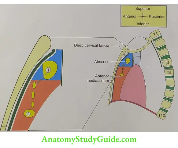
(1) Superior mediastinum anterior to pretracheal fascia;
(2) Anterior mediastinum
Mediastinum Anatomy
Middle Mediastinum Pericardium
Middle Mediastinum Pericardium Introduction: It is the widest of all the mediastina and is occupied by the heart and the
1. Middle Mediastinum Pericardium Boundaries
- Superiorly: Horizontal plane from sternal angle to lower border of 4th thoracic vertebra.
- Inferiorly: By the diaphragm.
- Anterior: By fibrous pericardium
- Posteriorly: By fibrous pericardium
2. Middle Mediastinum Pericardium Contents
Heart and the related structures
- Pericardium,
- Deep cardiac plexus,
- Structures entering pericardium,
- Four pulmonary veins, and
- Superior vena cava
- Structures leaving the pericardium
- Ascending aorta, and
- Pulmonary trunk.
Trachea and related structures
- Right bronchus,
- Left bronchus, and
- Inferior tracheobronchial lymph node.
Other structures
- Phrenic nerve, and
- Pericardiacophrenic vessels.
Mediastinum Anatomy
Posterior Mediastinum (longest)
Posterior Mediastinum Introduction: It is the longest part of the inferior mediastinum.
1. Posterior Mediastinum Boundaries
- Superiorly: By the plane extending from sternal angle to lower border of 4th vertebra.
- Inferiorly:The diaphragm.
- Anteriorly: From above downward
- Bifurcation of trachea,
- Pulmonary vessels,
- Fibrous pericardium, and
- Posterior surface of the diaphragm.
Posteriorly
- Bodies of lower 8 thoracic vertebrae,
- Intervertebral discs, and
- Anterior longitudinal ligament.
On each side: Mediastinal pleura.
2. Contents Longitudinal structures
Descending thoracic aorta DATES VP
Azygos vein,
Thoracic duct,
Esophagus, and
Splanchnic nerve.
Vagus nerve,
Posterior mediastinal lymph nodes

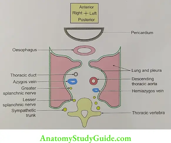
Transverse structures
- Superior and inferior hemiazygos veins,
- Posterior intercostal vein, and
- Posterior intercostal artery.
3. Posterior Mediastinum Applied anatomy: Abscess or bleeding between prevertebral and pretracheal fascia enters superior mediastinum and enters the posterior mediastinum.
Mediastinum Anatomy
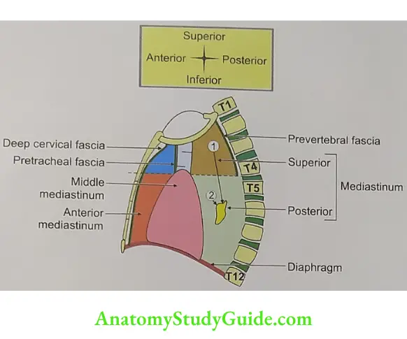
(1) The superior mediastinum posterior to pretracheal fascia and anterior to prevertebral fascia;
(2) Posterior mediastinum
Leave a Reply