Mixed Dentition Space Analysis Introduction
Adequacy of space is the first criterion for a well-aligned dental arch.
Table of Contents
Early correction of the space discrepancies prevents unfavorable events such as crowding of teeth.
The difference between the amount of space available in the arch perimeter and the amount of space required to accommodate erupting permanent teeth has to be measured to identify these space discrepancies.
Read And Learn More: Paediatric Dentistry Notes
This is done by space analysis. Space analysis constitutes an assessment of the arch length, arch perimeter, arch width, and tooth material.
It is an ideal tool that measures the space available for erupting teeth and the space required for their normal positioning in the dental arch.
Mixed Dentition Analysis
It is used to predict, anticipate and prevent malocclusion or reduce its severity.
The space analysis can be done during the mixed dentition period and permanent dentition period.
Mixed dentition space analysis is significant for endodontic practice as it involves primary teeth and newly erupted young permanent teeth.
Mixed dentition space analysis is done when the permanent lower incisors and fist molars are present in the arch.
This happens by around 7–8 years of age and marks the end of the first transitional phase.
The space available for the erupting canines and premolars can be calculated at this stage.
The main aim of mixed dentition space analysis is to predict the availability of required space for these unerupted canines and premolars at the earliest opportunity.
It is indicated to assess the space discrepancy in the arch and initiate appropriate preventive or interceptive orthodontic procedures for prevention or early correction of anticipated malocclusions.
The main parameter required to perform the analysis is the calculation of space required and space available.
Dentition Space Requirement And Availability
The space requirement or availability can be predicted as early as the first transition phase as mentioned earlier.
If not, it should be measured before the commencement of the third transition phase, which happens by around 11–12 years of age when the crucial exchange of posterior teeth and their settlement in occlusion take place.
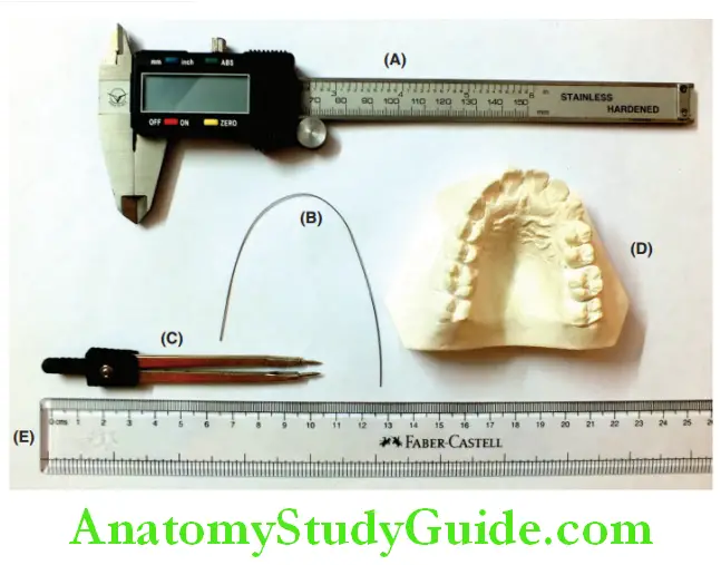
Measurement Of Space Available:
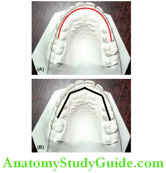
Space available is the distance along the arch perimeter from the mesiobuccal of the first permanent molar on one side to the mesiobuccal of the first permanent molar on the other side.
Mixed Dentition Analysis
This line passes along the buccal cusps and edges of incisors. The armamentarium for measuring space is available.
The space availability can be measured by the following methods:
1. Using a brass wire: A brass wire is contoured along the arc of the arch on a dental cast.
Markings are made on the wire at the mesial surface of the first permanent molars on either side.
The wire is straightened and the length between the two markings is measured to give the value of space available.
2. Using dividers: A fie-line divider can be used to measure space available. The dental arch is divided into four straight segments.
Space Analysis In Mixed Dentition
The segments extend on both sides of the arch from the distal surface of the second primary molar to the mesial surface of the canine and from the mesial surface of the canine to the midline of the arch.
Each straight segment is measured and summed up.
3. Using computer algorithm: Space available can be calculated on the computer by digitizing the dental cast.
A line is drawn along the curvature of the dental arch between the mesial surfaces of both sides’ first permanent molars.
The length of this line is measured as space available. The line is measured from the designated endpoint on the distal aspect of the second primary molars on both sides.
Mixed Dentition Analysis
The space available for posterior teeth during the third transition phase does not increase further.
It rather decreases due to the migration of the first permanent molars into leeway space.
Measurement Of Space Required:
Space required is the sum of mesiodistal widths of all the permanent teeth in the arch mesial to the first permanent molars.
They are the premolars, canines, and incisors of both sides. The width of the incisors is measured directly from the dental cast.
The width of canines and premolars can be measured from the cast if they have erupted.
It has to be predicted if the space required is measured before their eruption, as the premolars and canines would not have erupted during this first transition period.
The width of these teeth can be predicted with the help of prediction analysis, discussed in the following part of this chapter.
Space required = Width of four incisors + Predicted width canines and premolars.
If the measure of space available is adequate for the space required, the permanent teeth are likely to be well aligned in the arch.
If the space available is, they are likely to be spaced.
If the space required is more than that available, the clinician anticipates various malocclusions depending upon the severity of the shortage of space.
Mixed Dentition Analysis
Space deficiency can result in malaligned permanent teeth, proclamation, crowding, or even impaction of teeth.
Dentition Space Prediction Analysis
Any space analysis or mixed dentition analysis can be described as a prediction analysis when it involves an element of prediction.
Prediction charts or derivations are used to perform the analysis instead of physical measurements.
The prediction of the width of unerupted canines and premolars is of utmost importance in endodontic practice, as this parameter is required for the calculation of space requirement.
The permanent lower incisors and the permanent first molars are the early arrivers in the dental arch.
They erupt by 6–8 years of age. The canines and premolars erupt by 10–12 years, being the late arrivers.
Adequate space is essential for the eruption of these teeth in the arch and their settlement in occlusion.
If the width of the unerupted canines and premolars is predicted, the availability or requirement of space for the teeth yet to erupt can be decided.
Space Analysis In Mixed Dentition
This prediction enables the clinician to be aware of potential malocclusions.
By periodic observation and analysis, the clinician can indicate appropriate preventive and interceptive orthodontic procedures respectively.
The mesiodistal widths of the unerupted canines and premolars can be correlated with the mesiodistal widths of the mandibular incisors.
The maxillary incisors do not serve this purpose as the maxillary lateral incisors are unpredictable in size and presence.
The earliest opportunity to predict the space for unerupted canines and premolars is when the mandibular incisors have erupted.
Many researchers have advocated certain guidelines for predicting the mesiodistal width of unerupted premolars and canines.
Some researchers used models and radiographs only, while others derived their own prediction charts.
The prediction chart tabulated the predicted width of unerupted canines and premolars corresponding to the measured width of mandibular incisors.
Space Analysis In Mixed Dentition
The most commonly practiced prediction analyses are as follows:
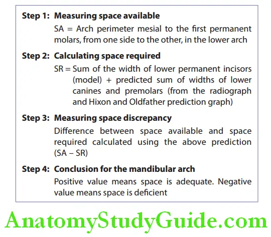
- Tanaka and Johnston mixed dentition analysis using model only
- Moyer’s mixed dentition analysis – using model + prediction chart
- The radiographic method by Huckaba – using model+ radiograph
- Hixon and Oldfather prediction graph – using model + prediction chart + radiograph
Tanaka And Johnston Mixed Dentition Analysis:
Tanaka and Johnston’s analysis is the simplest prediction analysis.
This method neither requires a prediction chart nor a radiograph.
According to this method, the width of lower permanent incisors is proportional to the combined width of permanent canines and premolars in both the upper and lower arches.
The width of lower incisors can be measured as early as 7 years of age.
Method:
Space available (SA) is the distance between the mesial surfaces of the first permanent molars on either side of the arch.
In the case of crowding of incisors, the space required for decrowding is subtracted from the space available.
Mesiodistal widths of the erupted four permanent mandibular incisors are measured from the model cast.
This value is divided by 2 to get an average width of two incisors on each side. The value is assumed as ‘A’.
According to Tanaka and Johnston, a constant of 11 and 10.5 is added to ‘A’, to get the sum of the widths of canines and premolars in the maxillary and mandibular arch, respectively.
Space Analysis In Mixed Dentition
The space required (SR) can be calculated using this.
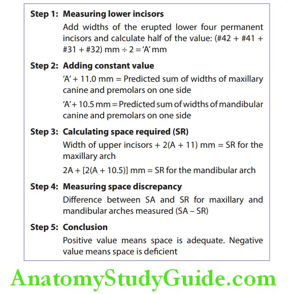
The advantages of this method are the easy and simple calculations.
There is no need for prediction charts. But the universal applicability of this method for all ethnic populations has not been proved.
Moyer’S Mixed Dentition Analysis:
Moyer’s concept is similar to that of Tanaka and Johnston.
According to Moyer, there exists a correlation between the widths of permanent lower incisors, permanent canines, and premolars.
If the mesiodistal width of permanent lower incisors is known, the sum of widths of unerupted canine and premolars can be predicted from Moyer’s prediction chart.
Space Analysis In Orthodontics
The chart gives different sets of predicted sizes for a given width of lower incisors. The chart also gives a percentage value to each of the sets.
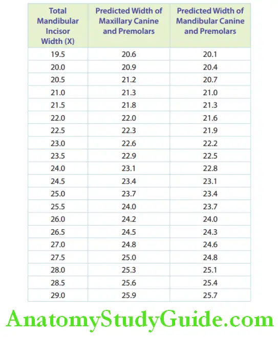
This means that the prediction value is applicable to 75% of the population.
The 75% prediction value set is generally used for predicting the sizes of unerupted teeth, with the hope that the reliability is higher.
In any case, it is better to overestimate and be forewarned of higher demand than be at the comfort of smaller demand only to get perturbed later upon realizing that the demand had actually been more.
Space Analysis In Orthodontics
Method:
The sum of the mesiodistal widths of lower incisors is assumed as ‘X’.
From the prediction chart, the corresponding value for the sum of widths of permanent canines and premolars for maxillary and mandibular arches is found.
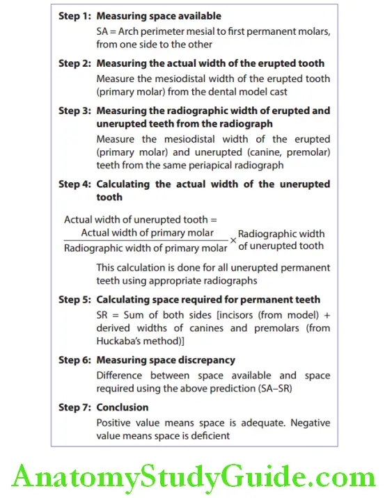
Radiographic Method Recommended By Huckaba:
Huckaba used intraoral radiographs to predict the width of unerupted teeth.
An intraoral radiograph of the posterior region of the dental arch is taken (Right angle technique) at around 8 years of age.
This reveals the image of unerupted canine and premolars and the erupted molar in the same vicinity.
The actual width of the erupted molar can be measured from the study model, while its radio graphic width can be measured from the radiograph.
Space Analysis In Orthodontics
The difference between the two measurements is the magnification factor.
This factor will be the same for the erupted molar and the unerupted canines and premolars in the same firm.
Hence, the ratio in dimensional change will also be the same.
The width of the unerupted teeth can be predicted from their radiographic image in this way.
Method:
The space available is calculated as explained above.
The true or actual width of the erupted tooth is measured from the model cast.
The radiographic or apparent width of the same tooth is measured from a periapical view radiograph.
The radiographic width of the unerupted teeth is also measured from the same periapical radiograph.
The idea of this proportional change is used to find the true width of the unerupted tooth.
This is a fairly reliable technique, which uses a model and a radiograph.
It can be used for maxillary and mandibular arches and on all groups of the population.
The radiographic image has to be clear. It is essential that the images are measured from the same film depicting both the unerupted teeth and the erupted tooth.
Obtaining an undistorted image of a canine is difficult due to its position.
This makes the measurement of its radiographic width difficult. Also, rotated teeth cannot be properly measured in radiographs
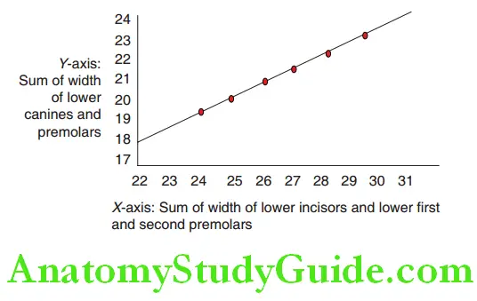
Hixon And Oldfather Prediction Graph:
Obtaining an undistorted radiographic image of saline and therefore prediction of its width from the radiograph was difficult.
Hixon and Oldfather predicted the relative width of unerupted permanent lower canines using a dental model, periapical radiographs, and a prediction graph.
The method was later revised by Staley and Kerber.
Space Analysis In Orthodontics
The X-axis of the prediction graph depicted the sum of the widths of the permanent lower incisors and lower fist and second premolars.
The widths lower canine and premolars are depicted on the Y-axis.
The width of the lower permanent incisors is measured from the study model.
The width of unerupted premolars is predicted from the periapical radiograph.
With these measurements, the width of the lower permanent canine can be deciphered from the prediction graph formulated by Hixon and Oldfather.
In the graph, the line connecting the red dots is the prediction line.
The given sum of lower incisors and premolars on the X-axis is projected onto this prediction line and the corresponding Y-axis value is read as the sum of the lower permanent canines and premolars, from which the size of the permanent canine is deciphered.
The values are applicable to the northern European population, with questionable reliability for other ethnic groups.

Tweed’s Method
Tweed suggested a modification in obtaining the space available (SA) for space analysis.
According to Tweed, the axial inclination of lower incisors on the mandibular basal bone plays a major role in treatment planning and that space analysis should include space needed for correction of inclination.
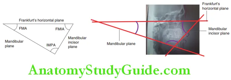
Method:
Tweed introduced a triangle widely known as ‘Tweed’s triangle’ formed by three planes, namely the Frankfurt horizontal plane (passing through portion to orbitale), the mandibular plane (passing through gonion and mention), and the mandibular incisor plane (long axis of the lower incisor).
Th angles formed are called Frankfurt-mandibularangle(FMA), incisormandibular plane angle (IMPA) and Frankfurt mandibular incisor angle.
The established relationships derived from Tweed’s research are as follows:
1. When FMA is between 21° and 29°, the FMIA should be 68°
2. When FMA is 30° or higher, the FMIA should be 65°
3. When FMA is 20° or lesser, the IMPA should be 92° Patient’s lateral cephalogram is traced.
The axial inclination of the lower incisor is checked and if found to not correspond with the relationship as said by Tweed, then an objective lower incisor axial line is drawn according to the patient’s FMA, as suggested.
Space Analysis In Orthodontics
The distance between the actual axial line of the lower incisor and the objective line is measured along the occlusal plane.
Double this distance is required for correction of incisor proclination (for the right and left sides of the arch) and is reduced from the space available.
This method not only took the teeth into consideration but also the facial balance and stability in planning treatment.
Total Space Analysis:
Total space analysis of the lower arch was developed by Levern Merrifield.
All the space analyses discussed previously in this chapter measure the magnitude of space discrepancy in the dental arch.
The total space analysis measures the magnitude of space discrepancy and also reveals the exact location of discrepancy in the arch.
The dental arch is divided into anterior, middle, and posterior areas.
The analysis is done separately for each of these areas to localize the specific area of space discrepancy in the lower arch.
For further information about this ‘total space analysis’, the reader is advised to refer to appropriate orthodontic books.
Mixed Dentition Space Summary
1. Space analysis calculates the difference between ‘space available’ and ‘space required’.
2. A mixed dentition space analysis involves the prediction of the mesiodistal widths of unerupted premolars and canines and then using those values to predict space availability.
3. Mixed dentition model analyses are as follows:
Tanaka and Johnston mixed dentition analysis:
- Half the sum of the width of the lower four permanent incisors + 11 mm = Predicted sum of widths of maxillary canine and premolars.
- Half the sum of the width of the lower four permanent incisors + 10.5 mm = Predicted sum of widths of mandibular canine and premolars.
Moyer’s mixed dentition analysis:
If the mesiodistal width of permanent lower incisors is known, the sum of widths of unerupted canine and premolars can be predicted from Moyer’s prediction chart (75th percentile value is generally considered).
Radiographic method:
True width of unerupted premolar = True width of primary molar ÷ Radiographic width of unerupted premolar x Radiographic width of the primary molar.
Hixon and Oldfather’s (revised by Staley and Kerber) prediction graph predicts the relative width of unerupted lower canines, using both model and periapical radiographs.
Tweed’s method: Lower incisors are given importance.
4. Total space analysis – It is an analysis of the magnitude of the discrepancy of space and the exact location of the identified discrepancy.
Leave a Reply