Morphology of Primary Teeth Notes
Primary Dentition
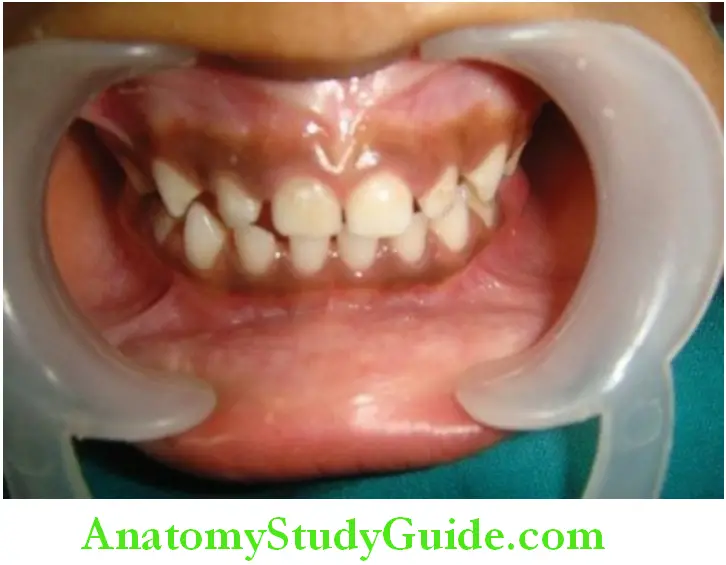
Primary teeth or deciduous teeth are commonly known as baby teeth, temporary teeth and milk teeth. The first primary tooth erupts into the oral cavity around 6 months of age. The dentition is completed by 2.5–3 years of age. They are 20 in number which includes two incisors, one canine and two primary molars per quadrant. The clinical features of primary or deciduous dentition are
Table of Contents
General characteristics:
- Both arches are half round in shape/ovoid
- Almost no curve of spee
- Shallow cuspal interdigitation
- Slight overjet and overbite (1–2 mm)
- Vertical inclination of the incisors
- Little/no crowding
Read And Learn More: Oral Anatomy Notes
Spacing – two types of primary dentition occurs.
1. Spaced dentition:
The spaces in between the teeth in spaced dentition can be utilized for Adjustment of permanent incisor which is always larger compared to Deciduous teeth.
Spaces present are of two types:
Primate spaces:
These spaces are very prominent. In humans, these exist between the Upper lateral incisors and canines and lower canines and first Deciduous molars.
These spaces are called anthropoid spaces or simian Spaces.
Physiologic/developmental space:
These spaces are present between the primary teeth and play an
Important role in normal development of permanent dentition. Total
Space present may vary from 0 to 8 mm with an average of 4 mm and
From 1 to 7 mm with an average of 3 mm.
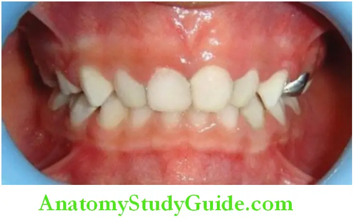
2. Nonspaced dentition:
Primary teeth are present without any spaces in between the teeth. This
Lack of space may be due to the narrowness of dental arches or teeth
Are wider than usual. This type of dentition usually indicates to
Crowding in developing permanent dentition. But it is not always the
Case. It may depend on individual’s jaw growth.
Primary Molar Relationship
The relationship of the distal surface of maxillary to mandibular second primary molars is one of the key factors that influence future occlusion of the permanent dentition.
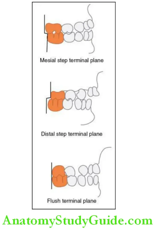
As the distal surface of second primary molar guides the first permanent molars into occlusion, mesiodistal relation between distal surface of upper and lower second primary molars usually can be classified into
- Flush terminal or vertical plane:
The distal surface of upper and lower second primary molars are in a straight plane and therefore situated on the same vertical plane. Usually, it is a favourable relation to guide permanent molars. - Mesial step
The distal surface of lower second primary molar is more mesial to that Of upper; invariably it is favourable to guide permanent molars into Class I molar relation. - Distal step:
The distal surface of lower second primary molar is more distal to that Of upper. This relationship guides the permanent molars into distal Occlusion.
Morphology Of Individual Primary Teeth
There are 20 primary teeth which start erupting at the age of 6 months and completely erupt into oral cavity by 2.5–3 years of age.
The different primary teeth present are incisors, canines, and molars.
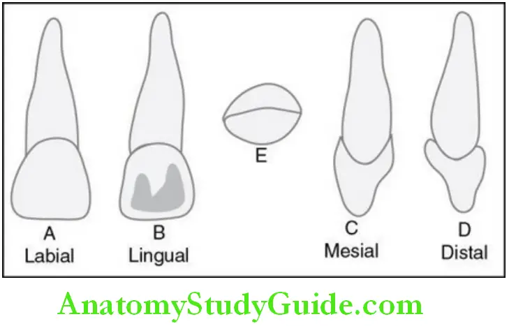
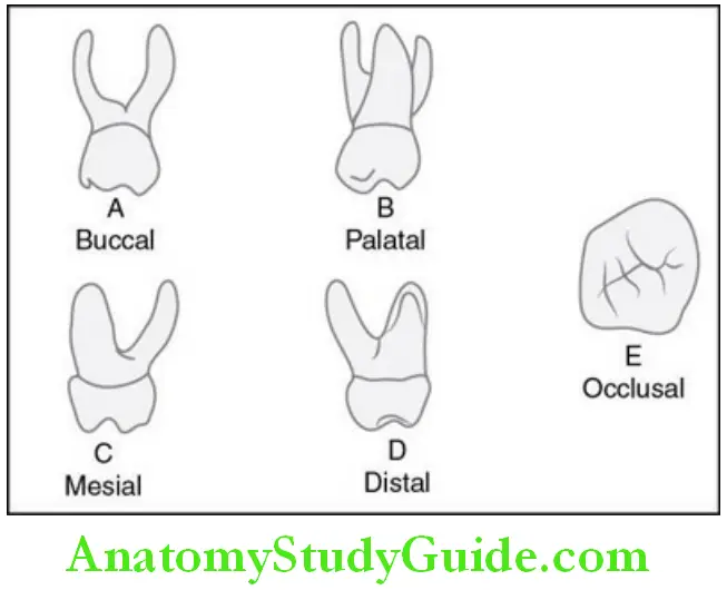
Maxillary central incisors:
Crown:
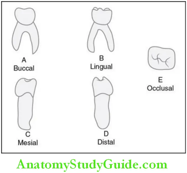
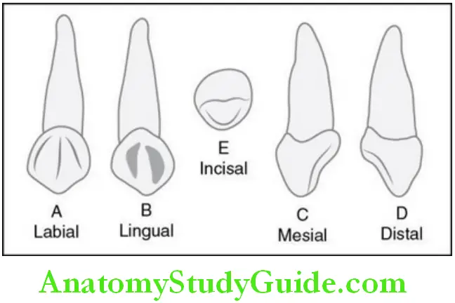
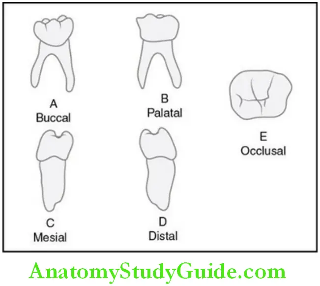
Maxillary central incisor has five surfaces, namely labial, lingual, mesial, distal and incisal.
Labial aspect:
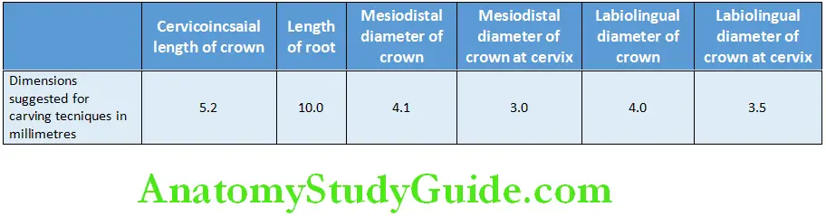
- The mesiodistal diameter of the crown of the primary central incisor is more than the cervicoincisal length (as oppose to permanent central incisors).
- The labial surface is very smooth, with a straight incisal edge.
- Usually, developmental lines are not evident.
- The root is cone-shaped having even tapered sides.
- The length of the root is more in comparison with the length of the crown.
Lingual aspect:
- The marginal ridges and the cingulum are highly developed in lingual aspect of the crown.
- The cingulum divides the lingual concavity into a mesial and distal fossa.
- The root narrows lingually.
- A ridge for its full length is present in comparison with a flatter surface labially.
Mesial and distal aspects:
- For the primary maxillary central incisor, the mesial and distal aspects are very similar.
- The measurement of the cervical third of the crown shows broad to its total length.
- The average measurement is only about 1 mm less than the entire crown length cervicoincisally.
- As the crown is short then its labiolingual measurement, it appears thick at the middle third and even down towards the incisal third.
- A distinct cervical line which represents cementoenamel junction is present which curves towards the incisal ridge.
- The cervical curvature distally is less than the curvature mesially.
- The root is more blunt from this aspect than the labial and lingual aspects.
- But it is still of an even taper with a shape of a long cone.
- At the apex, it is blunt.
- Usually, the root of the mesial surface will have a developmental groove or concavity, whereas distally, the surface is convex.
Incisal aspect:
- The incisal edge is centred over the crown and is approximately straight.
- From the incisal edge, the labial surface appears broader and also smoother than the lingual surface.
- The lingual surface tapers towards the cingulum.
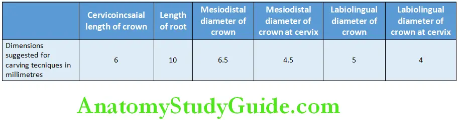
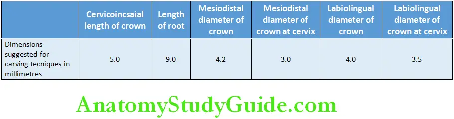
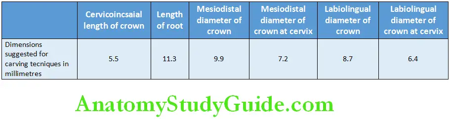
- The proximal surfaces of this tooth are somewhat broad. The measurements of the deciduous maxillary central incisor are given in table.
Measurement of deciduous maxillary central incisor:
Maxillary lateral incisor:
In general, the maxillary lateral is similar to the central incisor from all aspects, but its dimensions differ.
From all directions, the crown is smaller.
The cervicoincisal length of the crown is more than its mesiodistal width.
The distoincisal angles of the crown are more rounded than those the central incisor.
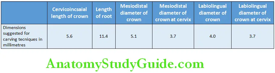
The roots of lateral incisor are similar in shape as of central incisor, but it is much longer in proportion to its crown. The measurements of deciduous maxillary lateral incisor are given in table .
Measurement of deciduous maxillary lateral incisor:
Maxillary canine:
Crown:
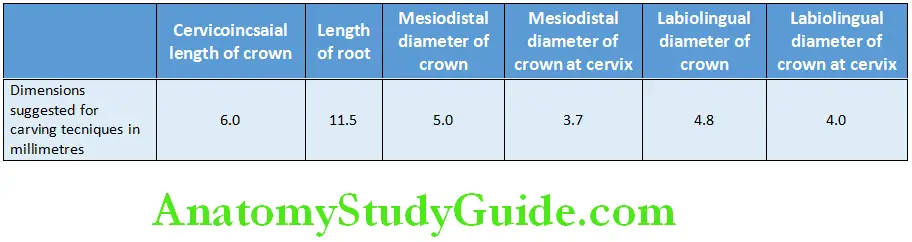
- It is the largest of all anterior teeth.
- This tooth is long and sharp.
- The crown is constricted at cementoenamel junction
Labial aspec:
- The labial side of the maxillary canine does not resemble either the central or the lateral incisor.
- Mesiodistally, the crown is more constricted at the cervix, and the mesial and distal surfaces appear to be convex.
- A well-developed sharp cusp is present instead of an incisal edge.
- As compared with the permanent maxillary canine, the cusp on the primary Canine is much longer and sharper.
- Also mesially, the crest of the contour is not as far down towards the incisal portion.
- When a line is drawn through the contact areas of the deciduous canine, it will bisect the line drawn from the cervix to the tip of the cusp whereas, the contact areas are not at the same level in the permanent canine.
- The mesial slope of the cusp of the primary canine is longer than the distal slope when the cusp is intact.
- The primary canine root is long, slender, and tapering and is more than twice the crown length.
Lingual aspect:
- The enamel ridges present lingually are pronounced that merge with each other.
- There are the cingulum, mesial and distal marginal ridges, and incisal cusp ridges, besides a tubercle at the cusp tip.
- The tubercle is a continuation of the lingual ridge which connects the cingulum with cusp tip.
- The lingual surface is divided into shallow mesiolingual and distolingual fossae by the lingual ridge.
- The root is tapered lingually.
- Above the middle third, the root is usually inclined distally.
Mesial aspect:
- From the mesial aspect, the outline form is similar to that of the lateral and central incisors. However, the difference in its proportion is evident.
- The measurement labiolingually at the cervical third is much greater.
Distal aspect:
The distal outline is the opposite of the mesial aspect of this tooth.
No notable differences may be noted except for the curvature of the cervical line towards the cusp ridge which is less on the mesial surface.
Incisal aspect:
- From the incisal aspect, the crown is essentially diamond-shaped.
- The angles that are present at the mesial and distal contact areas; the cingulum On the lingual side; and the cervical third, or enamel ridge, on the labial surface are more pronounced and less rounded in effect as compared to the permanent canines.
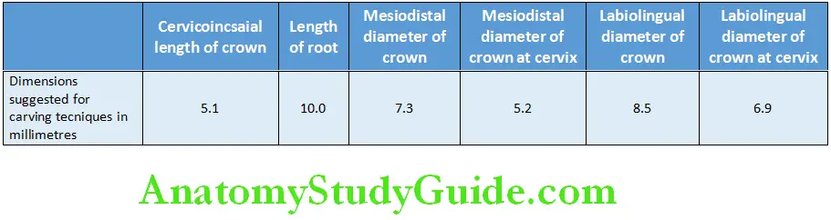
- The tip of the cusp is distal to the centre of the crown, and the slope of the mesial cusp is longer than the slope of the distal cusp. The measurements of deciduous maxillary canine are given in table.
Measurement of deciduous maxillary canine:

Maxillary first molar:
Crown:
- The tooth presents with well-defined surfaces, namely the buccal, lingual, mesial, distal and occlusal.
Buccal aspect:
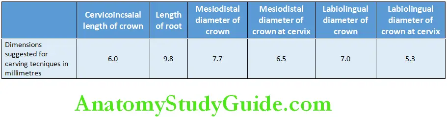
- The crown of maxillary first molar is broadest at the contact areas mesially and distally.
- From there, the crown converges towards the cervix, with the measurement Being 2 mm less than the measurement at the contact areas.
- This dimensional arrangement gives a narrower look to the cervical portion of the crown and root of the primary maxillary first molar as opposed to the permanent maxillary first molar.
- The occlusal line is slightly scalloped but with no definite cusp form.
- The buccal surface is smooth with less evidence of developmental grooves.
- The primary maxillary first molar is much smaller in all dimensions than the second maxillary molar.
- The maxillary first molar roots are slender, long and they spread widely.
- All three roots may be visible from this aspect.
- The distal root is considerably short as compared with the mesial root.
- At cervical line (cej), the bifurcation of the roots begins almost immediately.
Lingual aspect:
- The general outline of the lingual aspect and buccal aspect of the crown is similar.
- The crown converges more in a lingual direction, which makes the lingual Portion measure less mesiodistally as with the buccal portion.
- The mesiolingual cusp is most prominent cusp. It is longest and the sharpest cusp.
- The distolingual cusp is poorly defined. It is small and rounded when it is present.
- From the lingual aspect, the distobuccal cusp may be seen, since it is longer and better developed than the distolingual cusp.
Mesial aspect:
- From this aspect, the dimension at the cervical third is greater than the dimension at the occlusal third.
- It is true for all molars, but it is more pronounced on primary than permanent teeth.
- The mesiolingual cusp is longer and sharper compared to the mesiobuccal cusp.
- On the buccal outline of the cervical third, a pronounced convexity is evident which is characteristic of this tooth.
- Mesially, the cervical line shows some curvature in the direction of the occlusal surface.
- From the mesial side of this tooth, the mesiobuccal and lingual roots are visible only when looking from a point directly opposite of the contact area.
- The distobuccal root is unseen behind the mesiobuccal root.
- The lingual root appears long and slender and extends lingually to a marked degree from this aspect. It curves steeply in a buccal direction above the middle third.
Distal aspect:
- From this aspect, the crown is narrower distally than mesially, and it tapers significantly towards the distal side.
- Distobuccal cusp is long and sharp with a poorly developed distolingual cusp.
- The prominent bulge seen from the mesial aspect at the cervical third does not continue to the distal aspect.
- The cervical line from the buccal surface to the lingual surface may curve occlusally, or it may extend straight across.
- All three roots may be viewed from this side, but the distobuccal root is overlying on the mesiobuccal root so that only the buccal surface along with the apex of the mesiobuccal root may be seen.
- As described earlier, the point of bifurcation of the distobuccal root and the lingual root is near the cej which is typical.
Occlusal aspect:
- The outline of the crown converges lingually.
- A central fossa is present on occlusal surface.
- A mesial triangular fossa is just inside the mesial marginal ridge, with a mesial pit in this fossa and a sulcus with its central groove connecting the two fossae.
- A well-defined buccal developmental groove divides the mesiobuccal cusp and the distobuccal cusp.
- Sometimes the primary maxillary first molar has a well-defined triangular ridge connecting the mesiolingual cusp with the distobuccal cusp called the oblique ridge.
- The distal marginal ridge is thin and poorly developed in comparison with the mesial marginal ridge. The measurements of the deciduous maxillary first molar are given in table.
Measurement of deciduous maxillary first molar:
Maxillary second molar:
Buccal aspect:
- The primary maxillary second molar resembles the permanent maxillary first molar, but it is smaller in dimensions.
- Buccally, two well-defined buccal cusps with a buccal developmental groove is seen between them.
- Like all primary molars, the crown is narrow at the cervix as compared with its mesiodistal measurement at the contact areas. The crown is much larger as compared to first primary molar.
- Buccally, the roots appear slender but are much longer and heavier as compared with the maxillary first molar.
- The bifurcation point between the buccal roots is near to the cervical line of the crown.
- The size of the two buccal cusps is more nearly equal.
Lingual aspect:
- From this aspect, three cusps can be seen:
-
- The mesiolingual cusp, which is well developed and large.
- The distolingual cusp, which is again well developed (even more Than that of the primary first molar).
- A third supplemental cusp, which is apically located on the mesiolingual cusp and also called the tubercle of carabelli, or the fifth cusp. This cusp is developed poorly and just acts as a buttress or supplement to the bulk of the mesiolingual cusp.
- Sometimes, some traces of developmental lines or ‘dimples’ remain if tubercle of carabelli seems to be missing.
- All three roots are seen from this aspect. The lingual root is large and thick as opposed to the other two roots.
Mesial aspect:
- From the mesial aspect, the crown has a standard molar outline that resembles that of the permanent molars very much.
- The crown appears to be short as its width is more buccolingually in comparison to its length.
- The mesiolingual cusp with its supplementary fifth cusp is large in comparison with the mesiobuccal cusp.
- The mesiobuccal cusp from this side is relatively short and sharp
- From this side, the mesiobuccal root is broad and flat.
- The lingual root has almost the same curvature as that of the lingual root of the maxillary first deciduous molar.
- The mesiobuccal root extends lingually beyond the outline of the crown.
- The point of bifurcation between the mesiobuccal root and the lingual root is 2 or 3 mm apical to the cervical line of the crown.
- The mesiobuccal root presents itself as being wider from the mesial aspect.
- The mesiolingual cusp is directly below their bifurcation.
Distal aspect:
- The distobuccal cusp and the distolingual cusp are about the same in length.
- The cervical line is approximately straight.
- From this side, all three roots are seen, although only outline part of the mesiobuccal root may be seen as it is overlying by the distobuccal root.
- The distobuccal root is shorter and narrower as compared with the other roots.
- The point of bifurcation between the distobuccal root and the lingual root is more apically present in location as compared with any of the other points of bifurcation.
Occlusal aspect:
- From the occlusal aspect, this tooth resembles the permanent first molar.
- It is rhomboidal in shape and has four well-defined cusps along with one Supplemental cusp.
- The cusps include mesiobuccal, distobuccal, mesiolingual, distolingual and fifth cusps.
- The buccal surface is flat.
- The developmental groove between the cusps is less marked than that found on the first permanent molar.
- The occlusal surface has a central fossa with a central pit.
- It also has a well-defined mesial triangular fossa, just distal to the mesial marginal ridge, with a mesial pit at its centre.
- A well-defined developmental groove called the central groove is also at the bottom of a sulcus, connecting the mesial triangular fossa with the central fossa.
- The buccal developmental groove extends buccally from the central pit, Separating the triangular ridges, which are occlusal continuations of the Mesiobuccal and distobuccal cusps.
- Supplemental grooves often radiate from these developmental grooves.
- The oblique ridge is prominent and connects the mesiolingual cusp with the distobuccal cusp.
- Distal to the oblique ridge, the distal fossa is found, which harbours the distal developmental groove.
- The distal and mesial marginal ridges are well developed. The measurements of the deciduous maxillary second molar are given in table .
Measurement of deciduous maxillary second molar:

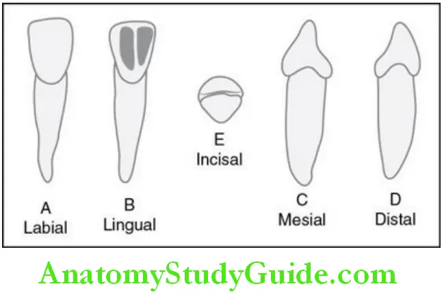
Mandibular central incisor:
Crown:
- They are narrow and are the smallest incisors in the oral cavity.
- It is smaller and narrower than lateral incisor and also the smallest of the primary teeth.
Labial aspect:
- The labial aspect of this crown has a flat face with no developmental grooves.
- From the mesial and distal sides, the crown is evenly tapered from the contact areas, the measurement being less at the cervix.
- The crown is wide in proportion as compared to its length.
- The root is long and evenly tapered towards the apex and is pointed.
- The root is almost twice the length of the crown.
Lingual aspect:
- On the crown of the lingual surface, the marginal ridges and the cingulum may be located easily.
- At the middle and incisal third of the lingual surface, the crown may have a slight concavity present, called the lingual fossa.
- The lingual side of the crown and root converges so that it is narrower towards the lingual and not the labial surface.
Mesial aspect:
- The mesial aspect shows the typical outline of an incisor tooth, even though the measurements are small.
- The incisal ridge is centred at the centre of the root.
- The convexity of cervical contours labially and lingually at the cervical third is pronounced.
- The labiolingual measurement is only about a millimetre less than that of the primary maxillary central incisor.
- The mesial surface of the root is almost flat and evenly tapered.
- The apex presents a more blunt appearance than is found with the lingual or labial aspects.
Distal aspect:
- From the distal aspect, the outline of this tooth is the opposite of that found from the mesial aspect.
- A slight difference can be noted between these aspects, except that the cervical line of the crown is less curved towards the incisal ridge than on the mesial surface.
- Often, a developmental depression is evident on the distal side of the root.
Incisal aspect:
- The incisal ridge is almost straight which bisects the crown labiolingually.
- A definite taper is evident towards the cingulum on the lingual side.
- The labial surface from this view presents a flat surface that is slightly convex, whereas the lingual surface presents a flattened surface that is slightly concave.
- The measurements of the deciduous mandibular central incisor are given in Table.
Measurement of deciduous mandibular central incisor:
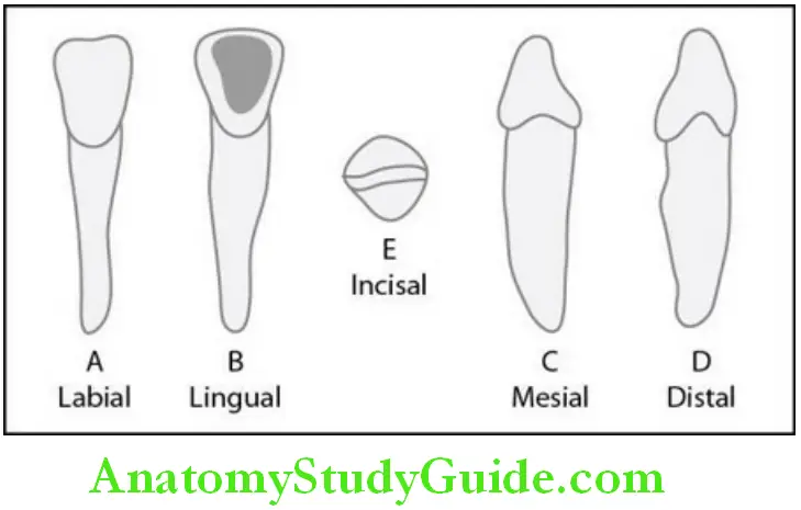
Mandibular lateral incisor:
- The basic features of the primary mandibular lateral incisor are very similar to the primary central incisor.
- Functionally, they both supplement each other.
- The lateral incisor is large in all measurements except for labiolingual, where these two teeth are identical.
- The cingulum may be slightly more generous as that of the central incisor.
- Also, a tendency for the incisal ridge to slope downward distally exists.
- This design lowers the distal contact area apically so that proper contact area can be achieved with the mesial surface of the primary mandibular canine. The measurements of the deciduous mandibular lateral incisor are given in table
Measurement of deciduous mandibular lateral incisor:
Mandibular canine:
- Little difference in functional form is evident between the mandibular canine and the maxillary canine.
- The difference is mainly in the dimensions.
- The crown is perhaps 0.5 mm shorter, and the root is at least 2 mm shorter.
- The mesiodistal measurement of the mandibular canine at the root trunk is greater when compared with its mesiodistal measurement at the contact areas than is that of the maxillary canine.
- The labiolingual measurement reflects the outstanding variation in size between the two deciduous canines.
- The deciduous maxillary canine is much larger labiolingually.
- The cervical ridges labially and lingually are not quite as pronounced as those found on the maxillary canine.
- The greatest variation in outline form when one compares the two teeth is seen from the labial and lingual aspects.
- The distal cusp slope is longer than the mesial slope. The measurements of the deciduous mandibular canine are given in table.
Measurement of deciduous mandibular canine:
Mandibular first molar:
The mandibular first molar does not resemble any of the other teeth.
Because of so much variation, it appears strange and primitive.
Buccal aspect:
- Buccally, the mesial outline of the crown of the primary mandibular first molar is almost straight from the contact area to the cervix.
- The constriction of the crown is very little at the cervix.
- The distal outline, however, converges towards the cervix more than usual, and so the contact area extends distally to a marked degree.
- The distal portion is shorter than the mesial portion of the crown, with the cervical line dipping apically where it joins the mesial root.
- The two buccal cusps are very distinct, with no developmental groove seen between them.
- The mesial cusp is larger than the distal cusp.
- A developmental depression dividing them (not a groove) extends over to the buccal surface.
- The roots are long and slender, and they spread considerably at the apical third beyond the outline of the crown.
- From the buccal aspect, if a line is drawn from the bifurcation of the roots to the occlusal surface, the tooth will be evenly divided mesiodistally. However, the mesial portion represents a tooth with a crown almost twice as tall as the distal half, and the root is again a third as long as the distal one. Two complete teeth are represented, but their dimensions differ considerably.
Lingual aspect:
- The crown and root converge lingually to a marked degree on the mesial
Surface. - Distally, the opposite arrangement is true for both crown and root.
The distolingual cusp is rounded. - The mesiolingual cusp is long and sharp more than any of the other cusps.
- The mesial marginal ridge is well developed.
- Part of the two buccal cusps may be seen from this angle.
- From the lingual aspect, the crown length mesially and distally is more uniform than seen from the buccal aspect.
- The cervical line is straight.
Mesial aspect:
- The most noticeable detail from the mesial aspect is the extreme curvature buccally at the cervical third.
- Except for this, the crown outline of this tooth from mesial aspect resembles the mesial aspect of the primary second molar.
- Both the mesiobuccal cusp and the mesiolingual cusp are seen from this aspect along with well-developed mesial marginal ridge.
- As the mesiobuccal crown length is more than the mesiolingual crown length, the cervical line tilts upward buccolingually.
- The buccal and lingual outlines of the root drop straight down from the crown.
- They are approximately parallel for more than half their length, with slight taper at the apical third.
- The root end is flat and almost square.
- A developmental depression usually extends almost the full length of the root on the mesial side.
Distal aspect:
- The distal side of the mandibular first molar differs from the mesial side in several ways.
- The cervical line does not drop buccally.
- The length of crown buccally and lingually is more uniform.
- The cervical line extends almost straight across buccolingually.
- The distobuccal cusp and the distolingual cusp are not as long or as sharp as the two mesial cusps.
- The distal marginal ridge is not as straight and well defined as the mesial marginal ridge.
- The distal root is rounder and shorter and tapers more apically.
Occlusal aspect:
- The general outline of the occlusal aspect is rhomboidal.
- The prominence present mesiobuccally is noticeable from this aspect.
- The mesiolingual cusp is the largest and well developed of all the cusps.
- The buccal developmental groove of the occlusal surface evenly divides the two buccal cusps.
- This developmental groove is short, extending from between the buccal cusp ridges to a point in the centre of the crown outline at a central pit.
- The central developmental groove merges it at this point which extends mesially separating the mesiobuccal and the mesiolingual cusp.
- The mesiobuccal cusp exhibits a well-defined triangular ridge on the occlusal surface.
- The lingual developmental groove extends lingually from this point, separating the mesiolingual cusp and the distolingual cusp. The measurements of the deciduous mandibular first molar are given in table .

Measurement of deciduous mandibular first molar:
Mandibular second molar:
The primary mandibular second molar has characteristics that resemble those of the permanent mandibular first molar, although its dimensions differ.
Buccal aspect:
- From this aspect, the primary mandibular second molar has a narrow Mesiodistal measure at the cervical portion of the crown compared with the Measurement mesiodistally.
- Also, mesiobuccal and distobuccal developmental grooves divide the buccal surface of the crown occlusally into three cuspal portions which are of almost equal in size.
- This forms a buccal surface having a mesiobuccal, buccal and distobuccal cusp.
- The roots of this aspect are slender and long and possess a unique flare Mesiodistally in the middle third and apical third.
- The roots may be of twice as long as the crown.
- The point of bifurcation of the roots is just below the cej of crown and root.
Lingual aspect:
- From the lingual aspect, two cusps of almost equal dimensions can be seen.
- The two lingual cusps are not quite as broad as the three buccal cusps which narrow the crown lingually.
- The cervical line is almost straight.
- Lingually, the mesial portion of the crown seems to be a little higher than the distal portion of the crown.
- Each of the three buccal cusps can be seen from this aspect.
- From this aspect, the roots give almost the same appearance as from the buccal aspect.
Mesial aspect:
- From the mesial aspect, the outline of the crown resembles that of the Permanent mandibular first molar.
- The crest of contour buccally is more prominent on the primary molar, and the tooth seems to be more constricted occlusally because of the flattened buccal surface above this cervical ridge.
- The crown is poised over the root of this tooth in the same manner as all mandibular posteriors; its buccal cusp is over the root and the lingual outline of the crown extending out beyond the root line.
- The marginal ridge is high, a characteristic that makes the mesiobuccal cusp and the mesiolingual cusp appear rather short.
- The lingual cusp is longer, or higher, than the buccal cusp.
- The cervical line is regular, although it extends upward buccolingually, making up for the difference in length between the buccal and lingual cusps.
- The mesial root is unusually broad and flat with a blunt apex that is sometimes serrated.
Distal aspec:
- The mesiobuccal and distobuccal cusps can be seen from this aspect.
- The distolingual cusp appears well developed.
- The distal marginal ridge is shorter buccolingually than the mesial marginal ridge.
- The cervical line of the crown is regular.
- The distal root is as broad as the mesial root.
- The distal root tapers more at the apical end than does the mesial root.
Occlusal aspect:
- The primary mandibular second molar shapes like rectangular from the occlusal aspect.
- The three buccal cusps are almost of similar sizes.
- The two lingual cusps are also of equal size.
- However, the total mesiodistal width of the lingual cusps is less than that of the total mesiodistal width of the three buccal cusps.
- Triangular ridges are well defined, which extend occlusally from each one of these cusp tips.
- The mesial triangular fossa is well defined than distal triangular fossa.
- The mesial marginal ridge is more developed and pronounced as compared With the distal marginal ridge.
- The crown outline converges distally.
- An outline following the tips of the cusps and the marginal ridges conforms to the outline of a rectangle more closely than does the gross outline of the crown in its entirety. The measurements of the deciduous mandibular second molar are given in table.
Measurement of deciduous mandibular second molar:
Primary Dentition Synopsis
- Primary teeth or deciduous teeth, commonly known as baby teeth, temporary teeth and milk teeth. The first primary tooth erupts into the oral cavity around 6 months of age. The dentition is completed by 2.5–3 years of age. They are 20 in number which includes two incisors, one canine and two primary molars per quadrant.
- The clinical features of primary or deciduous dentition are that both the arches are half round in shape/ovoid, almost no curve of spee, shallow cuspal interdigitation, slight overjet and overbite (1 2 mm), vertical inclination of the incisors, little/no crowding.
- Spacing: Two types of primary dentition occur: The spaces in between the teeth in spaced dentition can be utilized for adjustment of permanent incisor which is always larger compared to deciduous teeth.
- Spaces present are of two types. Primate spaces – spaces are very prominent. In humans, these exist between the upper lateral incisors and canines and lower canines and first deciduous molars. These spaces are called anthropoid spaces or simian spaces. Physiologic/developmental spaces are present between the primary teeth and play an important role in normal development of permanent dentition. Total space present may vary from 0 to 8 mm with an average of 4 mm and from 1 to 7 mm with an average of 3 mm.
- Primary teeth are present without any spaces in between the teeth. This lack of space may be due to the narrowness of dental arches or teeth are wider than usual. This type of dentition usually indicates to crowding in developing permanent dentition. But it is not always the case. It may depend on individual’s jaw growth.
- There are 20 primary teeth which start erupting at the age of 6 months and completely erupt into oral cavity by 2.5–3 years of age.
- The different primary teeth present are incisors, canines and molars.
Leave a Reply