Muscles in the Medial Compartment of the Thigh
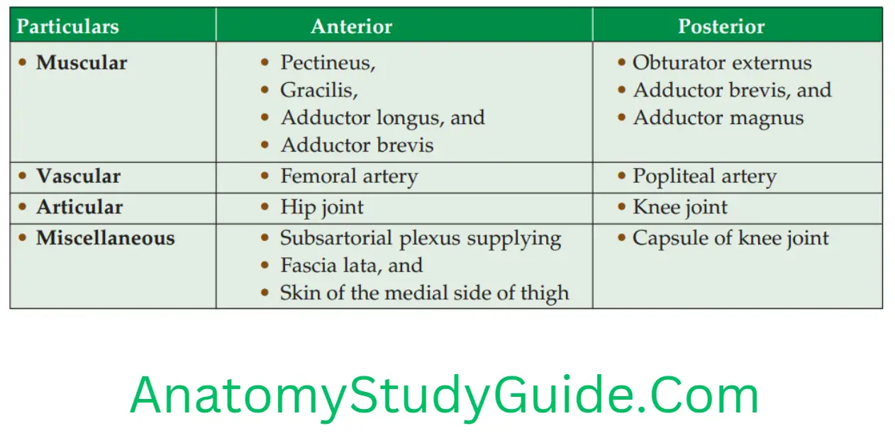
Enumerate the muscles of the Adductor Compartment
Muscles are grouped as
1. Intrinsic
- Adductor longus,
- Adductor brevis,
- Adductor Magnus,
- Gracilis, and
- Pectineus.
2. Extrinsic: Obturator externus lies deep in this region.
Read And Learn More: Anatomy Notes And Important Question And Answers
Enumerate the muscles supplied by the obturator nerve
It has two divisions
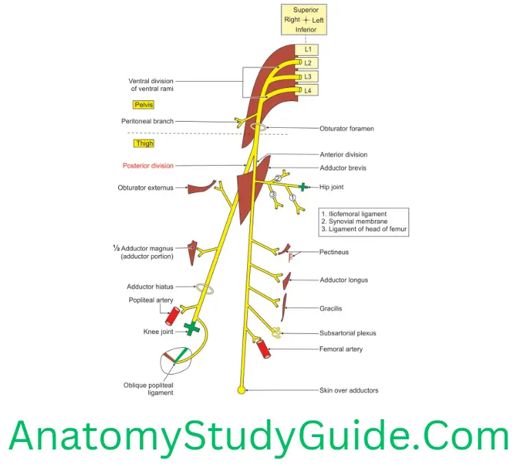
1. Anterior division supplies
- Pectineus,
- Adductor longus,
- Gracilis, and
- Adductor brevis.
2. Posterior division supplies
- Obturator externus,
- Adductor brevis, if not supplied by anterior division, and
- Adductor Magnus.
Describe the obturator nerve under the following heads
1. Obturator Nerve Root value,
2. Obturator Nerve Course,
3. Obturator Nerve Branches,
4. Obturator Nerve Relations, and
5. Obturator Nerve Applied anatomy.
Obturator Nerve Introduction: It is a nerve of the adductor compartment of the thigh. It supplies the adductor muscles and the skin over the medial side of thigh.
1. Obturator Nerve Root value: It arises from the ventral division of ventral rami of.
2. Obturator Nerve Course: It extends from the pelvis to knee joint.
1. In the pelvis, it
- Is formed within the substance of psoas major muscle,
- Passes on the medial border of psoas major,
- Lies behind the common iliac vessels,
- Runs lateral to the internal iliac vessels,
- Passes along the lateral wall of pelvis, and
- Passes through the obturator foramen and divides into anterior and posterior divisions in the obturator notch.
2. In the thigh
- The adductor brevis is sandwiched between anterior and posterior divisions of obturator nerve.
- The anterior division runs in front of obturator externus muscle in the upper part. It lies in front of adductor brevis muscle in the lower part. It runs behind the pectineus and adductor longus.
- The posterior division perforates the upper border of obturator externus and gives a branch to it. It lies deep to the posterior surface of adductor brevis. It runs in the thigh vertically on adductor magnus.
3. Obturator Nerve Branches: They are grouped as branches from trunk, anterior division and posterior division.
1. Branches from the trunk in the pelvis are
1. Peritoneal branch to lateral pelvic wall.
2. Branch to the hip joint: It springs from the main nerve within the obturator canal. It passes laterally and gives many twigs to the
- Pubofemoral ligament,
- Synovial membrane, and
- Ligament of the head of the femur.


4. Obturator Nerve Relations

5. Obturator Nerve Applied anatomy
1. The obturator nerve supplies both hip and knee joints. So, pain of one joint gives referred pain to other joint.
2. Injury to obturator nerve is uncommon. A penetrating wound may injure obturator nerve and results in the weakness of adduction of the hip joint.
3. Peritonitis in the lateral pelvic wall may irritate the obturator nerve. The pain in such conditions may be referred to the medial side of the
- Hip joint,
- Distal thigh, and
- Back of knee joint.
4. Obturator nerve may be involved with femoral nerve in retroperitoneal tumours.
5. A nerve entrapment syndrome leading to chronic pain on the medial side of thigh may occur in athletes with big adductor muscles.
6. There is a spasm of the adductors of thigh in certain intractable cases of spastic paraplegia. This may be relieved by surgical division of the obturator nerve.
7. Compression of the obturator nerve in the obturator canal may occur due to obturator hernia or osteomyelitis. There is pain and later weakness of the adductors of the thigh.
8. Scissors gait in cerebral palsy due to adductor spasm is treated by partial severance (cutting) of obturator nerve.
Leave a Reply