Muscles of the Gluteal Region
Ischial Tuberosity
1. It is a tuberosity present on the lower part of the dorsal surface of the ischium.
Table of Contents
2. Ischial Tuberosity Morphology: It is an example of traction epiphysis.
3. Ischial Tuberosity Relations
- In a standing position, it is covered by the gluteus maximus. In the sitting position, the muscle slips laterally so that weight is taken directly on the bone.
- It forms the lateral wall of the ischiorectal fossa.
Read And Learn More: Anatomy Notes And Important Question And Answers
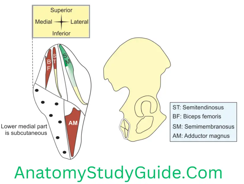
4. Ischial Tuberosity Attachments: It is rough and divided by a transverse ridge into upper quadrilateral and lower triangular.
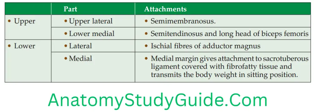
Apart from hamstring muscles, the following structures are attached to the ischial tuberosity
- Inferior gemellus from the upper margin of the ischial tuberosity
- Quadratus femoris arises from the lateral border below the inferior gemellus
- Obturator internus
- Superficial transverse muscles of the perineum
- Sacrotuberous ligament arises from the medial border of the ischial tuberosity.
Ischial Spine
5. Ischial Tuberosity Applied anatomy
- Weaver’s bottom: It is the inflammation of bursa. It lies between gluteus maximus and ischial tuberosity. It is inflamed and enlarged in persons whose occupation invites long periods of sitting.
- In debilitated old people, pressure can cause discomfort while sitting.
- It is one of the sites of bedsores in paralyzed patients and in immobilized patients.
- Nelaton’s line joins the anterior superior iliac spine to the ischial tuberosity. It should normally lie above the greater trochanter.
- The line passing through or below the trochanter indicates shortening at the head or neck of the femur.
- The surface markings of the sciatic nerve can be represented by a line drawn between the posterior superior iliac spine and ischial tuberosity. It curves outwards and downwards. It passes through a point midway between the greater trochanter and ischial tuberosity.
- Pudendal nerve block: In the pudendal nerve block, the needle is inserted into the pudendal canal along the medial side of the tuberosity. The canal lies 1″ deep to the ischial tuberosity.
Piriformis is the key muscle in understanding the anatomy of the gluteal region.
Actions of Gluteus Maximus
1. Powerful extensor of the hip joint.
2. Powerful lateral rotator of the thigh.
3. It is important in rising from sitting position or in climbing stairs.
4. Supports the extended knee.
5. Upper fibres are abductor of the thigh.
Actions of Gluteus Medius
1. It is powerful abductors of the thigh.
2. Its anterior fibres are medial rotator.
3. Its most important action is to maintain the balance of the body when opposite foot is off the ground as in walking and running.
Bones Under Cover Of Gluteus Maximus
Bones
1. Elements of hip bone
- Ilium, and
- Ischium.
2. Sacrum and coccyx, and
3. Upper end and greater trochanter of femur.
The Muscles Under Cover Of Gluteus Maximu
Key muscle is piriformis.
½ muscle is reflected head of the rectus femoris.
The single (1) muscle is the quadratus femoris.
The muscles in pair (2) are
1. Gemelli
- Superior, and
- Inferior
2. Obturator
- Internus, and
- Externus.
The muscles in the trio (3) are
- Gluteus maximus is excluded as we are describing structures under the same,
- Gluteus medius, and
- Gluteus minimus.
The muscles in group of (4) are
- Semimembranosus,
- Semitendinosus,
- Ischial fibres of adductor magnus, and
- Biceps femoris.
The Nerves Under Cover Of Gluteus Maximus
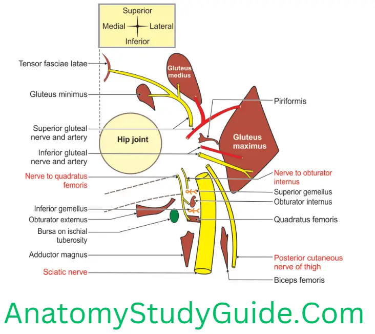
Nerves: The key nerve in this region is sciatic nerve. The other nerves under cover of gluteus maximus are the nerves supplying the muscles present in this region.
1. Key nerve is sciatic nerve.
2. Nerve to quadratus femoris,
3. Nerve to obturator internus,
4. Superior gluteal nerve,
5. Inferior gluteal nerve,
6. Pudendal nerve,
7. Posterior cutaneous nerve of thigh, and
8. Perforating cutaneous nerve branch of posterior cutaneous nerve of thigh.
The Vessels Under Cover Of Gluteus Maximus
Vessels can be remembered by the mnemonic
Superior gluteal artery.
Inferior gluteal artery.
Ascending branch of medial and lateral circumflex femoral arteries (profunda femoris).
Trochanteric anastomosis: The arteries taking part in trochanteric anastomosis are the arteries beginning with 1st and 3rd letters of the key word “sciatica”.
Internal pudendal vessels.
Cruciate anastomosis: The arteries are above the trochanteric anastomosis and artery below the cruciate anastomosis.
Ascending branch of 1st perforating artery.
Gluteus Maximus
Gluteus Maximus Introduction: This is the largest and the most superficial of the gluteal muscles. It is characterized by its large fibre bundles. It is quadrilateral muscle.
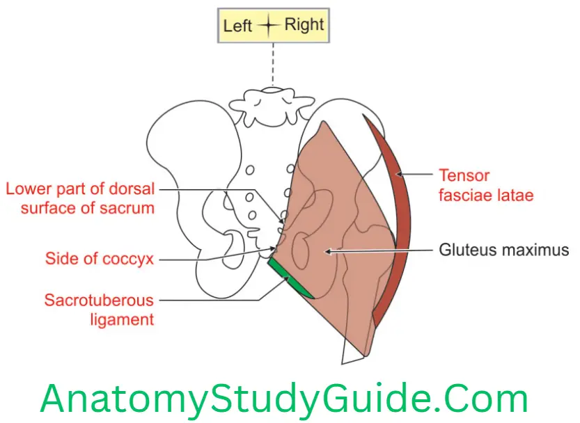
1. Gluteus Maximus Origin
1. Gluteus Maximus Aponeurotic: Aponeurosis of erector spinae.
2. Gluteus Maximus Bony
- Posterior gluteal line.
- Area posterior to the posterior gluteal line.
- Outer sloping area of the dorsal segment of the iliac crest.
- Dorsal surface of lower 2 pieces of the sacrum
- Side of coccyx
3. Gluteus Maximus Ligamentous: Sacrotuberous ligament.
4. Gluteus Maximus Fascial: Fascia covering the gluteus medius.
2. Gluteus Maximus Insertion
- Superficial major part (3/4th part) is inserted into the iliotibial tract.
- Deep, lower 1/4th part is inserted into the gluteal tuberosity.
3. Gluteus Maximus Features
1. It is
- Rhomboidal, that is to say, it is oblong whose opposite borders are parallel, but whose angles are not right-angled.
- Most powerful and bulkiest muscle in man.
- Largest and the most superficial of the gluteal muscles,
- Most fasciculated (clustered together) muscle in the body.
- Largely responsible for the prominence of the gluteal region.
- Known as the deltoid of the hip joint.
4. It has a coarse texture due to the presence of a large number of fibrous septa in its substance.
5. Gluteus Maximus Blood supply
1. Arterial
- Superior gluteal, branch of the posterior division of internal iliac artery and
- Inferior gluteal arteries, branch of the anterior division of internal iliac artery.
2. Gluteus Maximus Venous drainage: Veins form a plexus.
6. Gluteus Maximus Nerve supply: Inferior gluteal nerve
7. Gluteus Maximus Actions
- It is a powerful extensor of the hip joint.
- It is a powerful lateral rotator of the thigh.
- It acts in rising from a sitting position and in climbing the staircase.
- It supports the extended knee.
- It is an abductor of the thigh.
- The upper fibres of the muscle are a strong abductor of the thigh.
8. Gluteus Maximus Bursae related: There are usually three bursae beneath the muscle. They are bursa over
- Ischial tuberosity,
- Greater trochanter, and
- Vastus lateralis.
9. How to feel the muscle by oneself: If you climb stairs and put your hand on your buttock, you will feel the gluteus maximus contract strongly.
10. Gluteus Maximus Muscle testing: Patient lies prone. The right hand of the physician presses the patient’s right leg downwards. Patient is requested to extend his hip against resistance. Physician feels the contracting gluteus maximus muscle by left hand.
11. Gluteus Maximus Applied anatomy
- The sciatic nerve is explored by exposure at the lower border of gluteus maximus.
- The bursae associated with the gluteus maximus are prone to inflammation and are painful.
- The trochanteric bursa separates superior fibres of the gluteus maximus from the greater trochanter. The trochanteric bursa is commonly the largest of the bursae formed in relation to bony prominences and is present at birth. Other such bursae appear to form as a result of postnatal movement.
- The ischial bursa separates the inferior part of the gluteus maximus from the ischial tuberosity; it is often absent.
- The gluteofemoral bursa separates the iliotibial tract from the superior part of the proximal attachment of the vastus lateralis, a thigh muscle.
- Gluteus muscle is paralysed in muscular dystrophy.
- Climbing on one’s self: The patient with gluteus maximus paralysed, cannot standup from a sitting posture without support. Such patients rise gradually. He supports his hands first on legs and then on the thighs; and lastly, they stand. This is called climbing on oneself. This can be seen in pseudohypertrophy with muscular dystrophy syndrome (Duchenne muscular dystrophy).
Describe the structures under cover of Gluteus Maximus
1. Gluteus Maximus Bones,
2. Gluteus Maximus Muscles,
3. Gluteus Maximus Nerves,
4. Gluteus Maximus Vessels,
5. Gluteus Maximus Joints,
6. Gluteus Maximus Ligaments, and
7. Gluteus Maximus Bursae.
1. Gluteus Maximus Bones:
1. Elements of hip bone
- Ilium, and
- Ischium.
2. Sacrum and coccyx, and
3. Upper end and greater trochanter of femur.
2. Gluteus Maximus Muscles:
Key muscle is piriformis.
½ muscle is reflected head of rectus femoris.
The single (1) muscle is quadratus femoris.
The muscles in the pair (2) are
1. Gemelli
- Superior, and
- Inferior
2. Obturator
- Internus, and
- Externus.
The muscles in the trio (3) are
- Gluteus maximus is excluded as we are describing structures under the same,
- Gluteus medius, and
- Gluteus minimus.
The muscles in group of (4) are
- Semimembranosus,
- Semitendinosus,
- Ischial fibres of adductor magnus, and
- Biceps femoris.
3. Gluteus Maximus Nerves: The key nerve in this region is sciatic nerve. The other nerves under cover of gluteus maximus are the nerves supplying the muscles present in this region.
1. Key nerve is sciatic nerve.
2. Nerve to quadratus femoris,
3. Nerve to obturator internus,
4. Superior gluteal nerve,
5. Inferior gluteal nerve,
6. Pudendal nerve,
7. Posterior cutaneous nerve of thigh, and
8. Perforating cutaneous nerve branch of posterior cutaneous nerve of thigh.
4. Gluteus Maximus Vessels:
Vessels can be remembered by mnemonic
Superior gluteal artery.
Inferior gluteal artery.
Ascending branch of medial and lateral circumflex femoral arteries (profunda femoris).
Trochanteric anastomosis: The arteries taking part in trochanteric anastomosis are the arteries beginning with 1st 3rd letters of the key word “sciatica”.
Internal pudendal vessels.
Cruciate anastomosis: The arteries are above the trochanteric anastomosis and artery below the cruciate anastomosis.
Ascending branch of 1st perforating artery.
5. Gluteus Maximus Joint: Hip and sacroiliac joints.
6. Gluteus Maximus Ligaments
- Sacrotuberous,
- Sacrospinous, and
- Ischiofemoral ligament.
7. Gluteus Maximus Bursae: Under cover of gluteus maximus
- Trochanteric bursa: Bursa present at greater trochanter.
- Bursa over ischial tuberosity.
- Bursa between gluteus maximus and vastus lateralis.
Gluteus Medius
Gluteus Maximus Introduction: It is a fan-shaped muscle that covers the lateral surface of the bony pelvis.
1. Gluteus Maximus Features
- It is fan-shaped,
- The deep fascia over the gluteus medius is thick, dense, opaque and pearly white, and
- It is a post-axial muscle.
2. Gluteus Maximus Proximal Attachment: Gluteal surface of ilium between anterior and posterior gluteal lines.
3. Gluteus Maximus Distal attachment: Lateral surface of greater trochanter of femur along the oblique line that slopes downward and forward. Its distal attachment has a strong expansion that crosses the capsule of hip joint.
4. Gluteus Maximus Nerve supply: Superior gluteal nerve. It lies in the interval between gluteus medius and minimus; it divides into superior and inferior branches. The superior branch supplies the gluteus medius and the inferior branch supplies the gluteus medius, gluteus minimus and tensor fasciae latae.
5. Gluteus Maximus Relations: Structures deep to the gluteus medius are
- Superior gluteal nerve,
- Deep branch of the superior gluteal artery,
- Gluteus minimus, and
- Trochanteric bursa of the gluteus medius.
6. Gluteus Maximus Actions
- The most important action is to maintain the balance of the body when the opposite foot is off the ground, as in walking and running. They do this by preventing the opposite side of the pelvis from tilting downwards under the influence of gravity.
- The gluteus medius and minimus muscles are powerful abductor of the thigh at the hip joint when the limb is free to move. It occurs in all positions of the lower limbs.
- The anterior fibres of gluteus medius and minimus with the help of tensor fasciae latae produce medial rotation. It is restricted by tension in the lateral rotators and the Ischiofemoral ligament.
7. Gluteus Maximus Testing the gluteus medius: It is performed while the person is prone with the leg flexed to a right angle. The person abducts the thigh against resistance. The gluteus medius can be palpated inferior to the iliac crest, posterior to the tensor of the fascia lata. It is also contracts during abduction of the thigh.
8. Gluteus Maximus Applied anatomy
Intramuscular injection is given in the upper and lateral quadrant of the gluteal region. The needle enters gluteus medius.
Gluteus medius and minimus are supplied by the superior gluteal nerve. Injury to the superior gluteal nerve results in characteristic motor loss. It results in a gluteus medius limp.
Gluteus medius and minimus and poliomyelitis: The gluteus medius and minimus are supplied by the superior gluteal nerve. They may be paralysed when poliomyelitis involves the lower lumbar and sacral segments of the spinal
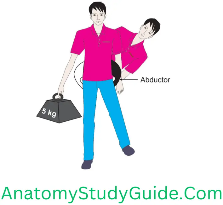
cord. Paralysis of these muscles seriously interferes with the ability of the patient to tilt the pelvis when walking.
The stability of the hip joint, when a person stands on one leg, depends on three factors.
- The gluteus medius and minimus must be functioning normally,
- The head of the femur must be located normally within the acetabulum, and
- The neck of the femur must be intact and must have a normal angle with the shaft of the femur.
- If any one of these factors is defective, then the pelvis sinks downward on the opposite, unsupported side, the patient is then said to exhibit a positive Trendelenburg’s sign.
Anterior approach of surgical exposure of the hip joint: It passes between gluteus medius and minimus laterally and sartorius medially. It then divides the reflected head of rectus femoris and exposes the anterior aspect of the hip joint. More room may be obtained by detaching these glutei from the external aspect of the ilium.
Cruciate Anastomosis
1. Cruciate Anastomosis Site: Cross-like anastomosing arteries are seen in the lower part of the gluteal region. They are present at the root of the greater trochanter between quadratus femoris and adductor magnus. To be precise, cruciate anastomosis is present at the middle of lesser trochanter.
2. Cruciate Anastomosis Arteries taking part
1. Transverse branch of
- Medial circumflex femoral artery, and
- Lateral circumflex femoral artery.
2. Descending branch of the inferior gluteal artery, and
3. Ascending branch of first perforating artery.
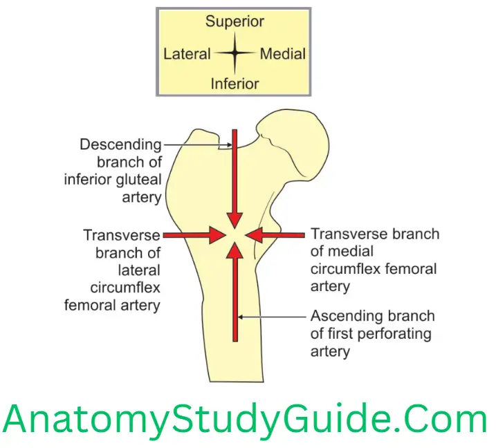
3. Cruciate Anastomosis Applied anatomy: This establishes the collateral circulation between internal iliac artery and profunda femoris artery. This is established in the case of ligature of the femoral artery proximal to the attachment of profunda femoris artery.
Leave a Reply