General Anatomy Muscles Notes And Important Questions With Answers
What is a Sarcomere?
Table of Contents
Cardiac Muscle Tissue
1. Sarcomere Features
- A sarcomere is segment of the fibre between successive Z bands.
- It is the smallest structural and functional contractile unit of the muscle.
- Repeating contractile units are seen along the entire length of each myofibril.
- Highly characteristic features of the sarcoplasm of skeletal and cardiac muscle fibres. It is 2.5 long.
- It contains the A” band and 1/2 of the “I” band.
- Myofibril is the finer structural and functional unit of the sarcomere.
2. The sarcomere has a central dark and peripheral light band.
3. The dark staining band contains thick myosin filaments.
Read And Learn More: Anatomy Notes And Important Question And Answers
4. The light staining band contains thin actin filaments.
5. Actin and myosin filaments are precisely aligned.
6. They are stabilised within individual myofibrils. The sarcomeres are stabilised by
accessory proteins.
7. The thin actin filaments are bound to the protein called -actinin. This binds them to the dense Z line (band).
8. The thick myosin filaments are anchored to the Z line by the very large protein called titin. Titin keeps myosin filament in the centre of the Z line. It acts like a spring between the end of the myosin filament and the Z line.
Pennate Muscles
1. Unipennate: The fleshy fibres have a linear or narrow origin. The appearance is that of 1/2 of a feather;
1. Pennate Muscles Examples
1. Extensors
- Extensor digitorum longus
- Peroneus tertius
2. Flexor: Flexor pollicis longus
3. Adductor: Palmar interossei
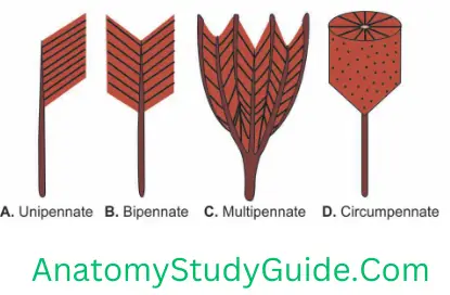
2. Bipennate: The fleshy fibres arise from a long broad surface, e.g.,
1. Muscle of thigh: Rectus femoris
2. Muscles of sole:
- Flexor hallucis longus,
- Dorsal interossei, and
- Peroneus longus
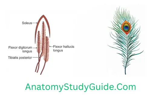
3. Multipennate: The septa extend into the origin and insertion. It gives the appearance is that of many feathers, e.g.
- Subscapularis
- Deltoid (acromial fibres)
4. Circumpennate e.g. tibialis anterior
Prime Movers (Agonists)
Prime Movers (Agonists) Introduction: The muscles which initiate and carry out the desired action by active contractions (shortening) are called prime movers, e.g. in the flexion of elbow joint, the biceps muscle is prime mover.
1. A muscle may perform all the 4 roles under different situations. Flexor carpi ulnaris is a prime mover in flexion at the wrist and an antagonist in extension. It acts as a fixator in flexion of the thumb and is a synergist in extension of the thumb.
2. Contraction of muscle may be isotonic or isometric.
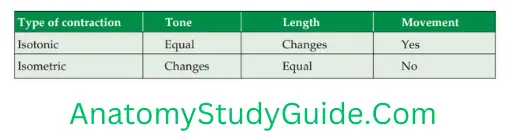
3. Isotonic (iso—equal, tonic—tone): During contraction, the length of the muscle decreases to 1/3rd and the tone of the muscle remains unchanged. It is associated with the movement, e.g. in flexion of the elbow joint, length of biceps is decreased but the tone remains unchanged.
Cardiac Muscle Tissue
4. Isometric (iso—equal, metric—measurement): During contraction, the length of muscle fibre remains unchanged but the tone of the muscles changed, e.g. after semi-flexion of the elbow joint against resistance, the length is not decreased but tone is changed.
5. Following are the forces opposing the action of the muscle
- Antagonist
- Gravity: It is a valuable aid to some movements, depending upon the position of the limb of the body, e.g. on raising the arm from the side, the deltoid is a prime mover at the shoulder and the gravity (certainly if there is a weight in the hand) is sufficient to lower the arm.
- Connective tissue resistance.
- Active resistance.
6. The range and force of the movement is directly proportional to
- 1. Length of the fibres, and
- 2. Number of the fibres.
7. Law of Sherrington: When the agonists or prime movers are active, antagonists are inhibited by reciprocal innervation. When a muscle receives a nerve impulse to contract, its antagonist muscle receives simultaneously an impulse to relax. Flexion of wrist joint is opposed by the extensor.
8. Action of paradox: When a prime mover helps opposite action by active controlled lengthening against gravity, it is known as action of paradox, e.g. putting a glass on a table is assisted by gravity but controlled by gradual, active lengthening of biceps.
Antagonists
Antagonists Introduction: They oppose the prime movers. They help the prime movers by actively controlling fractional relaxation so that the desired movement is smooth and precise.
Antagonistic Definition
- Thus, the antagonists cooperate rather than oppose the prime movers. This is due to reciprocal innervation of the opposite groups of muscles, regulated by the spinal cord through the stretch reflex. Antagonist’s muscles pass over the opposite side of the axis of rotation.
- Flexion of the wrist joint is done by flexor carpi radialis and ulnaris, which is opposed by extensor carpi radialis longus, brevis and extensor carpi ulnaris.
- Flexion of the digit is done by efficient extension of the wrist joint. This is done by flexor digitorum superficialis and profundus helped by extensor digitorum, which is the antagonist.
Fixators
Fixators Introduction: The accessory muscles which steady the proximal joint to bring the desired action on the joint under consideration.
In flexion of wrist joint, the rotator cuff (subscapularis, infraspinatus and teres minor) fix the shoulder joint, to have the smooth movement at the wrist joint.
Define Synergists
Synergists Introduction: Muscles that assist prime movers are called synergists. When a prime mover passes over more than 1 joint, certain muscles are required to steady the unstable joint such muscles are called synergists. Muscles cross the same site of axis of rotation.
1. During flexion of elbow, biceps muscle brings flexion of elbow assisted by brachialis which is synergist.
2. Flexion of digit is efficiently done by efficient extension of wrist joint. This is done by flexor digitorum superficialis and profundus helped by extensor digitorum which is antagonist.
3. In most instances, motion at a joint is initiated by one set of synergistic muscles and brought to close by the antagonists. For example, controlled flexion of the forearm at the elbow joint is initiated by flexor muscles and slowed or stopped at any desired position by extensor muscles.
4. Simultaneous contraction of both synergists and antagonists produces maximal joint stability (dynamic stability) with a little or no movement.
Leave a Reply