Nose and Paranasal Sinuses Anatomy Notes And Important Questions With Answers
Question: Why are the boils of nose and ear painful?
Table of Contents
Answer: Nose and ear are firmly attached to cartilages. There is no space for expansion of infection or pus. Hence, the cutaneous nerves are irritated and cause pain.
Question 2: What is epiphora?
Answer: It is an abnormal overflow of tears down the cheek. It is mainly due to stricture of the passage. It is also called ill lacrimation.
Read And Learn More: Face Anatomy Notes And Important Questions
Question 3: Describe nasal septum under the following heads:
1. Formation,
2. Blood supply,
3. Nerve supply, and
4. Applied anatomy
Answer: It is usually deviated to one side and each nasal cavity some what asymmetrical.
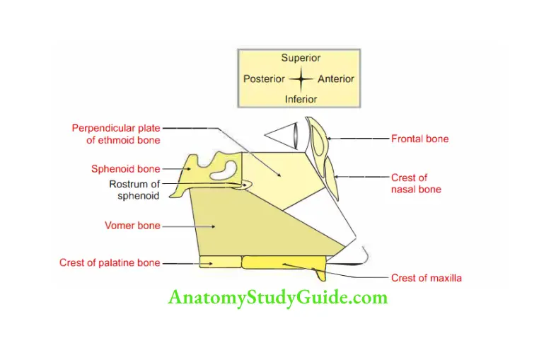
1. Formation: Partly by bones and partly by cartilage.
1. Bony part
1. Major part is formed by
- Vomer: Below and behind.
- Perpendicular plate of ethmoid.
2. Small bones taking part are
- Nasal spine of frontal bone,
- Anterior nasal spine of maxilla, and
- Rostrum of sphenoid
3. Crests of
- Nasal bone,
- Maxilla, and
- Palatine bone.
2. Cartilaginous part is fonned by
- Septal cartilage.
- Septal process of inferior nasal cartilage.
3. Cuticular part is formed by fibrofatty tissue.
2. Blood supply
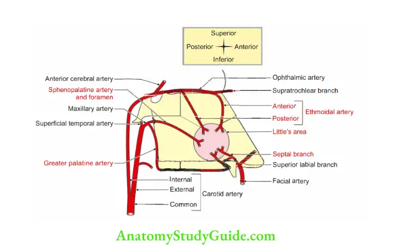
1. Arterial supply
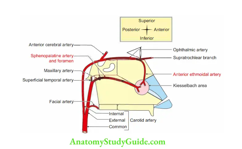
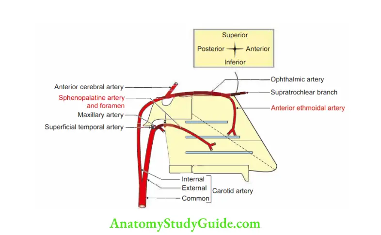
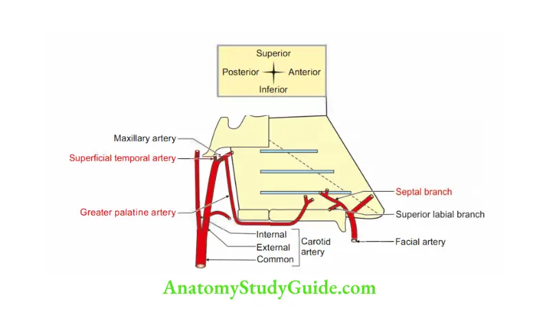
1. The main arteries of the nasal septum are
- The anterior ethmoidal artery, a branch of the ophthalmic artery, supplies the anterior superior part.
- The sphenopalatine artery, a branch of the maxillary artery, supplies the posterior inferior part.
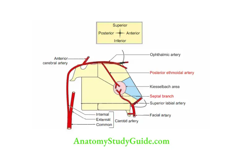
2. Less important arteries are
- Superior labial, a branch of the facial artery,
- Greater palatine artery, a branch of the maxillary artery, and
- Posterior ethmoidal artery, a branch of ophthalmic artery.
Note:
- All the arteries supplying the nasal septum are branches of the external carotid artery except anterior and posterior ethmoidal arteries which are branches of ophthalmic artery which is a branch of the internal carotid artery.
- All the arteries give septal branches which form important anastomosis at the anteroinferior quadrant of nasal septum.
It is called Kiesselbach plexus.
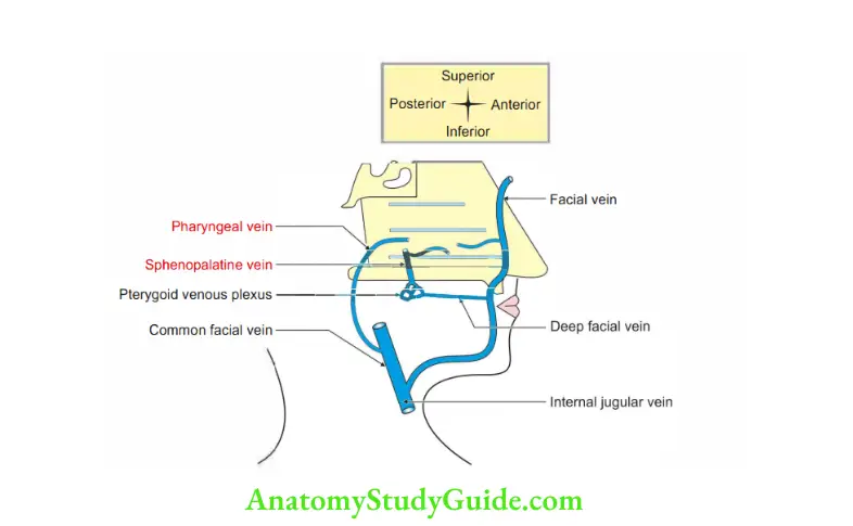
2. Venous drainage
- Anterosuperior part is drained by superior ophthalmic vein, which opens into cavernous sinus.
- Posteroinferior part is drained into pterygoid venous plexus.
- Upper part of the septum is drained into inferior cerebral vein.
- Lower mobile part of septum drains into facial vein which drains into internal jugular vein.
An infection from this part may extend into cavernous sinus via
- Deep facial vein, and
- Pterygoid venous plexus.
This belongs to dangerous area of face
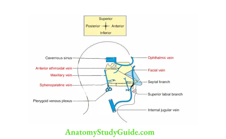
3. Lymphatic drainage
- Anterior half: To the submandibular nodes.
- Posterior half: To the retropharyngeal and deep cervical nodes.
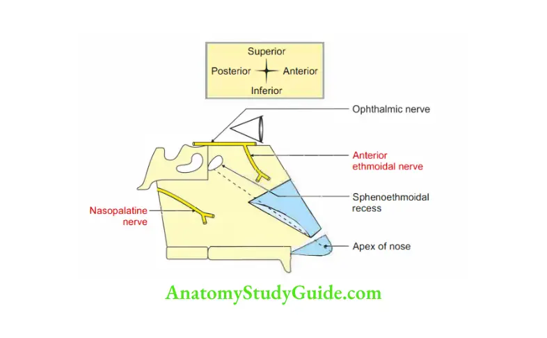
3. Nerve supply
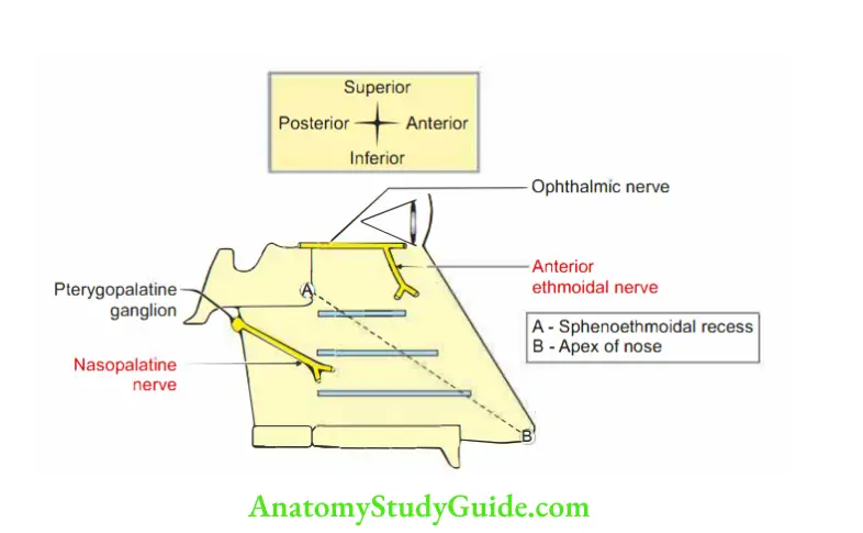
1. General sensory nerves.
- Anterosuperior part: Anterior ethmoidal nerve, a branch of ophthalmic nerve
- Posteroinferior part: Nasopalatine branch of pterygopalatine ganglion.
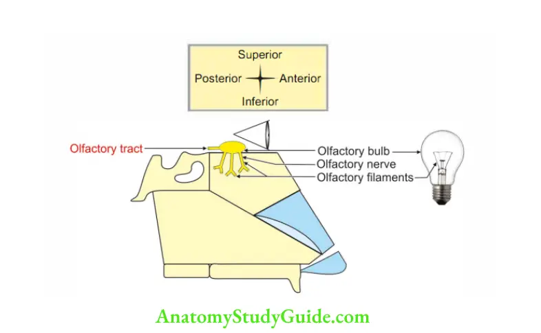
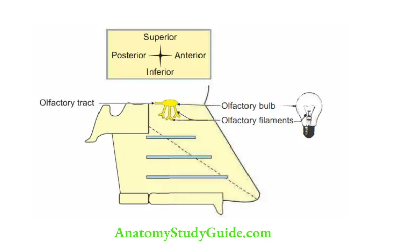
2. Special sensory nerves are olfactory nerves.
4. Applied anatomy
1. Deviation of nasal septum may be due to cartilage or bone. It may be due to
- Birth injury (most common cause).
- Congenital malformation.
- Excessive nasal deviation produces unilateral nasal obstruction and is treated by submucous resection of the septum (SMR)-septoplasty
2. Little’s area or Kiesselbach’s area for epistaxis.
Little’s area or Kiesselbach’s area
Introduction: It is an area on anteroinferior part of nasal septum.
The nose bleeding is relatively common because nasal mucosa is highly vascular.
The mild epistaxis results from nose pricking which tears the veins in the vestibule.
1. The profuse bleeding occurs due to rupture of one of the following arteries:
- Septal branch of anterior ethmoidal artery (branch of ophthalmic artery).
- Septal branch of the superior labial artery, a branch of facial artery.
- Septal branch of sphenopalatine artery .
- Septal branch of greater palatine artery } (maxillary artery).
2. The anastomosis formed by these arteries is called “Kiesselbach’s plexus”.
Even a small ulcer affecting this area can cause profuse bleeding.
3. Septal branch of sphenopalatine artery is the longest and tortuous. It is also called ur “artery of nose bleeding” or “rhinologist’s artery.
4. Sudden and severe nasal bleeding in elderly hyprtensive patient may be due to rupture of the venous communication.
It acts as nature’s safety procedure to reduce increased intracranial vascular pressure.
Question4: Describe lateral wall of nose under the following heads:
1. Formation,
2. Features,
3. Blood supply,
4. Nerve supply, and
5. Applied anatomy
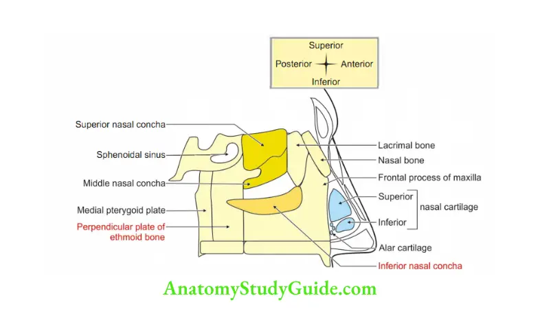
Answer:1. Formation
1. Bony part is formed by
- Conchae of
- Superior } concha of ethmoid bone
- Inferior nasal concha
- Lacrimal
- Maxilla
- Nasal
- Perpendicular plate of palatine bone.
- Medial pterygoid plate of sphenoid bone.
2. Cartilaginous part is formed by
- Upper nasal cartilage,
- Lower nasal cartilage, and
- Alar cartilage.
3. Cuticular part is formed by fibrofatty tissue
2. Features
1. The epithelium of lateral wall is as follows:
- Above superior concha: Olfactory epithelium.
- Below superior concha: Respiratory mucosa.
- Below inferior concha: Erectile tissue.
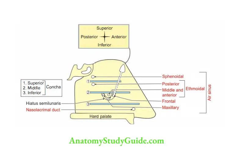
2. Lateral wall shows bony projections called nasal conchae. These are:
- Superior concha: A projection of ethmoid bone.
This is the smallest concha,situated above the middle concha.
It encloses a space called superior meatus. - Middle concha: A projection of ethmoid bone. It encloses middle meatus.
- Inferior concha: It is independent bone. It encloses inferior meatus.
3. Blood supply
1. Arterial supply

2. Venous drainage
- Anterior veins form a plexus and drain into facial vein.
- Posterior veins drain into pharyngeal plexus of veins.
- Middle part drains into pterygoid plexus of veins.
3. Lymphatic drainage
- Anterior ½ drains into submandibular lymph nodes.
- Posterior ½ drains into retropharyngeal and deep cervical lymph nodes.
4. Nerve supply
- Special sensory nerve : Olfactory (!)-upper part.
- General sensory nerve : Trigeminal (V).

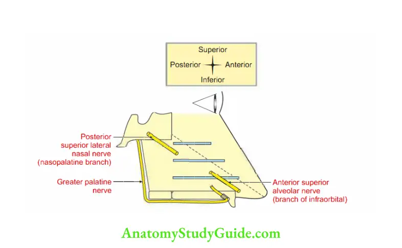
5. Applied anatomy
- Common cold is the commonest viral infection of nose.
- Paranasal air sinus may get infected from the infection of nose.
- Hypertrophy of mucosa over the inferior nasal concha is a common feature of allergic rhinitis presenting as sneezing, nasal blockage and excessive watery discharge.
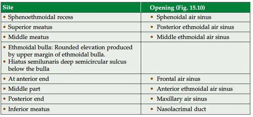
Question 5: Ethmoidal air sinuses
Answer:Introduction: They are small numerous spaces present in the labyrinth of the ethmoid bone.
1. Features:
1. They are completed from
- Above by the orbital plate of the frontal bone,
- Behind by the
- Sphenoidal conchae and
- Orbital process of the palatine bone, and
3. Anteriorly by the lacrimal bone.
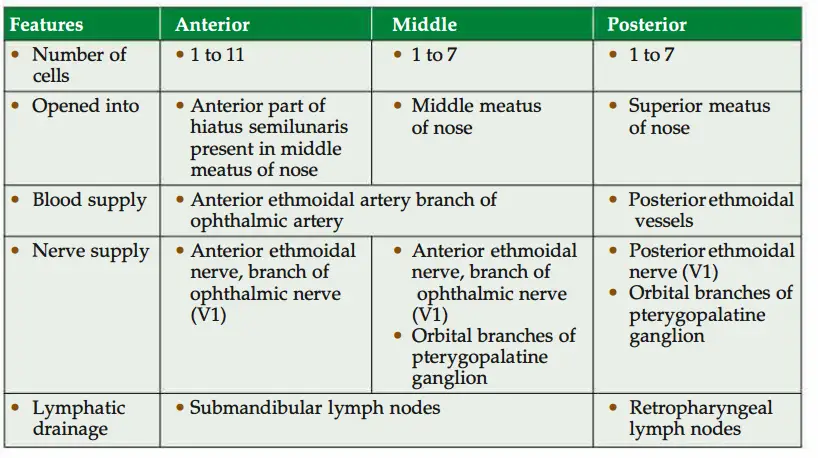
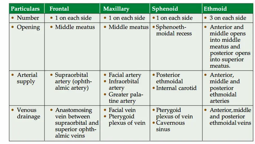
2. The sinuses are divided as given in Table 15.4.
3. Applied anatomy
- Fracture of the medial wall of the orbit may damage ethmoidal air sinus.
- Malignant tumour of ethmoidal sinus may erode the orbit and results in exophthalmos.
- If nasal drainage is blocked, infections of the ethmoidal cells may break through the fragile medial wall of the orbit.
- Severe infections from ethmoidal air sinus may cause blindness. It is because of close proximity with optic canal, which gives passage to the optic nerve and ophthalmic artery.
- Spread of infection from ethmoidal cells can also affect the dural nerve sheath of the optic nerve. It results in optic neuritis.
Frontal sinus
1. Morphology
- Number: There are two frontal air sinuses. Each is situated between the inner and outer tables of frontal bone.
- Situation: It is deep to supraorbital margin of fontal bone.
- Shape. triangular.
- Symmetry There is an oblique septum separating the right and left sinuses.
Hence, they are asymmetrical. - Dimensions
Vertical: 3 cm
Transverse: 2.5 cm
Anteroposterior: 1.8 cm. - Age changes: It is rudimentary at birth.
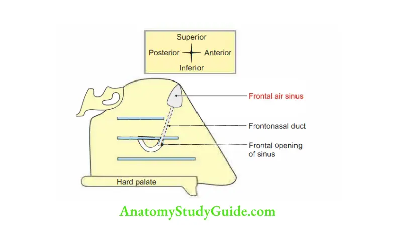
- Communications: It opens into anterior part of hiatus semilunaris.
This is ½ moon ( shaped depression present in middle meatus part of lateral wall of nose.
2. Features and relations
- Anterior wall of the sinus is thick and is related to the skin of the forehead.
- The posterior wall is thin and related to the meninges and frontal lobe of brain.
- Its inferior wall forms the roof of the orbit.
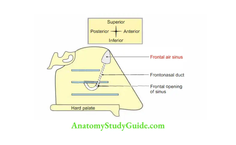
- The frontonasal duct begins in the opening of the sinus, which is located in the floor of the sinus.
3. Blood supply
1. Arterial supply
- Supraorbital artery, branch of ophthalmic artery.
- Anterior ethmoidal artery, a branch of ophthalmic artery arising from main trunk.
2. Venous drainage Supraorbital vein drains into angular vein-facial veininternal jugular vein.
4 Lymphatic drainage: Submandibular nodes.
5.Nerve supply: Supraorbital nerve, branch ofophthlmic divisionoftrigeminal nerve.
6. Development: Frontal air sinuses are the only sinuses not present at birth, they appear during the 2nd year.7.
7. Surface anatomy: Join following three points
- Point at nasion
- Point 3 cm above the nasion
- Point at the supraorbital margin at the junction of its medial one-third and lateral two-thirds.
8. Applied anatomy
- Pain of frontal sinusitis may be referred to the skin of the forehead and adjacent skin since they have same nerve supply.
- Frontal air sinus communicates with maxillary air sinus through infundibulum. It is located at higher level.
It tracks down into the maxillary air sinus. - The patients with frontal sinusitis nearly always have a maxillary sinusitis.
Why is headache the commonest presentation in involvement of nose, paranasal sinuses, teeth, gums, eyes (refractory error) and meninges?
1. The dura is sensitive to stretching. Pain arising from the dura is generally referred,perceived as a headache. It arises in cutaneous or mucosa! regions. It is supplied bythe cervical nerve or division of the trigeminal nerve.
2. The trigeminal nerve has three branches-ophthalmic, maxillary and mandibular.
3. The ophthalmic nerve is sensory nerve of eyeball.
4. The maxillary nerve is the sensory nerve of nose (common cold, boils), paranasal air sinuses (sinusitis), gums and teeth of upper jaw.
5. The mandibular nerve is the sensory nerve of gums and teeth of the lower jaw.
6. Hence, headache is a uniformly common symptom in
- Refractive errors of the eyes,
- Nose (common cold, boils) and the paranasal air sinuses (sinusitis),
- Infections and inflammations of teeth and gums, and
- Infection of the meninges as in meningitis.
Question 6: What are the junctions of paranasal sinuses?
Answer: Frontal air sinus and maxillary air sinus in the middle meatus in the lateral part of nose.
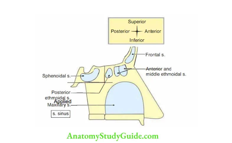
Paranasal sinuses
Introduction: These are air-filled spaces within the bones present around air passage.
1. Functions
- They add resonance to the voice.
- They improve the timbre of the voice.
- They condition the inhaled air by adding humidity.
2. Applied anatomy
- Infection of the sinus is known as sinusitis. It causes headache and persistent,thick, purulent discharge from the nose.
Diagnosis is done by X-ray.
A diseased sinus is opaque. - Frontal sinusitis and ethmoidal sinusitis can produce a brain abscess in the frontal lobe.
- Carcinoma of the maxillary sinus produces symptoms depending upon the growth.

Question 7: What is the clinical importance of maxillary sinus?
Answer:1. It may become infected either from the
- Nasal cavity, or
- Caries of the upper molar teeth.
2. Antral puncture is carried out using a trocar and cannula. It is passed through the nasal cavity in an outward and backward direction. The trocar passes below the inferior concha.
3. Caldwell-Luc operation: It is removing a portion of the medial wall of the sinus below the inferior concha. It is done to facilitate the drainage.
4. The carcinoma of the maxillary sinus produces.
- Obstruction of the nares and epistaxis by medial invasion.
- Blockage of the nasolacrimal duct by invasion of duct.
- Diplopia by invasion of the orbit.
- Facial pain by the invasion of infraorbital nerve.
- Ulceration in the palatal roof by invasion of sinus.
- Swelling of the face by spreading laterally.
- Posterior spread may involve th palatine nerves and produce severe pain referred to the teeth of the upper jaw.
Maxillary air sinus (antrum of Highmore)
Introduction: The largest and important paranasal air sinus present in maxilla, lined
by ciliated columnar epithelium.
1. Importance
- Helps in conditioning of the air by adding humidity.
- Acts as resonating chamber for production of sounds.
- Increases the quality of voice (timbre).
- Reduces the weight of the skull.
2. Gross anatomy: Shape is pyramidal;&
- Base is formed by nasal surface of body of maxilla and forms lateral wall of nose.
- Apex: Towards the zygomatic bone.
3. Boundaries
- Superior wall or roof: Orbital surface of maxilla.
- Inferior wall or floor: Alveolar surface of maxilla.
- Anterior wall: Anterior surface of maxilla.
- Posterior wall: Posterior surface of maxilla.
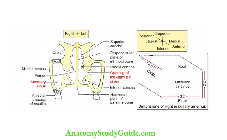
4. Dimension
- Vertical: 3.5 cm.
- Transverse: 2.5 cm
- Anteroposterior: 3.25 cm.
5. Opening: Maxillary air sinus opens in the middle meatus by two openings.
1. Upper opening is present in lower part of hiatus semilunaris. The opening is at higher level and is called antrum of Highmore (dental surgeon). The dimensions “of opening are reduced
- Superiorly by
Uncinate process of ethmoid bone
Descending part of lacrimal bone - Inferiorly by: Inferior nasal concha.
- Posteriorly by: Perpendicular plate of palatine bone.
- Internally by: Thick mucosa.
- 2. Lower opening is present at posterior end of hiatus.
6. Relations of maxillary air sinus
1. Anterolaterally related to
- Infraorbital nerves
- Infraorbital vessels
- Origin of muscles of upper lip
- Anterior superior alveolar vessels and nerves.
2. Posteriorly
- Infratemporal fossa,
- Pterygopalatine fossa,
- Posterior superior alveolar vessels and nerves.
4. Floor is related to roots of teeth especially 2nd premolar and 1st molar.
4. Roof is related to the floor of the orbit and eyeball.
5. Medially, it is related to lateral wall of nose.
6. Laterally, it is related to cheek.
7. Development: Developed from splitting of maxilla.
It is the 1st paranasal air sinus to develop.
It develops in the 4thmonth of intrauterine life.
It grows rapidly during 6years and reaches full size after the eruption of all permanent teeth.
8. Blood supply
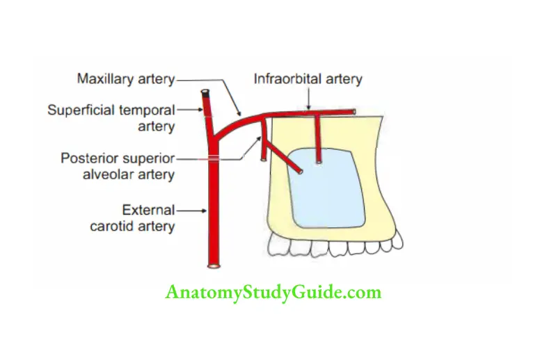
1. Arterial supply
- Infraorbital artery
- Greater palatine artery
- Posterior superior alveolar artery } Branches of maxillary artery
2. Venous drainage
- Infraorbital vein drains into angular vein.
- Greater palatine vein drains into pterygoid venous plexus.
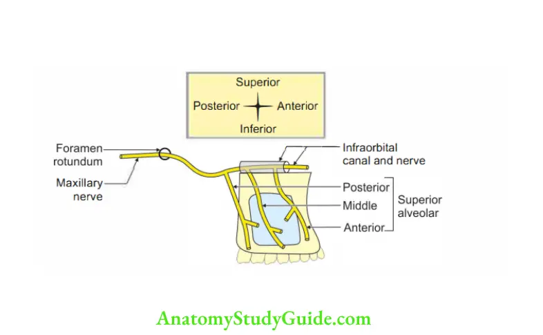
9. Nerve supply
- Infraorbital nerve (continuation of maxillary nerve).
- Greater palatine nerve (pterygopalatine ganglion)
10. Lymphatic drainage: By submandibular lymph node.
11. Applied anatomy
- Sinusitis: Inflammation of maxillary air sinus presents headache, which is maximum at 12:00 noon.
At 12:00 noon, there is maximum expansion of the air which is not accommodated in the maxillary air sinus.
Hence, it passes to the I frontal air sinus. - It can be visualised by taking X-rays of paranasal sinus.
- Abscess of maxillary air sinus is drained by antral puncture.
- The opening of maxillary air sinus is located much higher than the floor.
There is poor natural drainage. Hence, the maxillary sinus is most commonly infected. - Maxillary sinus becomes infected either from the
Nasal cavity or from
Caries of the upper molar teeth. - The maxillary sinus is sometimes called the ‘secondar reservoir’ of the frontal air sinus.
The frontal sinus drains into the hiatus semilunaris in the middle meatus, via infundibulum.
It is close to the opening of maxillary sinus. - The mucus or pus from maxillary sinus cannt be drained into the nasal cavity when the head is erect, until it is filled up to the top.
- In severe cases, drainage of maxillary sinus may require a surgical intervention.
The ‘antral puncture’ is done by passing a trocar and cannula through the nasal cavity.
It is directed in an outward and backward direction below the inferior nasal concha. It produces a hole in the lower part of the lateral wall of the nasal cavity. - For more adequate drainage, a portion of anterior wall of the sinus below the inferior nasal concha is removed.
Or, the sinus is fenestrated in the region of gingivolabial fold (Caldwell-Luc operation).
Applied anatomy of relations of maxillary air sinus
1. A tumour in the sinus may push
- Orbital floor and displace the eyeball.
- Project into the nasal cavity causing nasal obstruction and bleeding.
- Protrude into cheek causing numbness and swelling when the infraorbital nerve is damaged.
- It spreads back into infratemporal fossa, causing restriction of mouth opening due to pterygoid muscle damage and pain.
- Or spread down in th mouth looseni of th teethndmalocclusion of th teeth.
Pterygopalatine ganglion
Introduction: Ganglion is a collection of cell bodies.
It is the largest peripheral parasympathetic ganglion.
It is present in the course of parasympathetic nerve.
It is situated in the peripheral part of cranium.
1. Structurally, it belongs to trigeminal nerve since it is suspended from the maxillary nerve.
2. Functionally, it is related to greater petrosal nerve (facial nerve).
Eponym -Meckel’s ganglion.
1. Gross
1. Situation: Pterygopalatine fossa.
2. Relations
- Medially: Pharyngeal artery.
- Laterally: Artery of pterygoid canal.
- Superiorly: Maxillary nerve.
- Posteriorly: Pterygoid canal.
2. Connections
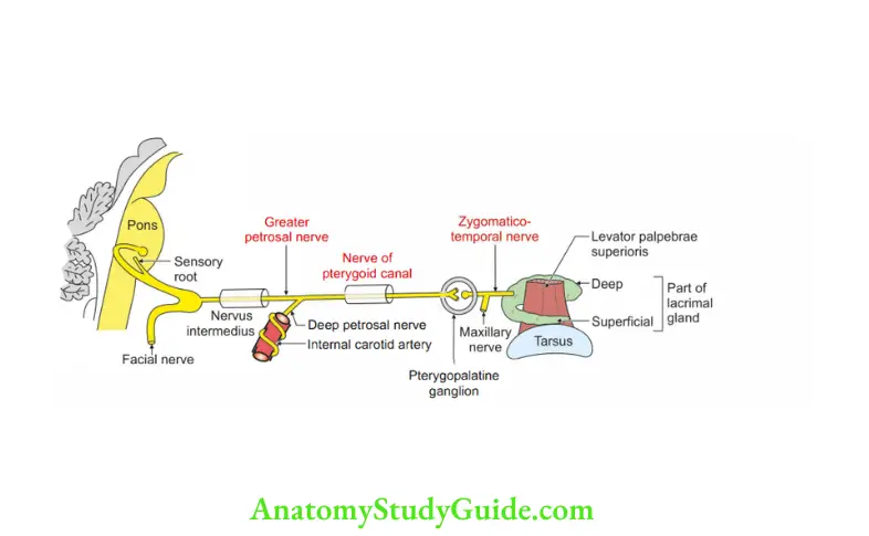
1. Parasympathetic (motor root)
- Preganglionic fibres arise from lacrimatorynucleus (pons)-facial nerve geniculate ganglion-greater petrosal nerve-deep petrosal nerve (plexus around internal carotid artery)-nerve of pterygoid canal-pterygopalatine ganglionrelay.
- Postganglionic fibers-maxillary nerve-zygomatic branch-zygomatic otemporal branch-communicating branch to lacrimal nerve.
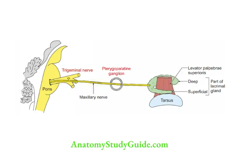
2. Sensory: From maxillary nerve passes through pterygopalatine ganglion without relay
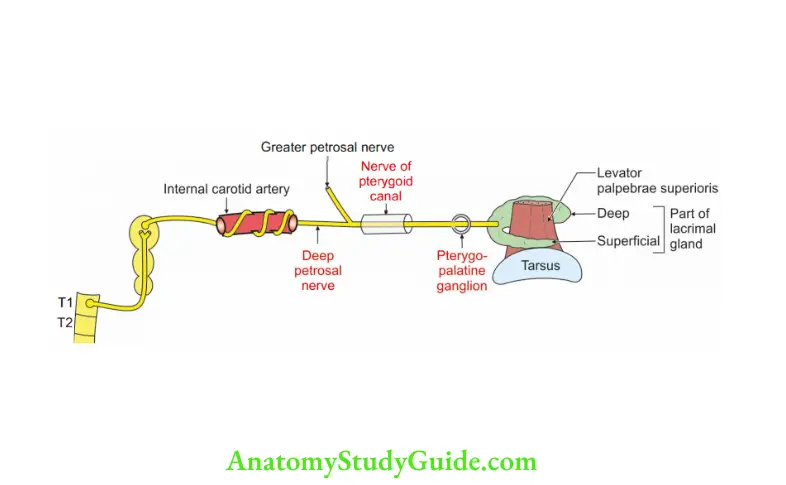
3. Sympathetic,
- Preganglionic fibres-spinal nerve-superior cervical sympathetic ganglion.
- Postganglionic fibres-plexus around internal carotid artery (deep petrosal nerve) passes without interruption.
3. Branches: They are virtually derived from maxillary nerve.
1. Orbital branch supplies
- Orbitalis muscle,
- Mucous membrane of sphenoid, and
- Mucous membrane of posterior ethmoidal sinus.
2. Palatine
- Greater palatine nerve-supplies mucous membrane of lateral wall of nose.
- Lesser petrosal nerve-supplies mucous membrane of soft palate and palatine tonsil.
3. Nasal branches
- Posterior superior lateral nasal.
- Medial nasal.
4. Applied anatomy
- It is called ganglion of hay fever and produces running of nose and eyes.
- Injection ofalcohol is occasionally employed inntractablecases ofallergic rhinitis.
Question 8: Describe maxillary nerve under
1. Embryology,
2. Course, and
3. Branches and distribution of the branches
Answer:Introduction: It is the 2nd division of trigeminal nerve (5th cranial nerve). It is purely completely sensory. It innervates meninges,
1. Skin of
- Temporal region,
- Scalp,
- Lower eyelid,
- Side of nose,
- Nasal septum,
- Cheek, and
- Upper lip
2. Air sinus
- Ethmoidal air sinus,
- Maxillary air sinus,
3. Teeth of upper jaw,
4. Upper gingivae and adjoining part of cheek,
5. Lateral wall of nose,
6. Floor of nasal cavity,
7. Adjoining part of nasal septum,
8. Lacrimal gland, and
9. Hard palate.
1. Embryology: It is said to be the pretrematic branch of trigeminal nerve. It supplies the derivative of the maxillary process.
2. Course: The nerve runs in the lateral wall of cavernous sinus, below the ophthalmic nerve. In the middle cranial fossa, it has a short course. It gives a meningeal branch.
- It leaves the skull via foramen rotundum and leads directly into posterior wall of pterygopalatine fossa.
It gives two larger ganglionic branches containing fibres to nose, palate and pharynx. - It inclines on the posterior surface of palatine bone and reaches on posterior surface of maxilla, runs through inferior orbital fissure.
It lies outside orbital periosteum and gives zygomatic and posterior superior alveolar branch.
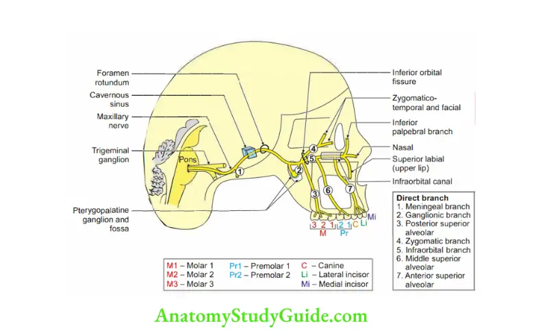
3. Branches and distribution of the branches-My good pretty zoology instructor
1. Branches from the trunk of maxillary nerve
- Meningeal: It is 1st branch arising from the trunk of mandibular nerve. It is given in the middle cranial fossa. It supplies the dura mater of anterior part of middle cranial fossa.
- Ganglionic branches: They are two in number. They suspend the ganglion.
- Posterior superior alveolar nerves (posterior superior dental): They are usually thr in number. The branches are given in the terygopalatine fossa. They pass downwards and laterally though pterygomaxillary fisure to reach posterior surface of maxilla. Here, they divide into numerous small branches. They enter th maxilla through posterior superior alveolar foramina. They supply
Maxillary sinus,
Upper molar teeth, and
Adjacent gum of the vestibule. - Zygatic: It arises from the trunk of maxillary nerve just before maxillary nerve enters the inferior orbital fissure. It enters the inferior orbital fissure and divides into
- Zygomaticotemporal: It is one of the terminal branches of zygomatic nerve.
It traverses through a canal in the zygomatic bone to emerge into anterior part of temporal fossa. It supplies - Skin above zygomatic arch.
Communicating twig to lacrimal nerve. - Zygomaticofacial: It is one of the terminal branches of zygomatic nerve. It passes through zygomaticofacial foramen. It supplies skin over the bone.
Infraorbital nerve: It is a terminal branch of maxillary nerve. It leaves pterygopalatine fossa and passes through the inferior orbital fissure. It passes forward along the floor of the orbit, sinks into groove. It enters infraorbital canal and emerges on the face through infraorbital foramen.
2. Branches on the face: They lie between levator labii superioris and levator anguli oris. They are described in two groups.
Branches given in the infraorbital canal
- Middle superior alveolar nerve: It is not always present. Whn present, most frequently arises as a branch of the infraorbital nerve. It supplies upper premolar
- Variations:
- It may directly arise from the maxillary nerve in the pterygopalatine fossa.
- It may arise as a branch of the anterior superior alveolar nerve.
- Anterior superior alveolar nerve: It arises at midpoint of infraorbital canal and enters the fine sinuous canal which passes downwards in the maxilla. It supplies
- Incisor and
- Canine teeth.
- It gives a nasal branch which supplies
- Anteroinferior quadrant of the lateral wall of nasal cavity.
Floor of the nasal cavity - Adjoining part of nasal septum.
Branches outside the infraorbital canal. They are divided into three groups.
- Palpebral branches: They supply skin in the lower eyelid
- Nasal branch: It supplies
- Skin of the
- Side of nose
- Movable part of nasal septum.
- Superior labial branches: They are large and numerous. They supply
- Skin of the
- Upper part of cheek
- Upper lip.
Branches frm the pteryopalatine ganglion: There are five branches that are distributed to nose, palate and nasopharynx.
Every branch carries sensory, secretomotor and sympathetic fibres.
1. Orbital branches: It enters the inferior orbital fissure and supply
- Orbital periosteum,
- Sphenoidal air sinus,
- Ethmoidal air sinus,
- Orbital branches join branches of carotid plexus. The plexus supplies
- Orbitalis, and
- Lacrimal gland.
2. Nasal branches: These enter the sphenopalatine foramen and divide into
- Posterior superior medial nasal nerves: They are 2 to 3 in number. One of the largest branches is called nasopalatine nerve.
As the word suggests, they supply medial part of nose, i.e. - Nasal septum
- Mucosa of posterior part of roof
- Nasopalatine (long sphenopalatine): It is the largest branch of posterior superior medial nasal branch.
It enters the posterior part of septum and turns down through incisive fossa and reaches the anterior part of hard “palate. As the name suggests, it supplies - Lower part of nasal septum
- Anterior part of hard palate.
- Posterior superior lateral nasal nerve (short sphenopalatine): As the name suggests, it supplies
- Posterior superior quadrant of lateral wall of nose which includes
- Superior concha and superior meatus,
- Middle concha, middle meatus, and
- Posterior ethmoidal sinus.
3. Palatine branches: They pass downwards. They are greater palatine and lesser palatine nerves.
- Greater palatine nerve (anterior palatine nerve) descends through greater palatine canal and emerges on hard palate and runs forwards up to the incisor teeth. It supplies
- Gingivae
- Mucosa and glands of hard palate and communicate with the terminal filaments of the nasopalatine nerves.
- In the greater palatine canal, it gives
- Posterior superior nasal branch which pierces the perpendicular plate of palatine bone and supply
- Mucous membrane of posterior superior quadrant of the lateral wall of nose.
- Outside the greater palatine canal, it gives branches to both surfaces of adjacent part of soft palate.
- Lesser palatine nerves: They are much smaller than greater palatine.
- They are descending through greater palatine canal and emerge through lesser palatine foramen. They innervate.
- Uvula,
- Tonsil, and
- Soft palate.
- Pharyngeal branches: It leaves the ganglion posteriorly. It passes through palatovaginal canal and supplies mucosa of nasopharynx behind pharyngotympanic tube.
- Lacrimal branches: They carry secretomotor fibres to the lacrimal gland. They travel through zygomaticotemporal branch of zygomatic nerve.

Question9: Describe sphenoidal air sinus under the following heads:
1. Morphology,
2. Relations,
3. Communication,
4. Blood supply,
5. Lyphatic drainage,
6. Nerve supply,
7. Development, and
8. Applied anatomy (Sphenoid-wedge-like)
Answer: Introduction: These are paired sinuses located within the body of sphenoid bone.
1. Morphology
1. Situation: Above and behind the nasal cavity.
2. Dimensions
- Vertical: 2 cm
- Anteroposterior: 2 cm
- Transverse: 1.5 cm
3. Extent
Anteriorly: Roof of the orbit.
Posteriorly: Anterior margin of foramen magnum.
Laterally: Pterygoid canal.
4. Types
- Sellar: The commonest type, where the sinus extends for a variable distance beyond tuberculum sellae.
- Presellar: It doesn’t extend beyond tuberculum sellae.
- Concha: It is rarest type. Here, a small sinus is separated from the sella turcica.
2. Relations
1. Above
- Optic chiasma
- Hypophysis cerebri
2. Below Roof of nasopharynx.
3. On each side
- Cavernous sinus,and
- Internal carotid artery.
4. Behind
- Pons,and
- Medulla oblongata.
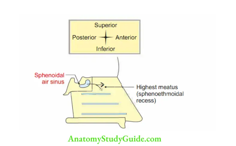
5. Front: Sphenoethmoidal recess.
3. Communication: Sphenoidal air sinus opens into sphenoethmoidal recess,present above superior concha of nose.
4. Blood supply: Posterior ethmoidal vessels (branches/tributaries of ophthalmic vessels).
5. Lymphatic drainage: Retropharyngeal nodes
6. Nerve supply
- Posterior ethmoidal nerve (branch of ophthalmic division of trigeminal nerve).
- Orbital branch of pterygopalatine ganglion.
7. Development: At birth, the sinuses are minute and main development occurs after puberty.
8. Applied anatomy
- Pituitary tumours are commonly approached via sphenoidal air sinus (transnasal approach).
The septal mucous membrane is elevated on both sides.
A part of the septal bones and cartilages are removed.
The nasal conchae are flattened against lateral nasal walls.
The anterior wall and the roof of the sphenoidal sinus are removed to expose the floor of the sella turcica.
Leave a Reply