Packed Cell Volume and Blood Indices Definition
- Packed cell volume (PCV) is the proportion of blood occupied by RBCs expressed in percentage.
- It is the volume of RBCs packed at the bottom of a hematocrit tube when the blood is centrifuged. It is also called hematocrit value or erythrocyte volume fraction (EVF).
Method Of Determination
- Blood is mixed with the anticoagulant EDTA or heparin and filled in hematocrit or Wintrobe’s tube (110 mm long and 3 mm bore) up to 100 mark.
- The tube with the blood is centrifuged at a speed of 3000 revolutions per minute (rpm) for 30 minutes.
- The RBC packed at the bottom is the packed cell volume and the plasma remains above this.
Read And Learn More: Medical Physiology Notes
Table of Contents
- In between the RBCs and the plasma, there is a white buffy coat, which is formed by white blood cells and platelets.
- In laboratories with modern equipment, hemato- crit is not measured directly but calculated indirectly by an autoanalyzer. It is determined by multiplying RBC count by mean cell volume.
- However, some amount of plasma is always trapped between the RBCs. So, accurate value is obtained only by direct measurement of PCV.
Significance Of Determining PCV
Determination of PCV helps in:
- Diagnosis and treatment of anemia
- Diagnosis and treatment of polycythemia
- Determination of extent of dehydration and recovery from dehydration after treatment
- The decision of blood transfusion.
Normal Values Of PCV
The normal PCV:
- In males = 40-45%.
- In females = 38-42%
Variations In PCV
Increase In Pcv: PCV increases in
- Polycythemia
- Dehydration
- Dengue shock syndrome dengue fever (a tropical disease caused by a flavivirus transmitted by the mosquito Aedes aegypti) of grade 3 or 4 severity.
Decrease In PCV: PCV decreases in
- Anemia
- Cirrhosis of liver
- Pregnancy
- Hemorrhage due to ectopic pregnancy (pregnancy in which the fertilized ovum is implanted in tissues other than the uterine wall-characterized by vaginal bleeding).
Blood Indices
Blood indices are specifically meant for erythrocytes. The number, shape, volume, and color of the RBCs indicate the quality of blood. So, these features are named as blood indices.
Importance Of Blood Indices
Blood indices have got diagnostic value in determining the type of anemia.
Different Blood Indices: Following are the various blood indices:
- Mean Corpuscular Volume (MCV)
- Mean Corpuscular Hemoglobin (MCH)
- Mean Corpuscular Hemoglobin Concentration (MCHC)
- Color Index (CI)
1. Mean Corpuscular Volume (MCV)
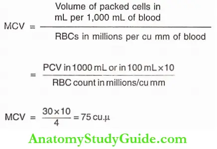
- MCV is the average volume of a single RBC and it is expressed in cubic microns (cu.μ). The normal MCV is 90 cu. μ(78-90 cuμ).
- When MCV is normal, the RBC is called normocyte. When MCV increases, the cell is known as a macrocyte and when it decreases, the cell is called microcyte.
- In pernicious anemia and megaloblastic anemia, the RBCs are macrocytic in nature. In iron deficiency anemia the RBCs are microcytic.
2. Mean Corpuscular Hemoglobin (MCH): It is the quantity or amount of hemoglobin present in one RBC. It is expressed in micro microgram or picogram (pg). The normal value of MCH is 30 pg.
3. Mean Corpuscular Hemoglobin Concentration (MCHC)
- This is the concentration of hemoglobin in one RBC. It is the amount of hemoglobin expressed in relation to the volume of one RBC.
- So, the unit of expression is a percentage. This is the most important absolute value in the diagnosis of anemia. The normal value of MCHC is 30% (30-38%).
- When MCHC is normal, the RBC is normochromic. When the MCHC decreases, the RBC is known hypochromic.
- In pernicious anemia and megaloblastic anemia, RBCs are macrocytic and normochromic or hypochromic.
- In iron deficiency anemia, RBCs are microcytic and hypochromic. A single RBC cannot be hyperchromic because the amount of hemoglobin cannot increase beyond normal.
4. Color Index (CI)
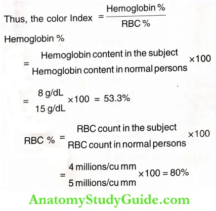
- This is the ratio between the percentage of hemoglobin and the percentage of RBCs in the blood.
- Actually, it is the average hemoglobin content in one cell of a patient compared to the average hemoglobin content in one cell of a normal person.
- The normal color index is 1.0 (0.8-1.2). It was widely used in olden days. However, it is useful in determining the type of anemia.
- It increases in macrocytic (pernicious) anemia and megaloblastic anemia. It is reduced in iron deficiency anemia. And, it is normal in normocytic normochromic anemia.
Calculation Of Blood Indices
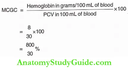
Blood Indices are calculated by using different formula. These calculations require the values of RBC count,
Hemoglobin content and PCV. For example, the values from the blood of a male subject are:
RBC count= 4 million/cu mm
Hemoglobin content = 8 g/dL = 30%
PCV = 30%
Color index, MCV, MCH, and MCHC are calculated as follows:
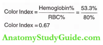
Color Index: This is calculated by dividing hemoglobin percentage by the RBC count percentage
By using these two, CI is calculated
Mean Corpuscular Volume
Mean Corpuscular Hemoglobin
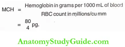
Thus, MCH = 20 pg. or micro microgram.
Mean Corpuscular Hemoglobin Concentration
Result
CI = 0.67 (Normal = 0.8-1 .2)
MCV = 75 cu.μ (Normal = 78-90 cu.μ)
MCH = 20 pg. (Normal = 27-32 pg.)
MCHC = 26.67% (Normal = 30-38%)
The results of these indices indicate that the person is suffering from microcytic hypochromic anemia, which commonly occurs during iron deficiency.
Leave a Reply